Báo cáo sinh học: "Neuronal remodeling on the evolutionary timescale" ppsx
Bạn đang xem bản rút gọn của tài liệu. Xem và tải ngay bản đầy đủ của tài liệu tại đây (140.26 KB, 3 trang )
Minireview
NNeeuurroonnaall rreemmooddeelliinngg oonn tthhee eevvoolluuttiioonnaarryy ttiimmeessccaallee
Ithai Rabinowitch and William Schafer
Address: MRC Laboratory of Molecular Biology, Hills Road, Cambridge CB2 0QH, UK. Email:
One of the hallmarks of the nervous system is its
exceptional capacity to remodel itself through a huge variety
of complex mechanisms occurring at multiple timescales.
Within an individual’s lifetime, parameters such as synaptic
efficacy, membrane excitability and micro-morphology can
undergo major changes during development or as a conse-
quence of learning and memory. Over the much longer
evolutionary timescale, more fundamental remodeling can
take place across species: the number of neurons can be
significantly modified, the gross anatomy can be re-
organized and the specializations of particular neurons and
neuronal circuits can be substantially altered. Given the
fundamental importance of behavior to an organism’s
survival and reproduction, understanding the mechanisms
by which evolutionary changes in brain circuitry modify
behavior is a major challenge in evolutionary biology.
Nematodes offer unique advantages for exploring neuronal
remodeling at the evolutionary timescale. They have
relatively simple nervous systems, typically consisting of
around 300 neurons, and ample information exists on the
phylogenetic relationships among nematode species. In
addition, a complete connectivity map is available for the
widely used model nematode Caenorhabditis elegans [1], and
a significant and increasing body of information exists
about the functional properties of particular neurons in this
organism. Perhaps most unusually, nematode nervous
systems are exceptionally stereotyped in their anatomy,
even across wide evolutionary distances. Not only is neuron
number remarkably consistent across diverse nematode
species; even the arrangement and anatomy of individual
neurons shows extensive conservation [2,3]. Remarkably,
the counterpart of an individual C. elegans neuron can
typically be identified in other nematodes to which C. elegans
is quite distantly related. Thus, evolutionary changes in
nervous system function appear to occur within a consistent
and well defined anatomical framework: all nematode
nervous systems seem to make use of the same complement
of cells in the same overall pattern of organization. The
problem of understanding behavioral evolution therefore
reduces to a much simpler, tractable question: how do
changes in the functional properties of particular neurons
lead to behavioral differences between species?
A new paper in BMC Biology by Srinivasan et al. [4] explores
these questions in the nociceptive circuits that mediate
avoidance of noxious stimuli. Nematodes contain poly-
modal sensory organs called amphids, which contain
ciliated neurons of varying morphologies. The anatomy and
sensory specialization of many of these neurons are
remarkably similar across nematode species [2,5]. In C.
elegans, the sensory modalities of the amphid neurons have
been assessed by cell ablation studies. Seven amphid
neurons extend cilia directly into the amphid channel and
AAbbssttrraacctt
Despite its remarkable capacity to undergo change at timescales ranging from a fraction of a
second to a lifetime, there are many aspects of the nervous system that can be modified only
at the enormously longer evolutionary timescale. A new study in
BMC Biology
using
nematodes illustrates such evolutionary neuronal remodeling.
Journal of Biology
2008,
77::
37
Published: 15 December 2008
Journal of Biology
2008,
77::
37 (doi:10.1186/jbiol102)
The electronic version of this article is the complete one and can be
found online at />© 2008 BioMed Central Ltd
are specialized for tasting soluble attractants or repellents,
three form wing-like cilia at the edge of the channel and are
specialized for olfaction, and one so-called finger cell
projects its cilium into the cuticle and appears to be thermo-
sensory. One neuron, ASH, is unusual in that it has the
morphology of a taste neuron, but is polymodal in its
response properties: ASH is a major neuron for detection of
both soluble and volatile repellents, as well as aversive
touch and osmotic stimuli. In other nematodes, similar
classes of neurons are observed, but fine inter-species differ-
ences in anatomy, such as the number of sensory processes
stemming from each neuron [2,5], as well as variation in
the responses of particular homologous neurons to a
specific stimulus, have been reported [6].
EEffffeecctt ooff nneeuurroonnaall aabbllaattiioonn oonn rreessppoonnssee ttoo nnooxxiioouuss
ssttiimmuullii
In their new study published in BMC Biology Srinivasan et al.
[4] systematically compared the neural circuits involved in
detecting noxious stimuli in six different nematode strains.
To characterize these circuits, they determined which single-
cell ablations affected avoidance of particular stimuli. For
example, nematodes of all species tested showed strong
avoidance of the odorant 1-octanol. In this case, all strains
showed similar ablation phenotypes: killing ASH strongly
impaired octanol avoidance, whereas ablation of other
amphid neurons had no significant effect. Likewise, light
mechanical stimulation of the nose produced comparable
avoidance responses in all species, although habituation
was much faster in one species, Cruznema tripartitum.
However, whereas three neuron types, ASH, FLP and OLQ,
affect nose touch avoidance in C. elegans, in a different
species (Caenorhabditis sp. 3) only ASH is important (Figure
1a). A similar but opposite effect was observed for osmotic
avoidance, which in C. elegans is mediated solely by ASH,
but was found to involve the ADL and ASH neurons in
Pristionchus pacificus (Figure 1b). Surprisingly, P. pacificus
was one of several species tested that responded more
weakly to the high osmotic stimulus despite the extra
neurons in its circuit. A clustering analysis based on the
avoidance responses of the various species in the study
revealed not only examples of correlation between
behavioral similarities and phylogenetic proximity, but also
cases of greater behavioral differences between closely
related species than between more distantly related ones.
Thus, evolutionary remodeling of these sensory circuits
might occur readily in response to natural selection.
What do ablation results tell us about how nociceptive
circuits have been remodeled during nematode evolution?
One possibility is that particular neurons might alter or
even lose functionality in the course of evolution. One
should be cautious, however, as the components of a neural
circuit are not necessarily limited to those neurons whose
ablation early in development impairs the circuit’s function.
During development, an ablated animal can sometimes
compensate for a missing neuron, for example by reorgani-
zing the remaining neurons in the circuit. Moreover, recent
examples demonstrate that it can be easier for a circuit to
37.2
Journal of Biology
2008, Volume 7, Article 37 Rabinowitch and Schafer />Journal of Biology
2008,
77::
37
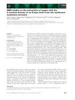
FFiigguurree 11
Evolutionary neuronal remodeling between nematode strains.
((aa))
In
C. elegans
three sets of neurons, ASH, FLP and OLQ, mediate aversion to light
mechanical stimulation of the nose (top). The same response was found to require ASH alone in
C.
sp. 3 (bottom).
((bb))
In
C. elegans
, only the ASH
neurons are necessary for sensing high osmotic stress (top). This response was sensed in
P. pacificus
by the ADL neurons in addition to the ASH
neurons (bottom). Arrows indicate the direction of the response.
(a) (b)
Nose touch avoidance Osmotic stress avoidance
FLP
ASH
FLP
ASH
ADL
OLQ
FLP
ASH
ADL
C.
OLQ
FLP
ASH
ADL
P.
OLQ
ASH
ADL
OLQ
ASH
C. elegans
FLP
ASH
OLQ
FLP
ASH
OLQ
FLP
ASH
C.
FLP
ASH
C. sp. 3
OLQ
ASH
ADL
P.
ASH
ADL
P. pacificus
C. elegans
compensate for a missing neuron than for an inactive one,
even when the neuron’s function is absent throughout
development [7,8]. Ablation studies can be said to define
the group of neurons whose functions are most critical for a
given behavior. Thus, if ablation of a neuron no longer affects
the function of a particular circuit, this might not indicate a
change in the overall function of the neuron, but might
indicate its importance or dispensability for the circuit.
Another recent study comparing feeding behavior in four
nematode species [9] provides some insight into how such
changes might occur. Nematodes feed by pumping food
through a muscular pharynx, which is controlled by the
pharyngeal nervous system. Three motor neurons (MC, M3
and M4) appear to have particularly important roles in
controlling pharyngeal contraction in all species. However,
in one species, Panagrolaimus sp. PS1159, a fourth motor
neuron, M2 (which has no known function in the other
species), has apparently acquired a role in controlling
contraction of the pharyngeal isthmus. Likewise, the M4
neuron controls contraction of the pharyngeal isthmus and
terminal bulb in most species; in C. elegans, however, it
appears to have lost the latter function. Interestingly, the
mechanism for this change in M4 function appears to
involve silencing of M4’s terminal bulb synapses during
evolution. It is possible that similar types of change might
occur in sensory circuits to reconfigure the roles of
individual neurons in particular sensory modalities.
Clearly, ablation studies are only a first step in understand-
ing how behavior evolves in nematodes. With modern
electron microscopy and computational methods, it should
be practical to reconstruct the neuroanatomies of other
nematodes at the single-cell level and compare the connect-
ivity patterns with those of C. elegans. With the develop-
ment of transgenesis protocols for other nematode species
[10], it will also be possible to use genetically encoded
sensors to probe the activity patterns of homologous neural
circuits in a range of nematodes. In the near future, there is
a real possibility of understanding the detailed genetic and
cellular mechanisms by which nematode nervous systems
are remodeled during evolution.
RReeffeerreenncceess
1. White J, Southgate E, Thomson J, Brenner S:
TThhee ssttrruuccttuurree ooff tthhee
nneerrvvoouuss ssyysstteemm ooff tthhee nneemmaattooddee
CCaaeennoorrhhaabbddiittiiss eelleeggaannss
Phil Trans
R Soc Lond
1986,
331144::
1-340.
2. Ashton FT, Li J, Schad GA:
CChheemmoo aanndd tthheerrmmoosseennssoorryy nneeuurroonnss::
ssttrruuccttuurree aanndd ffuunnccttiioonn iinn aanniimmaall ppaarraassiittiicc nneemmaattooddeess
Vet Parasitol
1999,
8844::
297-316.
3. Forbes WM, Ashton FT, Boston R, Zhu X, Schad GA:
CChheemmooaatt
ttrraaccttiioonn aanndd cchheemmoorreeppuullssiioonn ooff
SSttrroonnggyyllooiiddeess sstteerrccoorraalliiss
iinnffeeccttiivvee
llaarrvvaaee oonn aa ssooddiiuumm cchhlloorriiddee ggrraaddiieenntt iiss mmeeddiiaatteedd bbyy aammpphhiiddiiaall
nneeuurroonn ppaaiirrss AASSEE aanndd AASSHH,, rreessppeeccttiivveellyy
Vet Parasitol
2004,
112200::
189-198.
4. Srinivasan J, Durak O, Sternberg PW:
EEvvoolluuttiioonn ooff aa ppoollyymmooddaall
sseennssoorryy rreessppoonnssee nneettwwoorrkk
BMC Biol
2008,
66::
52.
5. Bumbarger DJ, Wijeratne S, Carter C, Crum J, Ellisman MH,
Baldwin JG:
TThhrreeee ddiimmeennssiioonnaall rreeccoonnssttrruuccttiioonn ooff tthhee aammpphhiidd sseenn
ssiillllaa iinn tthhee mmiiccrroobbiiaall ffeeeeddiinngg nneemmaattooddee,,
AAccrroobbeelleess ccoommpplleexxuuss
((NNeemmaattooddaa:: RRhhaabbddiittiiddaa))
J Comp Neurol
2009,
551122::
271-281.
6. Ketschek AR, Joseph R, Boston R, Ashton FT, Schad GA:
AAmmpphhiiddiiaall nneeuurroonnss AADDLL aanndd AASSHH iinniittiiaattee ssooddiiuumm ddooddeeccyyll ssuullpphhaattee
aavvooiiddaannccee rreessppoonnsseess iinn tthhee iinnffeeccttiivvee llaarrvvaa ooff tthhee ddoogg hhooookkwwoorrmm
AAnnccllyyoossttoommaa ccaanniinnuumm
Int J Parasitol
2004,
3344::
1333-1336.
7. Kindt KS, Viswanath V, Macpherson L, Quast K, Hu H, Patapoutian
A, Schafer WR:
CCaaeennoorrhhaabbddiittiiss eelleeggaannss
TTRRPPAA 11 ffuunnccttiioonnss iinn
mmeecchhaannoosseennssaattiioonn
Nat Neurosci
2007,
1100::
568-577.
8. Li W, Feng Z, Sternberg PW, Xu XZ:
AA
CC eelleeggaannss
ssttrreettcchh rreecceepp
ttoorr nneeuurroonn rreevveeaalleedd bbyy aa mmeecchhaannoosseennssiittiivvee TTRRPP cchhaannnneell hhoommoo
lloogguuee
Nature
2006,
444400::
684-687.
9. Chiang JT, Steciuk M, Shtonda B, Avery L:
EEvvoolluuttiioonn ooff pphhaarryynnggeeaall
bbeehhaavviioorrss aanndd nneeuurroonnaall ffuunnccttiioonnss iinn ffrreeee lliivviinngg ssooiill nneemmaattooddeess
J
Exp Biol
2006,
220099::
1859-1873.
10. Li X, Massey HC, Jr., Nolan TJ, Schad GA, Kraus K, Sundaram M,
Lok JB:
SSuucccceessssffuull ttrraannssggeenneessiiss ooff tthhee ppaarraassiittiicc nneemmaattooddee
SSttrroonnggyy
llooiiddeess sstteerrccoorraalliiss
rreeqquuiirreess eennddooggeennoouuss nnoonn ccooddiinngg ccoonnttrrooll eellee
mmeennttss
Int J Parasitol
2006,
3366
:671-679.
/>Journal of Biology
2008, Volume 7, Article 37 Rabinowitch and Schafer 37.3
Journal of Biology
2008,
77::
37