Báo cáo khoa học: "Comparison of Diagnostic Methods for the Detection of Parasites in Fish" pdf
Bạn đang xem bản rút gọn của tài liệu. Xem và tải ngay bản đầy đủ của tài liệu tại đây (141.7 KB, 9 trang )
Journal of Science and Development April 2008: 136-144 HANOI UNIVERSITY OF AGRICULTURE
Comparison of Diagnostic Methods for the Detection of
Parasites in Fish
Kim Van Van
*
& Dinh Thi Thuy
**
*
Faculty of Animal and Aquacultural Science, Hanoi University of Agriculture
**
Research Institute for Aquaculture II
Abstract
In recent years, Aquaculture has developed very rapidly. However, fish parasitic diseases in
fry and fingerling occur often. There are many methods which were used to diagnose fish
parasites. In this paper, fifty wild fish belonging to three fish species: roach (Rutilus rutilus),
perch (Perca fluviatilis) and bream (Blicca bjoerkna) were collected from Arreso Lake in
Copenhagen, Denmark in 2005 to diagnose parasites. Parasitological investigation was
implemented by normal observation, compression, digestion and PCR methods at the Fish
Disease Laboratory, Pathology Department, Life Science University, Copenhagen, Denmark. The
results show a high prevalence of eye fluke metacercaria in wild fish (100% of Blicca bjoerkna
infected by Diplostomum sp.). Each method has advantages and disadvantages. The classical
methods are simple, cheap and easy to apply in every fish laboratory. PCR methods produced
results rapidly, sensitively and exactly. But this method costs much for equipment, and
chemicals and needs exacting technique.
Keywords: Parasites, fish, diagnostic.
1. INTRODUCTION
World aquaculture production now
accounts for 32% of total fisheries production,
according to the FAO (2005). Globally, fish
provide about 15% of all the animal proteins
consumed, with variations from an average of
23% in Asia to approximately 18% in Africa
and around 7% in Latin America. Total world
fisheries production in 2003 was 132.5 million
tonnes, of which 42.3 million tonnes were from
aquaculture and 90.2 million tonnes were from
capture fisheries. Total fish production has
increased in recent years, mainly due to
improvements in the aquaculture industries.
However, intensive aquaculture systems with
high stocking densities are vulnerable to
infectious diseases.
Parasitic diseases in fish have become
increasingly prevalent during the past few
decades, in parallel with the growth and
development of aquaculture industries
throughout the world. Disease problems,
including hazards caused by parasitic organisms,
are the biggest threat to the continuing
development of the industry (Buchmann, 2001).
Fish parasitology is a rapidly expanding area, as
Gyrodactylus salaris was introduced to Norway
in the 1970s. Since its introduction in Norway
the parasite has spread to a total of 45 salmon
rivers. The affected salmon populations have
experienced a significant decrease as a result
(Buchmann, 2004).
The increasing importance of aquaculture
products, including farmed fish, has emphasized
the need for health control and proper fish
disease diagnosis. Parasitological methods are
vitally important for the parasitological study of
fish. There are a wide variety of parasitological
methods, and each method has its advantages
136
Comparison of Diagnostic Methods for the Detection of Parasites in Fish
and disadvantages, depending on the purpose
and target of study. For parasitological
investigation, the classical methods (the normal
observation, compression, and digestion
methods) have been applied. To find blood
parasites, the blood smear preparation or wet
blood method has been used. PCR (Polymerase
Chain Reaction) is a new method for parasite
diagnosis. The use of the PCR method has
allowed links to be elucidated between the
various developmental stages such as cercariae,
metacercariae and adults of specific trematodes
(Cribb et al., 1998; Jousson et al., 1998;
Anderson, 1999; Bartoli et al., 2000).
The objectives of the present study were to
investigate the use of different methodologies
in fish parasite studies. Thus, the aim is to
compare classical and molecular methods for
the diagnosis of fish parasites.
2. MATERIALS AND METHODS
2.1 Fish samples
Fish samples were collected during
November, 2005 from Arreso lake,
Copenhagen, Denmark by local fishermen.
Three species were used including roach
(Rutilus rutilus), perch (Perca fluviatilis) and
bream (Blicca bjoerkna). A total of 50 fish were
examined (Figure 1).
2.2 Dissection of the fish
Fish species were identified, anaesthetized
by MS 222 (100 ppm) and killed by cervical
dislocation. Each fish was weighed (gram),
measured (cm) and recorded. Gills, fins, the
nostril, and scales were taken off; the eyes were
removed from the fish and opened; then the lens
and vitreous humour were exposed. All these
organs were placed separately in petri dishes
with PBS (phosphate buffered saline pH 7.0).
The internal organs were exposed after a vertical
incision was made from the anal opening to the
lateral line and to the operculum (Buchmann &
Bresciani, 2001). Liver, gall bladder, spleen,
oesophagus, stomach, pyloric caeca, intestine,
gonads, swim bladder, and urine bladder were
cut and placed separately in petri dishes
containing PBS.
Figure 1. Fish samples used for parasitological
methods
2.3 Parasitological investigation
The normal observation
Before any dissection, the exterior of the
fish was observed under the dissecting
microscope at 7x-40x magnification. Scrapings
of the body surface were done with a cover slip
to remove epithelial cells and mucus with
parasites for examination in the compound
microscope (40x-1000x). The fins, gills, eye
lenses and vitreous humour were examined in
the dissecting microscope (7x-40x). All
parasites were recovered and placed in separate
vials with PBS.
The content of selected separate organs
(oesophagus, stomach, pyloric caeca and
intestine) was scraped from the lumen and
epithelial lining and inspected under the
microscope. Parasites were found and
transferred by pipettes, pincers or forceps to
separate glass vials with PBS. In addition,
137
Kim Van Van & Dinh Thi Thuy
parasites were kept in Eppendorf tubes with
ethanol (70%) or neutral formalin (4%).
The compression method
Different parts of the fish (muscles, fins,
gonad, liver, spleen, etc.) were taken. Each part
was compressed between 2 glass slides. Thus,
by applying a little pressure to the tissue it is
flattened until the presence of parasites is
revealed (Buchmann, 2005). The two glass
slides were placed under the dissecting
microscope (7x-40x magnification). Parasites
were observed, recovered and placed in
separate vials with PBS. For later study, all of
the parasites were kept in Eppendorf tubes with
ethanol (70%) or neutral formalin (4%).
The digestion method
For larger fish, different parts (e.g. fins,
muscles, and bone structures) were taken. For
small fish, the whole fish body or whole head
(except eyes) were used. Then, each different
part of each fish was weighed, ground in a
mortar with pestle and transferred into a beaker
(1:5 to 1:10 w/v) with pepsin solution (2%
pepsin, pH 2) at acid conditions. They were
mixed well and placed in a 37
o
C incubator for
2-3 hours (longer for hard tissues) with
occasional stirring. Samples were added to
saline water (0.85%), shaken, and allowed to
settle. Digest was poured through a 1x1 mm
mesh brass sieve, washed with saline and
settled until sediment was easily observed. The
supernatant part was discarded very carefully
and the sediment kept. This procedure was
repeated several times (typically between seven
and eight) or until the supernatant became clear.
The encysted metacercariae were found and
isolated. Then, these encysted metacercariae
were excysted using a trypsin solution at
slightly basic conditions (0.5% Bile: 0.25%
Trypsin: 0.5% Chymotrypsin; pH: 8.4), and
placed in a 37
o
C incubator for 5-10 minutes
(Buchmann, 2005). The metacercariae out of
the cyst were collected and placed in separate
vials with physiological saline. They were
observed and identified using a compound
microscope. Stretching of these metacercariae
was done by hot formalin for two minutes.
Then, they were kept in Eppendorf tubes with
neutral formalin (4%).
Diagnosis of parasitic infections
* Diagnosis based on morphological criteria
Morphological characteristics of parasites
are important values. Features observed were
shape, total length and width, external
structures of parasites (spines, lobes, etc.),
different appendices, sclerotinized structures
(hamuli, attachment hook, etc.), sex organs
(testes or ovaries). Parasite morphological
diagnosis followed the key of Bykhovskaya-
Pavlovskaya et al. (1964). Infection was
described by prevalence (the percentage of the
hosts which are infected with a certain parasite)
and mean intensity (the mean number of
parasites in the infected fish only) (Buchmann
& Bresciani, 2001).
* Diagnosis based on PCR techniques
Metacercariae of eye flukes were collected
from fish eyes, other metacercariae were
collected by digestion method in the fish
parasitological laboratory of KVL and
preserved in 70% ethanol.
- Extraction of total genomic DNA
Total genomic DNA was extracted using
commercial DNA extraction kits (QIAamp
DNA kit, Qiagen Inc., USA). The extracted
genomic DNA used as the template in PCR
reactions was diluted to a final concentration of
100-150 ng/µl, and the template for this
concentration was used in a normal PCR
reaction of 50 µl volume (25µl of master mix,
138
Comparison of Diagnostic Methods for the Detection of Parasites in Fish
2µl of each primer, 1 or 3 µl of template and 18
or 20 µl of water).
- Polymerase Chain Reaction (PCR)
A master mix was prepared for PCR in a 1.5
ml Eppendorf tube which included H
2
0, PCR
buffer (10X), dNTPs, Primer 1 (forward) NC2:
5’-TTAGTT TCT TTT CCT CCG CT-3’ and
Primer 2 (reverse) NC5: 5’-GTA GGT GAA
CCT GCG GAA GGA TCATT-3’(Maniatis et
al., 1989). All materials were kept on ice all the
time. The master mix was divided with 50 µl
going into each of the PCR tubes. One or 3 µl of
DNA was added. PCR was performed in the
PCR machine (Gene Amp PCR system 9700)
with an initial 95
o
C step for 5 minutes and 30
cycles of denaturation at 94
o
C for 30 seconds,
annealing at 55
o
C for 30 seconds and extension
at 72
o
C for 30 seconds; followed by a final
extension at 72
o
C for 7 minutes.
- Gel loading: 1.5% agarose in 10% TAE
buffer was placed in an erlenmeyer flask. Then it
was placed in the microwave on full power until
boiling (2 minutes). It was mixed again and
placed once again in the microwave on full
power until boiling. It was cooled to 45-50
o
C
(not hotter to avoid plastic deformation) on the
table and poured into the gel frame which had
been sealed at the ends with autoclave tape. The
gel comb making the wells was added. The gel
then polymerized. The combs were removed and
the gel was placed in the electrophoresis
chamber. One x TAE buffer was poured into the
chamber until the gel was covered. Each well
received 3 µl of loading buffer and 5 µl of the
digested product or 5 µl undigested PCR
product. The first and last lanes on the gel were
loaded with 6 µl size markers (100bp). The
samples were run for about 45 minutes at 100 V.
The gel was stained 20 minutes in TAE buffer,
which contained ethidium bromide (0.01%). The
DNA bands were visualized under UV
illumination and a photo of the gel was taken by
the machine. The gel was discarded in special
containers for toxic material. After that, the
banding pattern was analyzed.
3. RESULTS
Fish parasite prevalence
By the normal observation method, 32 fish
of the three species were examined. The
parasite prevalence was 59.4%. All bream
samples were infected with parasites, while
parasite prevalence of perch and roach samples
ranged from 53.3 to 57.1% (Table 1).
Table 1. The results of fish parasitological examination by normal observation (for all parasites).
Name of fish species
No. of fish
examined
Total body length
(cm)(SD)
Total body
weight (g) (SD)
No. of fish
infected with
parasites
Prevalence
parasites
Perch Perca fluviatilis 15 10.24 ± 4.59 14.61± 9.94 8 53.3 %
Bream Blicca bjoerkna 3 14.33 ± 6.66 18.67± 6.11 3 3/3
Roach Rutilus rutilus 14 10.66 ± 2.81 14.64 ±7.65 8 57.1 %
Total 32 11.77±4.69 15.97±7.9 19 59.4 %
Eighteen samples of the three fish species
were examined by the digestion method; the
prevalence of metacercaria infection was
27.8%. The prevalence between fish species
ranged from 2/8 to 1/3 (Table 2).
139
Kim Van Van & Dinh Thi Thuy
Table 2. The result of fish metacercariae testing by the digestion method (for all metacercaria).
Name of fish species
N
o
of fish
examined
Total body
length
(cm)(SD)
Total body weight
(g) (SD)
N
o
of fish infected
with
metacercariae
Prevalence
metacercaria
infection
Perch Perca fluviatilis 7 12.27± 1.68 21.71± 7.66 2 2/7
Bream Blicca bjoerkna 3 11.83± 0.76 15.23± 2.36 1 1/3
Roach Rutilus rutilus 8 15.30± 3.89 49.11± 4.12 2 2/8
Total 18 13.13±2.11 28.68±4.71 5
27.8%
Three fish species with a total of 6 samples
were tested by the compression method, no
parasites were found.
3.2 Results of parasitological examinations
Results from the normal observation
method are shown in Table 3. In perch and
roach, the prevalence was 13.3%, 28.6%
respectively (eye lens), and 26.7% and 42.9%
respectively (vitreous humour). In bream
samples, this value was in all samples (eye
lens) and 1/3 (vitreous humour). Diplostomum
sp. was identified from eye lenses, and
Tylodelphys sp. was identified from vitreous
humours of all three fish species. In addition,
other parasites were found including
tapeworm, crustaceans, roundworm,
metacercariae of trematodes. The crustacean
Argulus sp. was found on the skin of bream
and roach with a prevalence of 1/3 (bream)
and 7.1% (roach). Tapeworm was found in the
intestine of perch with a prevalence of 15%. A
roundworm Philometra sp. was found on roach
fins with a 7.1% prevalence. There was a 20%
prevalence of metacercariae1 (trematodes)
present in the abdominal cavity of perch. The
names of cestodes1 and metacercariae1 were
not determined. The mean intensity of eye
flukes of bream ranged from 13.3 to 16.0,
perch from 5.0 to 8.5 and in roach from 3.3 to
6.7. Intensities of other parasites had low
values, ranging from 1 to 4.
Table 3. Parasites recovered by normal observation method.
Fish Species Parasite species Infected
organs
Number of
fish examined
Number of
infected fish
Prevalence Mean
intensity
Diplostomum sp. Eye lens 15 2 13.3% 5.0
Tylodelphys sp. V. humour 15 4 26.7% 8.5
Metacercariae
1
A. cavity 15 3 20.0% 3.3
Perch
Perca
fluviatilis
Cestodes
1
Intestine 15 1 15.0% 1.0
Diplostomum sp. Eye lens 3 3 3/3 13.3
Tylodelphys sp. V. humour 3 1 1/3 16.0
Bream
Blicca
bjoerkna
Argulus sp. Skin 3 1 1/3 4.0
Diplostomum sp. Eye lens 14 4 28.6% 3.3
Tylodelphys sp. V. humour 14 6 42.9% 6.7
Philometra sp. Fins 14 1 7.1% 1.0
Roach
Rutilus rutilus
Argulus sp. Skin 14 1 7.1% 1.0
V. humour: Vitreous humour; A. cavity: Abdominal cavity; Metacercariae
1
, Cestodes
1
.
140
Comparison of Diagnostic Methods for the Detection of Parasites in Fish
With the use of the digestion method,
metacercaria 2 was found from fins. When
bream (whole fish, except fin) was digested, a
cyst1 was found; metacercariae were found in
roach at a 33.3% prevalence. No parasites were
found in perch. In roach, the metacercaria 3, 4
could be Opisthorchis sp The infection
prevalence with metacercariae 3 was 12.5%
(whole head), and 12.5% for metacercariae 4
(whole head except eyes) (Table 4). Mean
intensity of metacercariae ranged from 1 to 5
(Table 4).
Table 4. Parasites recovered by the normal observation method.
Fish Species Parasite species Infected organs
Number of
fish
examined
Number of
infected fish
Prevalence (%)
Mean
intensity
Bream
Blicca bjoerkna
Cyst Whole fish
(except fins)
3 1 33.3 3.0
Metacercariae Fins 8 2 25.0 2.0
Metacercariae Whole head
(except eyes)
8 1 12.5 1.0
Roach
Rutilus rutilus
Metacercariae Whole head 8 1 12.5 5.0
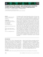
3.3 The use of the PCR method for
identification of trematodes with the NC2-
NC5 primer pair
The results from testing eye flukes by the
PCR method are shown in Figure 2. The NC2-
NC5 primer pair shows the difference between
metacercariae in vitreous humour of eyes (one
band) and metacercariae in lens of eyes (two
bands). No difference between 1 and 3 µl of
DNA was observed.
Figue 2. Testing of NC2-NC5 primer pair from
eye flukes lanes 1 & 3: metacercariae of
Tylodelphys sp.; lans 2 & 4: metacercaria of
Diplostomum sp.
4. DISCUSSION
The normal observation, compression and
digestion methods were the simplest methods
for examining parasite infections of fishes. In
this study, with the normal observation method,
each fish species attained over 50% parasite
prevalence. The parasites discovered were
found in different organs, such as eye lens,
vitreous humours, intestine, fins, skins and the
abdominal cavity of fish. The detected parasite
types include eye flukes, tapeworm,
roundworm, crustacean and metacercariae of
trematodes. However, names of cyst1,
metacecaria1,2 and cestodes1 were not
determined because their morphological
characteristics were damaged after the
examination. The mean intensity of eye flukes
was almost higher than the other parasite types.
Some advantages of the normal observation
method were recorded, it is easy to do and easy
to apply at a fish farm, cheap, no need for any
chemicals for examination. The compression
and digestion methods were also applied. Thus,
metacercariae of digeneans, third stage larvae
of nematodes, plerocercoids of cestodes, cysts
141
Kim Van Van & Dinh Thi Thuy
of myxosporeans may be hidden in different
types of tissues. The compression technique can
be used to obtain a fast and preliminary visual
impression (Buchmann, 2005). Some other
advantages were also recorded from this
technique. The exact location, or infection site,
of metacercariae can be determined. It is
economical, without the need to use expensive
reagents. Features of the host tissue wall
surrounding the metacercarial cyst can be useful
in identification, but this can be lost in
digestion.
The digestion method is also applied when
parasitic stages of various species are difficult
to discern, and a number of parasite forms are
located in fish tissue such as fins, flesh, skin,
etc. Cyst1, metacercaria2,3,4 were the parasitic
stages of various species identified by this
method. With this technique: a large number of
samples can be processed; metacercariae can be
isolated and collected; and exact numbers of
metacercariae can be prepared for experimental
infection. This method was previously used to
estimate the number of Cryptocotyle spp.
metacercariae in the skin of fish (Lysne, 1995).
Some encysted metacercariae were found and
were excysted by artificial digestion (trypsin
solution) using this technique. Morphology was
excellent and aided further identification.
It is often difficult to identify different
stages of trematodes based on morphology
(eggs, cercariae, metacercariae and adult
worms). Eggs in the faeces of the definitive
hosts have been difficult to identify due to the
fact that the eggs are very small and can not be
assigned to a specific species using light
microscopy (Pauly et al., 2003). To find a
relationship between metacercariae and adult
worms, it is often necessary to conduct an
infection experiment with sensitive final hosts.
Such work takes a lot of time and money. The
PCR method can help in this regard (Sirisinha
et al., 1991). DNA technology has had a major
impact in many areas of parasitology,
including the identification and classification
of parasites, the diagnosis of infections, the
epidemiology of parasites, the analysis of
population genetic structures, gene expression
and organization, the study of drug resistance
and vaccine development. In particular, the
advent of the PCR has revolutionized
parasitological research and has found broad
applicability, mainly because its sensitivity
permits the amplification of genes or gene
fragments from minute amounts of parasite
material. While specific determination of
larval stages by morphological traits is often
difficult and ambiguous, experimental
demonstration of the life history is frequently
unachievable due to the unidentified nature of
the specific intermediate or definitive host.
The use of molecular methodologies has
allowed links to be elucidated between the
various developmental stages as cercariae,
metacercariae and adults of specific
trematodes (Cribb et al., 1998; Jousson et al.,
1998; Anderson, 1999; Bartoli et al., 2000).
Currently the morphological characteristics of
either the metacercariae recovered from fish or
adult worms from humans are
indistinguishable, and limited information on
genetic studies is available. Up to now, the
detection of eggs, cercariae, metacercariae and
adult worms of certain species has been
implemented by the PCR method. PCR assays
have proven useful in demonstrating genetic
links between metacercariae and adult worms
of Heterophyidae species. These tools may be
used for early diagnosis as they were shown to
be sensitive in the identification of early
infection in fish and useful for studying
trematode life history. The ITS rDNAregion
have been utilized for species-specific
identification (Cribb et al., 1998; Jousson et
142
Comparison of Diagnostic Methods for the Detection of Parasites in Fish
al., 1998; Anderson, 1999). Our primer sets
were designed for identification of different
flukes and they were useful for detection of
eye flukes. Although the PCR method gives
rapid, sensitive and exact results, it is still a
new method in parasitic studies so many
things are still limited, such as primer design
or the PCR process, and this method requires a
lot of money to be spent on expensive
equipment and chemicals. Thus, it is difficult
to develop in poor countries. For parasitic
studies need to combine all the convenient
methods well.
5. CONCLUSIONS
During the time devoted to the practical
work of parasitological methods, three wild fish
species, with a total of 50 fish, were collected
and tested for parasites using the following
classical methods: normal observation,
compression, and digestion methods. These
methods are simple, cheap and easy to apply in
every fish laboratory. A new and model
method, PCR, has been implemented for
detection of metacercariae of Tylodelphys sp.
and Diplostomum sp. by the NC2-NC5 primer
pair.This method produced results rapidly,
sensitively and exactly. But until now, this
method has had some limitations due to the
primer design or PCR process for parasite
studying, which costs a lot of money.
6. ACKNOWLEDGEMENTS
Thanks to Kurt Buchmann for his help,
Kurt is not only a supervisor in this subject but
also a PhD supervisor. Thanks also to Henrik
Christensen for his help with the PCR method.
Thanks to DANIDA and FIBOZOPA for
funding, and finally, thanks to all the lecturers
and students in the Parasitological method
course.
7. REFERENCES
Anderson, G. R. (1999). Identification and
maturation of the metacercaria of
Indodidymozoon pearsoni. J Helminthol
73: 21-26.
Bartoli, P., O. Jousson, F. Russell-Pinto (2000).
The life cycle of Monorchis parvus
(Digenea: Monorchiidae) demonstrated
by developmental and molecular data. J
Parasitol 86(3): 479-489.
Buchmann, K., and J. Bresciani (2001). An
introduction to Parasitic Diseases of
Freshwater Trout. The Royal Veterinary
and Agricultural University, Denmark.
Buchmann, K. (2004). Diagnosis and Control of
Fish Diseases. SCOFDA workshop,
November 3 and 4, 2004. Frederiksberg
Bogtrykkeri, Frederiksberg.
Buchmann, K. (2005). An introduction to
Practical Methods in Fish Parasitology
Classical and Molecular Techniques.
KVL, Copenhagen.
Bykhovskaya- Pavlovskaya, I.E., A.V. Gusev,
M.N. Dubinina, N. A. Izyumova, T.S.
Smirnova, I. L. Sokolovskaya, G.A.
Shtein, S. S. Shul’man, V.M. Epshtein
(1964). Key to Parasites of Freshwater
Fish of the U.S.S.R. Academy of Science
of the U.S.S.R. Zoological Institute.
Israel Program for Scientific
Translations, Jerusalem, 1964.
Cribb, T. H., G. R. Anderson, R. D. Adlard, and
R. A. Bray (1998). A DNA based
demonstration of a three host life-cycle
for the Bivesiculidae (Platyhelminthes:
Digenea). Int J Parasitol 28: 1791-1795.
Jousson, O., P. Bartoli, L. Zaninetti, and J.
Pawlowski (1998). Use of the ITS rDNA
143
Kim Van Van & Dinh Thi Thuy
for elucidation of some life cycles of
mesometridae (Trematoda, Digenea). Int
J Parasitol 28: 1403-1411.
Lysne, D.A., W. Hemmingsen, and A. Skorping
(1995). Pepsin digestion reveals both
previous and present infections of
metacercariae in the skin of fish. Fish
Res 24: 173-177.
Maniatis, T., E.F. Fritsch, J. Sambrook (1989).
Molecular Cloning - A laboratory
manual. Press New York.
Pauly, A., R. Schuster, and S. Steuber (2003).
Molecular characterization and
differentiation of opisthorchiid
trematodes of the species Opisthorchis
felineus (Rivolta, 1884) and Metorchis
bilis (Braun, 1790) using polymerase
chain reaction. Parasitol Res 90: 409-414.
Sirisinha, S., R. Chawengkirttikul, R.
Sermswas, S. Amornpant, S.
Mongkolsuk, and S. Panyim (1991).
Detection of Opisthorchis viverrini by
monoclonal antibody-based ELISAand
DNAhybridization. Am J Trop Med Hyg
24: 833-43.
/>php. Fish Diseases II -Diagnosis and
Possible Cures.
http:// www.fishupdate.com: 9 December,
(2005). FAO report: China responsible
for two-thirds of world aquaculture
production.
144