Báo cáo sinh học : "How do terrestrial Antarctic organisms survive in their harsh environment" ppt
Bạn đang xem bản rút gọn của tài liệu. Xem và tải ngay bản đầy đủ của tài liệu tại đây (119.85 KB, 4 trang )
Minireview
HHooww ddoo tteerrrreessttrriiaall AAnnttaarrccttiicc oorrggaanniissmmss ssuurrvviivvee iinn tthheeiirr hhaarrsshh
eennvviirroonnmmeenntt??
David A Wharton* and Craig J Marshall
†
Addresses: *Department of Zoology and
†
Department of Biochemistry, University of Otago, PO Box 56, Dunedin 9054, New Zealand.
Correspondence: David A Wharton. Email:
Antarctic terrestrial organisms live permanently on the
continent (unlike penguins and seals that only breed there)
and survive in one of the harshest environments on Earth.
Sites that support life are largely limited to regions that are
ice free, for at least part of the year, and which receive
meltwater in spring and summer. Living at the limits of life,
these organisms may be particularly sensitive indicators of
climate change and are good models for studying how life
survives in extreme environments. Antarctic species show
high levels of endemicity and recent molecular studies
suggest that many terrestrial Antarctic organisms have
ancient origins, dating from before the break up of
Gondwana [1]. Although controversial, there is increasing
interest in bioprospecting amongst Antarctic organisms for
molecules with practical uses.
LLiiffee wwiitthhoouutt wwaatteerr
Although the most obvious stress faced by organisms in
Antarctica is cold and the risk of freezing, there are a variety
of other stressors that are significant [2]. The most
important factor determining their distribution is the
presence of liquid water, to which organisms must have at
least occasional access in order to grow and reproduce.
When liquid water is absent organisms survive in a dormant
state known as anhydrobiosis - life without water - in which
their metabolism comes reversibly to a standstill.
Anhydrobiosis is a feature of many organisms in habitats
where they are exposed to desiccation. Among animals, it is
found in rotifers, tardigrades, nematodes and some
arthropod larvae. Many species of nematodes are capable of
anhydrobiosis and nematodes have proved to be good
models for the study of this phenomenon. Anhydrobiotic
nematodes are important components of the Antarctic
terrestrial fauna [3].
The disaccharide trehalose has long been thought to be
important for anhydrobiosis; especially by acting as a
replacement for water, preserving the function of
membranes and proteins. More recently, other mechanisms
have been recognized. In particular, a group of proteins
called late embryogenesis abundant (LEA) proteins, first
identified from plant seeds, are associated with anhydro-
biosis in a number of animals. They may play a role in
preventing protein aggregation during desiccation [4].
However, focusing on specific adaptations, such as trehalose
AAbbssttrraacctt
Anhydrobiosis, or extreme desiccation tolerance, is one of the strategies that allows
terrestrial Antarctic organisms to survive in a harsh environment. A new study in
BMC
Genomics
analyses gene expression in an Antarctic nematode during desiccation, and sheds
new light on this phenomenon.
Journal of Biology
2009,
88::
39
Published: 29 April 2009
Journal of Biology
2009,
88::
39 (doi:10.1186/jbiol142)
The electronic version of this article is the complete one and can be
found online at />© 2009 BioMed Central Ltd
and LEA proteins, may result in important mechanisms
being overlooked. The construction and screening of cDNA
libraries and cDNA arrays have proved successful in identi-
fying freezing-responsive gene expression in a freezing-
tolerant frog, Rana sylvatica [5]. Similar approaches have
been used to study the responses of plants to a variety of
stressors, including desiccation, and have been applied to
desiccation survival and anhydrobiosis in nematodes [6].
An Arctic springtail (Collembola), Onychiurus arcticus, over-
winters by desiccating at low temperatures (cryoprotective
dehydration). An expressed sequence tag (EST) analysis has
indicated that a number of biochemical pathways are
associated with desiccation and recovery [7].
A recent paper in BMC Genomics describes the first EST
and differential expression analysis of the response of a
terrestrial Antarctic animal to environmental stress [8].
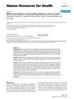
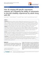
In this work, Adhikari et al. describe changes in gene
expression in response to desiccation in the free-living
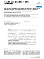
nematode Plectus murrayi (Figure 1). This nematode is
found in the Dry Valleys and coastal sites in the
McMurdo Sound region (Figure 2), and from several
other areas of continental East Antarctica, where it is the
most widely distributed and abundant free-living
terrestrial nematode. Despite this abundance, little is
known about its survival strategies.
DDeessiiccccaattiioonn iinndduucceedd ggeennee eexxpprreessssiioonn iinn
PP mmuurrrraayyii
In the study by Adhikari et al. [8] a total of 2,486 ESTs were
generated comprising 1,387 unique transcripts. The
Caenorhabditis elegans genome comprises about 20,000
genes and the P. murrayi genome is probably of a similar
size. The transcripts reported in this paper are therefore
likely to represent only a small proportion of those
expressed overall.
The unique transcripts from P. murrayi were compared
with known sequences. Of these, 38% were considered to
have homologs in C. elegans, 7% showed matches with
other nematode databases (typically C. briggsae), and a
further 11% of transcripts were similar to sequences from
other organisms. The remaining 44% did not match any
known sequence.
The breakdown of functions was assessed by Gene
Ontology (GO database) and by assignment to metabolic
pathways using the Kyoto Encyclopedia of Genes and
Genomes (KEGG) database. These analyses showed a wide
range of functions associated with the EST transcripts,
representing most of the functions that might be expected
of an eukaryotic organism. Analysis of abundant transcripts
suggested that metabolic genes and those associated with
the processing of environmental information were highly
expressed. The authors note that ribosomal protein
transcripts were abundant; findings consistent with protein
expression. In particular, the most abundant transcript
detected in the desiccation cDNA library was S28, a
ribosomal small subunit component. This is perhaps not
too surprising, as any increase in protein synthesis
associated with environmental stress is likely to be
associated with an increase in ribosome number, and
therefore the synthesis of ribosomal proteins (and RNA).
KEGG analysis also indicated that protein degradation was
active. In particular, a cathepsin-L-like protease was
identified that is implicated in protein turnover during
39.2
Journal of Biology
2009, Volume 8, Article 39 Wharton and Marshall />Journal of Biology
2009,
88::
39
FFiigguurree 11
The Antarctic nematode
Plectus murrayi
. Photo: DA Wharton.
50 µm
FFiigguurree 22
Lake Canopus in the Wright Valley, Dry Valleys area of East Antarctica
is one of the locations where
Plectus murrayi
is found, in the mat of
cyanobacteria at the edge of the lake. Photo: DA Wharton.
development and differentiation in C. elegans, especially in
molting.
In addition to sequencing a library of transcripts present in
desiccated nematodes, a library of differentially expressed
transcripts was also made. Two rounds of subtractive
hybridization using cDNAs from desiccated and hydrated P.
murrayi produced 80 sequences specific to the desiccated
sample (in subtractive hybridization, two samples are
hybridized to remove cDNAs present in equal amounts in
both samples). About a quarter (22) of these were
considered to be associated with metabolism, 15 were
involved in environmental information processing
(including a homolog of type II antifreeze protein from fish
(GenBank accession number FK670242)), 23 with genetic
information processing, 17 were considered to encode
novel proteins, and three matched hypothetical proteins of
unknown function.
Fourteen of the genes identified by subtractive hybridiza-
tion were further examined by real-time PCR. Many of these
showed a significant increase in mRNA abundance after
desiccation. Among this group were LEA, trehalose-6-
phosphate synthase (TPS), aldehyde dehydrogenase and
glycerol kinase. Both glycogen synthase and the clone
identified as an antifreeze protein homolog showed a
decrease in expression (along with an unidentified protein).
The heat-shock proteins Hsp70 and Hsp90 did not alter
expression during desiccation.
Water stress increases the formation of reactive oxygen
species so the production of antioxidants may be a part of an
anhydrobiotic response. Genes associated with antioxidant
production and that are stimulated during desiccation in P.
murrayi include superoxide dismutase, Ras-related protein
and glutathione S-transferase. An aquaporin (proteins that
regulate the flow of water across cell membranes) is one of
the most abundant transcripts in the cDNA library, which is
consistent with the need to control water flow as osmotic
strength changes during desiccation in P. murrrayi.
The use of subtractive hybridization imposes an inherent
limitation on the data. Only those genes that are
differentially expressed in desiccation are likely to be
detected. It is possible that some genes are constitutively
expressed, rather than induced by stress, but are nonetheless
important in desiccation (and perhaps freezing). For
example, in notothenioid fish, which are highly represented
in the Antarctic, Hsp70 is not induced by environmental
challenge but instead is constitutively expressed at a high
level [9]. The finding of Adhikari et al. [8] that Hsp70 and
Hsp90 expression is not increased by desiccation suggests
that this might be the case in P. murrayi.
FFuurrtthheerr tthhoouugghhttss aanndd ffuuttuurree ddiirreeccttiioonnss
The distribution of genes expressed during desiccation raises
a number of issues. Those genes that had homologs in other
animals suggest the usual range of metabolic activities that
might be expected of a nematode. Does this mean that
desiccation tolerance reflects subtle modifications to the
normal suite of metabolic processes? Alternatively, does the
as-yet unidentified 44% of the total EST library encode
components of new pathways that confer resistance to
desiccation in P. murrayi? It is not unusual for organisms
newly sequenced to reveal unique reading frames and
transcripts, and it is not always clear whether these new
transcripts are expressed and play a role. However, it is
likely that at least some of these unique sequences play
some interesting role in the metabolism of the organism
that is their host.
About a quarter of the transcripts identified by subtractive
hybridization belong in the category of new sequences, and
these may provide a fruitful set of candidates to answer this
question. Identifying the roles of these genes will prove
challenging. We will need some integrated molecular,
physiological and biochemical studies to see if some of the
potential mechanisms identified by EST and similar
approaches do in fact play an important role in anhydrobiosis.
The desiccation survival abilities of P. murrayi are ill defined.
Adhikari et al. [8] exposed the nematodes to a relatively mild
desiccation stress of 87% relative humidity at 23°C for 2
days. Are they capable of anhydrobiosis, which we may
practically define as surviving exposure to 0% relative
humidity? Are further mechanisms invoked when
nematodes are exposed to more severe desiccation? Are
different pathways involved during repair and recovery upon
rehydration? What effect does lowered temperature have and
is the response to freezing similar to that to desiccation?
As perhaps might be expected, enzymes involved in
trehalose synthesis (such as TPS) and LEA proteins are
upregulated during desiccation of P. murrayi. However, a
note of caution may be that C. elegans has several tps and lea
genes and yet is not particularly desiccation tolerant. It has
only been shown to survive exposure to 97% relative
humidity, which can hardly be considered anhydrobiotic.
Nematode species vary widely in their ability to survive
desiccation [10]. To identify the mechanisms that are
important in anhydrobiosis, rather than looking at changes
in gene expression in response to desiccation in a particular
species, it may be more instructive to compare the responses
to desiccation between species that are not capable of
anhydrobiosis, those that will survive anhydrobiotically if
they are dried slowly, and those that will survive immediate
exposure to severe desiccation. Nevertheless, this study is an
/>Journal of Biology
2009, Volume 8, Article 39 Wharton and Marshall 39.3
Journal of Biology
2009,
88::
39
important first step in understanding the survival mecha-
nisms of terrestrial Antarctic organisms.
RReeffeerreenncceess
1. Convey P, Stevens MI:
AAnnttaarrccttiicc bbiiooddiivveerrssiittyy
Science
2007,
331177::
1877-1878.
2. Convey P:
TThhee iinnfflluueennccee ooff eennvviirroonnmmeennttaall cchhaarraacctteerriissttiiccss oonn lliiffee
hhiissttoorryy aattttrriibbuutteess ooff AAnnttaarrccttiicc tteerrrreessttrriiaall bbiioottaa
Biol Rev
1996,
7711::
191-225.
3. Wharton DA:
TThhee eennvviirroonnmmeennttaall pphhyyssiioollooggyy ooff AAnnttaarrccttiicc tteerrrreess
ttrriiaall nneemmaattooddeess:: aa rreevviieeww
J Comp Physiol B
2003,
117733::
621-628.
4. Goyal K, Walton LJ, Tunnacliffe A:
LLEEAA pprrootteeiinnss pprreevveenntt pprrootteeiinn
aaggggrreeggaattiioonn dduuee ttoo wwaatteerr ssttrreessss
Biochem J
2005,
338888::
151-157.
5. Storey KB:
SSttrraatteeggiieess ffoorr eexxpplloorraattiioonn ooff ffrreeeezzee rreessppoonnssiivvee ggeennee
eexxpprreessssiioonn:: aaddvvaanncceess iinn vveerrtteebbrraattee ffrreeeezzee ttoolleerraannccee
Cryobiology
2004,
4488::
134-145.
6. Tyson T, Reardon W, Browne JA, Burnell AM:
GGeennee iinndduucc
ttiioonn bbyy ddeessiiccccaattiioonn ssttrreessss iinn tthhee eennttoommooppaatthhooggeenniicc nneemmaa
ttooddee
SStteeiinneerrnneemmaa ccaarrppooccaappssaaee
rreevveeaallss ppaarraalllleellss wwiitthh
ddrroouugghhtt ttoolleerraannccee mmeecchhaanniissmmss iinn ppllaannttss
Int J Parasitol
2007,
3377::
763-776.
7. Clark MS, Thorne MAS, Purac J, Grubor-Lajsic G, Kube M, Rein-
hardt R, Worland MR:
SSuurrvviivviinngg eexxttrreemmee ppoollaarr wwiinntteerrss bbyy ddeessiiccccaa
ttiioonn:: cclluueess ffrroomm AArrccttiicc sspprriinnggttaaiill ((
OOnnyycchhiiuurruuss aarrccttiiccuuss
)) EESSTT
lliibbrraarriieess
BMC Genomics
2007,
88::
475.
8. Adhikari BN, Wall DH, Adams BJ:
DDeessiiccccaattiioonn ssuurrvviivvaall iinn aann
AAnnttaarrccttiicc nneemmaattooddee:: MMoolleeccuullaarr aannaallyyssiiss uussiinngg eexxpprreesssseedd sseeqquueenncceedd
ttaaggss
BMC Genomics
2009,
1100::
69.
9. Buckley BA, Place SP, Hofmann GE:
RReegguullaattiioonn ooff hheeaatt sshhoocckk ggeenneess
iinn iissoollaatteedd hheeppaattooccyytteess ffrroomm aann AAnnttaarrccttiicc ffiisshh,,
TTrreemmaattoommuuss
bbeerrnnaacccchhiiii
J Exp Biol
2004,
220077::
3649-3656.
10. Wharton DA:
SSuurrvviivvaall ssttrraatteeggiieess
In
The Biology of Nema-
todes.
Edited by Lee DL. London: Taylor and Francis;
2002:389-411.
39.4
Journal of Biology
2009, Volume 8, Article 39 Wharton and Marshall />Journal of Biology
2009,
88::
39