Báo cáo sinh học: "The human spermatozoon – a stripped down but refined machine" ppt
Bạn đang xem bản rút gọn của tài liệu. Xem và tải ngay bản đầy đủ của tài liệu tại đây (439.85 KB, 4 trang )
Barratt et al.: Journal of Biology 2009, 8:63
Abstract
A recent paper in BMC Developmental Biology describes the
development of the annulus of the mouse sperm cell, but much
remains to be learnt about sperm cells despite their importance
in human fertility.
The global health problems of infertility and
sperm dysfunction
In a world in which population growth and its subsequent
control seem to be critical problems, it is counterintuitive
to think that one in six couples are subfertile (still trying
for a child after over 1 year of unprotected intercourse). In
fact, approximately 80 million couples throughout the
world are subfertile [1]. What is perhaps more surprising is
that male infertility accounts for at least 50% of these cases
and that sperm dysfunction (sperm being present in the
ejaculate but lacking ‘normal’ function) is the single most
common cause, affecting approximately one in 20 men.
This is a high proportion of the population compared with
other prevalent diseases; male subfertility is thus a very
significant global problem and, most worryingly, reports
suggest that its prevalence is increasing.
A recent report by Guan and colleagues in BMC
Developmental Biology [2] describes the development of a
specific but relatively poorly studied structure, the annulus,
in the formation of the mature spermatozoon. The annulus
is an electron dense ring structure at the junction of the
mid piece and principal piece. Its function is not clearly
established but it may constitute a diffusion barrier
between the two compartments and/or facilitate
mitochondria migration and alignment along the axoneme.
This study adds to what is a remarkably small amount of
knowledge about the human sperm cell. Here, we review
what we know about this highly specialized cell, the
spermatozoon, and what treatments are available for men
with sperm dysfunction.
Unfortunately, no treatments are available for sperm
dysfunction except for those that simply bring the sperm
closer to the egg. Despite the claims of a number of
authors, there is no drug a man can take to improve sperm
function. The only realistic treatment option for the man is
assisted conception (one of the treatments that are
collectively termed assisted reproductive technology or
ART), which in the majority of cases consists of in vitro
fertilization (IVF) or intracytoplasmic sperm injection
(ICSI). IVF is used if there are sufficient sperm available to
add to eggs in culture (about 500,000 sperm at about
50,000 per egg). ICSI, which is by far the more common
technique, is generally used when only a few sperm (<2-3
× 10
6
) are seen in the ejaculate and they are perceived to be
too weak or abnormal to penetrate the outer layers of an
egg. A single spermatozoon is then selected by the
embryologist and injected directly into the cytoplasm of
the egg. Both these techniques are expensive (an average of
about €7,000 per cycle in the UK), they are invasive, they
have limited success (in the overwhelming majority of
European Union countries, fewer than 25% of cycles
started produce a birth [3]), they carry significant risks and
they are not widely available. However, the number of ART
cycles is increasing in all areas of the world and shows no
signs of leveling off, probably because there are no
alternative treatments and also because couples are leaving
it later to try for children and ignoring the negative
consequences of age on gamete quality in men and women.
The composition of the spermatozoon
Developing effective rational non-ART therapy for sperm
dysfunction requires a clear understanding of the
biological, genetic, cellular and molecular mechanisms of
the production (spermatogenesis) and function of the
normal spermatozoon. However, until recently, progress
has been painfully slow. In part this is due to the
complexity and specialized function of the mature sperm,
which is dramatically different from all other cells in the
body (Figure 1). Sperm cells are characterized by their lack
of physiologically active transcription or translation, their
rapid motility, their tightly condensed DNA (in an almost
crystalline state, packaged by protamines) and the fact that
they are produced in excessive numbers (a man produces
about 1,000 sperm every heartbeat and only one is used
per conception). Although such attributes make the cells
Minireview
The human spermatozoon – a stripped down but refined machine
Christopher LR Barratt*, Vanessa Kay
†
and Senga K Oxenham*
Addresses: *Reproductive and Developmental Biology, Maternal and Child Health Science Laboratories, Centre for Oncology and Molecular
Medicine, Ninewells Hospital, University of Dundee, Dundee DD1 9SY, UK.
†
Assisted Conception Unit, Ninewells Hospital, Dundee DD1 9SY,
UK.
Correspondence: Christopher LR Barratt. Email:
63.2
Barratt et al.: Journal of Biology 2009, 8:63
particularly interesting to study, they present challenges to
the use of traditional methods of cell and molecular
biology, such as transfection and gene/protein expression
patterns.
In addition, a bedrock tool of understanding – knockout
mice – has limited use in this context because (i) there is
significant redundancy in the reproductive process, (ii) the
pathology of knockouts, although similar to that seen in
men with sperm dysfunction, is often not the same, and (iii)
fertilization in humans has several very specific differences
from that in mice. Consequently, successful examples of
identifying gene defects in subfertile men by screening for
genes knocked out in mice are rare. Usually no mutations in
a gene of interest are found. For example, knockout mice
lacking the Csnk2a2 gene (which encodes casein kinase IIa)
have round-headed sperm similar to those seen in subfertile
men with globozoospermia, but when six such men were
tested for mutations in the human homolog of the gene, no
mutations were found [4]. Thus, alternative strategies are
required. Recently, breakthrough results using other
techniques have appeared in three areas: the composition
of the spermatozoon, the packaging of sperm DNA and the
chemotaxis that leads the sperm to the egg.
The sperm toolkit
With no physiologically active transcription and
translation, spermatozoa are ideal cells to study from a
proteomic perspective – a true proteome can be established
[5]. The first indication of the complete sperm toolkit – the
human sperm proteome – is now appearing [6]. This
includes some surprising findings, such as that sperm have
a complete proteasome (used for degrading proteins for
reuse), which is at odds with the idea of a cell type that
does not require protein turnover. Data will probably be
available on specific regions of the cell, such as tail
proteins, membrane proteins and nuclear complexes,
which together will allow a comprehensive first draft of the
human sperm proteome. Comparing this with the
proteomes of sperm from other species will help to answer
fundamental questions, such as what basic machinery is
necessary to make a functionally mature male gamete.
Proteomics also provides the opportunity to examine key
dynamic processes involving post-translational protein
modifications, about which we know almost nothing; for
example, the development of fertilizing capacity
(capacitation). Key kinases are only just being identified,
and the first details of the identity and dynamics of
proteins phosphorylated during human sperm capacitation
have now emerged [7]. Such studies will not be confined to
phosphorylation, as the first, rough draft of the human
sperm S-nitroso proteome is now available, which shows
that over 240 proteins undergo post-translational
modification (S-nitrosylation) in response to stimulation
with nitric oxide, which enhances sperm motility [8].
Proteomic studies such as these [5-8] will allow a
comprehensive and unbiased comparison between the
normal spermatozoon and abnormal or dysfunctional cells,
providing insights into critical aspects of sperm function
and dysfunction. However, current case reports are limited
by rather crude and unconvincing clinical diagnosis, for
example ‘poor’ motility (which encompasses many causes),
combined with unsophisticated proteomics and
bioinformatics. Therefore, only a glimpse of the richness of
the data has yet been revealed. With refined diagnosis and
more sophisticated proteomics, such as quantitative
labeling, our understanding will increase substantially.
Packaging of the DNA and the consequences of
damage to sperm DNA
The chromatin packaging in the mature spermatozoon is
very different from that of somatic cells – it is very tightly
packed, and the DNA is resistant to nucleases and
sonication and cannot repair itself. The DNA is arranged
into a toroid structure in which protamines account for at
least 90% of the chromatin and histones for about 10%.
Abnormalities in packaging, such as abnormal ratios
between protamines 1 and 2, have been known for some
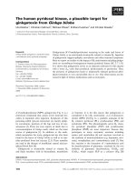
Nucleus Mitochondria
1µm
Mid piece Principal piece
Annulus
Figure 1
Montage transmission electron micrograph of a human sperm cell. The cell has a compact nucleus, conspicuous mitochondria, no
endoplasmic reticulum, minimal cytoplasm and a large tail (about 45 µm in length). Superfluous cytoplasm and associated machinery is
jettisoned when the sperm emerges from the testis, leaving a ‘stripped down’, minimalist cell.
63.3
Barratt et al.: Journal of Biology 2009, 8:63
time to have adverse reproductive consequences, such as
reduced success at ART. However, surprisingly little is
known about the histone fraction, which was previously
presumed to be a ‘leftover’ from remodeling during
spermiogenesis (the last stages of spermatogenesis when
the cell is transformed from a round haploid cell to a motile
cell). Recent data have changed our perception and
suggested a bias in gene localization, with the histone-
containing regions containing significantly more genes
encoding proteins involved in embryogenesis than other
regions [9]. In addition, the histones are susceptible to a
myriad of potential epigenetic changes through the histone
code, the normality of which could easily be disturbed in
dysfunctional cells. This is a level of complexity in the
sperm cell that has not yet been fully appreciated.
Of critical importance to men with sperm dysfunction is
the unambiguous relationship between poor-quality sperm
and high levels of DNA damage in the cells. This has
minimal relevance for natural conception as damaged cells
are unable to fertilize, but with ICSI cells with high levels
of DNA damage are often the only ones available and are
thus used for injection. Clinical data from many ART
programs using a variety of DNA-damage assays show a
strong negative correlation between DNA damage in the
sperm and rates of embryo development and implantation
and, importantly, a positive correlation with levels of
miscarriage [10]. However, the longer term consequences
of using damaged cells, such as the health of offspring, are
unknown. Data from mice suggest that caution is needed,
as using DNA-damaged sperm for ICSI was associated with
pathological changes in the resultant adult offspring, such
as changes in behavior, higher incidence of cancer and
premature aging [11].
We are only at the beginning of our understanding of
sperm chromatin and DNA and, when there is damage, we
do not know the point in the lifecycle of the cell at which
the damage originates, its causes (for example whether it is
oxidative in nature) or the nature of the damage (for
example, single- and/or double-strand breaks and/or DNA
crosslinking). Other basic questions remain unanswered.
1
2
4
3
5
6
7
8
(b)
Isthmus
Ovary
Oolemma
Cumulus cell
Zona pellucida
Matrix
Egg
(a)
(c)
Oviduct
Cervix
Uterus
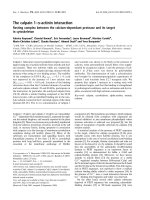
Figure 2
The remarkable journey of the sperm to the egg. (a) After intercourse, sperm enter the cervical mucus (1), where they begin capacitation.
During passage to the uterus (2), the sperm induce a host reaction. Leukocytes, which outnumber the sperm 100:1, engulf normal and
abnormal spermatozoa. In humans only one in 14,000,000 ejaculated human sperm reach the oviduct. Experiments in non-human mammals
have shown that the oviduct (particularly the isthmus, where the duct is narrowest) (3) acts as a sperm store, potentially involving intimate
contact between the sperm and the epithelial surface. Fertilization occurs in the oviduct (4). (b) In vitro experiments suggest that intracellular
Ca
2+
is low in the sperm that are attached to the isthmus epithelium (dark blue), and that this maintains their longevity and function. Before
ovulation, the sperm detach, possibly as a result of the expression of hyperactivated motility. Detached cells (red) have higher intracellular
Ca
2+
levels and more vigorous motility and find the egg by responding to chemotactic cues (not yet identified in humans). (c) When the
sperm reach the egg, hyperactivation enables penetration of the surrounding layer of cumulus cells (light blue) embedded in matrix (yellow)
and the sperm attaches to the zona pellucida (5). Upon attachment the sperm undergoes the acrosome reaction, in response to binding of
the zona pellucida (6), which, combined with hyperactivated motility, permits penetration of the zona (7) and fusion with the egg membrane
(oolemma) (8). Adapted from [12].
63.4
Barratt et al.: Journal of Biology 2009, 8:63
Does the origin and nature of the damage suggest less or
more severe consequences? Can the egg repair the damage
and, if so, is there a threshold of damage above which it
can no longer do so? This is an unusual field in which the
clinical data are relatively clear and consistent but have yet
to be accompanied with high quality scientific studies.
Searching for the egg
Sperm motility is essential for natural fertility, and one
key way to understand the cellular and molecular
mechanism of motility is to document the response of the
individual cell during chemotaxis (Figure 2). This process,
once thought to be the preserve of sea urchins and star
fish, has now been convincingly demonstrated in humans
using both artificial factors and egg-derived ones, such as
progesterone. With the advent of high-speed single-cell
imaging combined with controlled uncaging of fluorescent
molecules, the complex kinetics and signaling systems
involved in sperm motility are now beginning to be
explored. In humans, although cAMP, cGMP and calcium
are known to be important regulatory signals, the timing
of events is elusive. It is unlikely to be as simple as an
increase in cellular calcium resulting in a change in
direction following increased flagellar asymmetry.
Potentially, the development of hyperactivation (an excited
erratic movement of the sperm cell associated with
capacitation) may be a good model for studying the
calcium response of cells [12], particularly the involvement
of the release of calcium from stores and its association
with pH-sensitive calcium channels (CatSper channels).
This is clinically relevant, as failure to undergo
hyperactivation is significantly associated with reduced
fertilization success.
A fascinating aspect of internal fertilization in humans is
that sperm operate in a highly complex, mucus-based
environment, with different viscoelastic properties and
fluid mechanics from those of the simple media that are
universally used to study motility. For example, sperm
preferentially swim along surfaces and thus might crawl,
rather than swim, up the female reproductive tract. As a
result, experiments modeling and examining sperm
behavior in these physiologically relevant environments
are essential for obtaining an accurate analysis, as recently
demonstrated by Smith and colleagues [13].
In summary, there is an urgent need to develop a more
detailed understanding of the physiological, biochemical
and molecular functioning of the human sperm cell. We
can use this knowledge as a platform to improve the
diagnosis of male infertility with, for example, robust
biomarkers and the development of non-ART-based
therapies. The tools at our disposal have never been more
accessible, powerful and sophisticated, so, if they are used
wisely, it is likely that rapid progress will be made in the
very near future. Perhaps then a structure such as the
annulus – which has been known for over 100 years – will
reveal its secrets.
Acknowledgements
Work in the authors’ laboratories is sponsored by NHS Tayside,
Wellcome Trust, TENOVUS (Scotland), University of Dundee,
British Council and Scottish Enterprise. The authors thank all
patients and donors for providing samples for the Reproductive and
Developmental Biology research program in Dundee.
References
1. Boivin J, Bunting L, Collins JA, Nygren KG: International esti-
mates of infertility prevalence and treatment-seeking:
potential need and demand for infertility medical care. Hum
Reprod 2007, 22:1506-1512.
2. Guan J, Kinoshita M, Yuan L: Spatiotemporal association of
DNAJB13 with the annulus during mouse sperm flagellum
development. BMC Dev Biol 2009, 9:23.
3. Nyboe Andersen A, Goossens V, Bhattacharya S, Ferraretti AP,
Kupka MS, de Mouzon J, Nygren KG; European IVF-monitoring
(EIM) Consortium, for the European Society of Human
Reproduction and Embryology: Assisted reproductive tech-
nology and intrauterine inseminations in Europe, 2005:
results generated from European registers by ESHRE:
ESHRE. Hum Reprod 2009, 24:1267-1287.
4. Pirrello O, Machev N, Schimdt F, Terriou P, Ménézo Y, Viville S:
Search for mutations involved in human globozoospermia.
Hum Reprod 2005, 20:1314-1318.
5. Lefièvre L, Bedu-Addo K, Conner SJ, Machado-Oliveira GS,
Chen Y, Kirkman-Brown JC, Afnan MA, Publicover SJ, Ford
WC, Barratt CL: Counting sperm does not add up any more:
time for a new equation? Reproduction 2007, 133:675-684.
6. Baker MA, Reeves G, Hetherington L, Muller J, Baur I, Aitken RJ:
Identification of gene products present in the Triton X soluble
and insoluble fractions of human spermatozoa lysates using
LC-MS/MS analysis. Proteomics Clin Appl 2007, 1:524-532.
7. Platt MD, Salicioni AM, Hunt DF, Visconti PE: Use of differen-
tial isotopic labeling and mass spectrometry to analyze
capacitation-associated changes in the phosphorylation
status of mouse sperm proteins. J Proteome Res 2009,
8:1431-1440.
8. Lefièvre L, Chen Y, Conner SJ, Scott JL, Publicover SJ, Ford
WC, Barratt CL: Human spermatozoa contain multiple
targets for protein S-nitrosylation: an alternative mecha-
nism of the modulation of sperm function by nitric oxide?
Proteomics 2007, 7:3066-3084.
9. Hammoud SS, Nix DA, Zhang H, Purwar J, Carrell DT, Cairns BR:
Distinctive chromatin in human sperm packages genes for
embryo development. Nature 2009, 460:473-478.
10. Zini A, Sigman M: Are tests of sperm DNA damage clinically
useful? Pros and cons. J Androl 2009 30:219-229.
11. Fernández-Gonzalez R, Moreira PN, Pérez-Crespo M,
Sánchez-Martín M, Ramirez MA, Pericuesta E, Bilbao A,
Bermejo-Alvarez P, de Dios Hourcade J, de Fonseca FR,
Gutiérrez-Adán A: Long-term effects of mouse intracyto-
plasmic sperm injection with DNA-fragmented sperm on
health and behavior of adult offspring. Biol Reprod 2008,
78:761-772.
12. Publicover S, Harper CV, Barratt C: Ca
2+
i
signalling in sperm
– making the most of what you’ve got. Nat Cell Biol 2007,
9:235-242.
13. Smith DJ, Gaffney EA, Gadêlha H, Kapur N, Kirkman-Brown
JC: Bend propagation in the flagella of migrating human
sperm, and its modulation by viscosity. Cell Motil
Cytoskeleton 2009, 66:220-236.
Published: 7 August 2009
doi:10.1186/jbiol167
© 2009 BioMed Central Ltd