Báo cáo sinh học: "Physical forces in myelination and repair: a question of balance" potx
Bạn đang xem bản rút gọn của tài liệu. Xem và tải ngay bản đầy đủ của tài liệu tại đây (507.23 KB, 4 trang )
Bauer and ffrench-Constant: Journal of Biology 2009, 8:78
Abstract
A recent report in BMC Cell Biology examines how the balance
of extracellular forces and intracellular contractions regulate the
shape changes required for oligodendrocyte myelination. A
failure of remyelination such as seen in multiple sclerosis could
be caused by loss of this balance.
See related research article
The interplay between intracellular and
extracellular forces
During development, all cells undergo enormous changes
in cell shape. After a cell is ‘born’, it migrates to its final
destination, where it then changes its shape to assume its
final role. Very often, this involves the formation of cellular
processes, many of which have specific shapes and
functions that are characteristic to the individual cell types.
This process outgrowth and other changes in morphology
are supported internally by a sturdy network of specialized
structural proteins that form the cytoskeleton. In addition,
the surrounding extracellular environment, the extra-
cellular matrix (ECM), mediates the changes in cell shape
through its mechanical properties. The role of the ECM
becomes particularly apparent when adherent cells (cells
that are part of a tissue) are compared with non-adherent
cells (cells that are floating freely within a liquid, such as
blood). Although most adherent cells have a very particular
shape, non-adherent cell types are usually rounded but
change shape when they attach to surrounding tissue [1],
suggesting that adherent cells can sense and respond to
mechanical signals from the ECM.
These mechanosensory properties are mediated by special
adhesion sites, where the ECM binds to a family of receptor
proteins within the cell membrane. The most significant of
these are the integrins. Binding of ECM ligands to
integrins, with the associated varying degrees of mecha-
nical strain or stretch, promotes the recruitment and
linkage of part of the cytoskeleton, the actomyosin net-
work, to intracellular integrin domains and thus anchors
this network at the lipid membrane [2,3]. The actomyosin
network consists of two major components, actin filaments
and myosin molecules, which can slide along each other,
creating an intracellular contractile force (Figure 1a). The
relationship between this intracellular force and the
strength of cell adhesion (the extracellular force) could
then simply regulate shape such that stronger external
forces would pull the cellular membrane outwards,
whereas stronger internal forces would maintain a
rounded shape.
However, cellular events seem to be more complex than
this. First, recent findings have emphasized the importance
of functional actomyosin contractile mechanisms for the
regulation of a wide range of cell properties, including
tissue formation, cell migration and cell differentiation [4].
Second, in contradiction to this simple model, low
contractile forces generally yield membrane-rich, bulgy cell
types, whereas strong contractions lead to the formation of
highly structured cell shapes. Finally, different cell types
reportedly have different ECM rigidity preferences for the
induction of their particular shape. All this suggests that
cellular shapes are determined by a precisely regulated
interplay of intracellular contractile forces and extra-
cellular attachment.
Interplay of forces in myelination
A particularly striking example of this interplay is the
neural cell lineage, which gives rise to neurons, astrocytes
and oligodendrocytes in the central nervous system (CNS).
Developmentally, all three neural cell types develop from
the same multipotent stem cells. However, neurons, which
are generated first, prefer relatively soft surfaces for
elaboration and branching of axons and dendrites. These
softer substrates possibly correspond to the environmental
conditions at the time of initial pathfinding of neuronal
processes. In contrast, in recently published work in BMC
Cell Biology [5], Simons and colleagues show that the
myelin-forming oligodendrocytes that develop later form
their highly processed morphology and extensive myelin
sheets best on more rigid surfaces.
This seems logical if we take a closer look at the develop-
mental context of the formation of the insulating myelin
sheath around axons. Once the migratory oligodendrocyte
precursor cells (OPCs) have reached their destination and
start to establish contact with an axon, their processes
Minireview
Physical forces in myelination and repair: a question of balance?
Nina G Bauer and Charles ffrench-Constant
Address: MRC Centre for Regenerative Medicine, Centre for Multiple Sclerosis Research, The University of Edinburgh, Queen’s Medical
Research Institute, 47 Little France Crescent, Edinburgh EH16 4TJ, UK.
Correspondence: Nina G Bauer. Email:
78.2
Bauer and ffrench-Constant: Journal of Biology 2009, 8:78
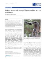
Figure 1
The role of force in myelination. (a) Molecular force generation. Cells bind ECM components through integrin receptors (represented by α
and β subunits), thus increasing extracellular adhesion. Integrin activation then triggers signaling cascades involving Fyn kinase, which
inhibits RhoA, thus activating ROCK and Myosin IIB. Activated Myosin IIB interacts with actin filaments and creates strong intracellular
contractions, which in turn enhances extracellular attachment and possibly mediates cell differentiation. ECM and cytosol color schemes
represent the force intensity generated by these molecular events, gray being weakest and red being strongest. (b) Hypothetical effects of
extracellular rigidity and intracellular contractions. Optimal myelination conditions require a balance between extracellular forces mediated by
matrix rigidity and intracellular forces based on actomyosin contractions (diagonal arrow). A softer matrix inhibits cell differentiation and
myelination (shift to the left), which can be counteracted by myosin IIB inhibition (cells return to being balanced). Gliosis, as it occurs in MS,
might represent a more rigid matrix (shift to the right), which would require stronger contractile forces to counteract.
Fyn
Rho
ROCK
MyosinIIB
MyosinIIB
P
α
β
MyosinIIB
P
MyosinIIB
P
MyosinIIB
P
y
P
P
s
α
β
α
β
ECM
Cytosol
Extracellular rigidity
Intracellular contractile force
Softer matrix
Gliosis
Myosin inhibition
(a)
(b)
Actin
Unknown target
78.3
Bauer and ffrench-Constant: Journal of Biology 2009, 8:78
change from exploring the unstructured extracellular
environment of the presumptive myelinated tract, as
required for migration, to the establishment of close
contact with the highly structured (and therefore
probably more rigid) surface of the axon, initiating the
process of wrapping it with membranous sheets that will
eventually become the compact myelin sheath [6]. An
increase in intracellular force would therefore be
necessary to enable the opposing forces to be matched
and promote the next stage of oligodendrocyte
development - the elaborate shape changes that
accompany myelination (Figure 1b).
The findings of Simons and co-workers [5] also provide
information about these intracellular mechanisms. Investi-
gation of the role of intracellular contractility in
differentiation and myelination identified myosin IIB, one
of the major components of the actomyosin cytoskeleton,
as a central player in generating intracellular force. In cell
culture experiments, softer surfaces inhibit process out-
growth, as would be predicted if oligodendrocyte differen-
tiation is normally associated with increased levels of
intracellular force to match the increased rigidity of the
axons. This effect can be overcome by pharmacological
inhibition of myosin IIB, which will reduce intracellular
contractions and thus better match intracellular force with
the lower extracellular attachment efficacy provided by less
rigid substrate (Figure 1b).
These findings are of particular interest for two reasons.
The first is that they offer a clue as to how one might
explain the rather surprising reported effects of myosin
IIB inhibition on myelination in culture. Wang et al. [7]
showed that myosin IIB inhibition in a neuron-oligo-
dendrocyte co-culture system significantly enhanced the
formation of the myelin sheath, a change that resulted
from individual oligodendrocytes forming more
wrapping processes than cells in untreated control
cultures. In complete contrast, inhibition of myosin IIB
in co-cultures of Schwann cells (the myelinating cells of
the peripheral nervous system) with neurons inhibited
myelination, and cellular morphology was characterized
by aberrant process outgrowth. In short, while Schwann
cells react as would be predicted, oligodendrocytes
exhibit a behavior that contradicts the conclusions
obtained from previous experiments. This might reflect
important differences in the biology of the Schwann cell
and the oligodendrocyte, in particular in respect to their
adjustment to in vitro conditions: the extracellular forces
on the Schwann cell appear to be similar in culture and
in vivo, whereas the extracellular forces on
oligodendrocytes in culture are potentially weaker than
in vivo. The presence of a basal lamina on the non-axonal
side of the Schwann cell but not the oligodendrocyte both
in vitro and in vivo might be one means of retaining such
an extracellular force.
Interplay of forces in multiple sclerosis
The second, and more important, reason for interest in the
findings of Simons and colleagues [5] is that they offer
explanations as to why remyelination might fail in the
demyelinating disease multiple sclerosis (MS) [8]. In MS,
unknown molecular triggers induce an inflammatory
reaction in the brain leading to an invasion and activation
of immune cells (B and T lymphocytes and macrophages)
and/or the produc tion of antibodies directed against
myelin components. These events lead to the damage and
degeneration of the myelin sheath. Remyelination does
occur in the early stages of the disease as intrinsic
mechanisms mediate the recruitment of OPCs, which then
align with the denuded axon and regenerate the sheath.
However, this repair mechanism eventually fails, for as-yet
unknown reasons. An implication of the results of Simons
and colleagues [5] is that increased rigidity in the scarred
brain may play a role by unbalancing the intracellular and
extracellular forces and inhibiting oligodendrocyte
differentiation (Figure 1b).
How might the rigidity of the chronically demyelinated
CNS be altered? Astrocytes, the third cell type derived from
the neural lineage, provide nutrients to neurons and
oligodendrocytes, give biochemical support to the cells
forming the blood-brain barrier and, in particular, mediate
the repair and scarring processes in the CNS following
traumatic injuries. They respond to pathological insults,
including inflammation and demyelination, with so-called
reactive gliosis. On a cellular level, this is characterized by
an upregulation of intermediate filament proteins, leading
to the formation of a prominent intermediate filament
network directly underneath the plasma membrane,
rendering the cellular texture more fibrous [9]. Further-
more, pronounced changes in expression of adhesion
molecule genes have been described, which would result in
an altered ECM composition compared with that of initial
myelination. In demyelinated plaques, reactive astrocytes
are the most abundant cellular component, and astroglial
scars have been described as being more rigid than their
surrounding tissue. These properties might alter force-
sensing integrin function in the oligodendrocyte, unbalan-
cing the cellular forces and inhibiting remyelination.
The main implications of the findings of Simons and
colleagues [5] are, therefore, that a particular balance of
extracellular adhesion, matrix rigidity and intracellular
contractile forces mediated by the oligodendrocyte
actomyosin cytoskeleton is required for successful
myelination and remyelination. One interesting prediction
implied by these data is that extracellular cues that do not
in themselves alter rigidity, but that do change the activity
of signaling molecules regulating intracellular force, could
also inhibit remyelination. As discussed above, the pre-
dominant pathway involved in the signaling mecha nisms
underlying mechanosensing and mechanotrans duction is
78.4
Bauer and ffrench-Constant: Journal of Biology 2009, 8:78
binding of ECM ligands to integrin receptors in the
membrane. The activation of integrins by mechanical forces
results in the recruitment of intracellular mediators that
signal through a pathway involving RhoA and its
downstream effector ROCK to activate force-generating
myosin II (Figure 1a). The observation that the inhibitory
effects of myelin debris on OPC differentiation, myelination
and remyelination are mediated by RhoA-ROCK signaling
[10] is consistent with this hypothesis [5]. Subsequent
pharmacological disruption of the ROCK pathway,
inhibiting myosin IIB and thus actomyosin contractility,
was able to enhance oligodendrocyte differentiation [10].
Clearly, the signaling molecules that regulate intracellular
force now provide an intriguing source of candidates for
drug discovery programs aimed at enhancing remyeli nation
(Figure 1b).
References
1. Discher DE, Mooney DJ, Zandstra PW: Growth factors, matri-
ces, and forces combine and control stem cells. Science
2009, 324:1673-1677.
2. Choquet D, Felsenfeld DP, Sheetz MP: Extracellular matrix
rigidity causes strengthening of integrin-cytoskeleton link-
ages. Cell 1997, 88:39-48.
3. Schewkunow V, Sharma KP, Diez G, Klemm AH, Sharma PC,
Goldmann WH: Thermodynamic evidence of non-muscle
myosin II-lipid-membrane interaction. Biochem Biophys Res
Commun 2008, 366:500-505.
4. Clark K, Langeslag M, Figdor CG, van Leeuwen FN: Myosin II
and mechanotransduction: a balancing act. Trends Cell Biol
2007, 17:178-186.
5. Kippert A, Fitzner D, Helenius J, Simons M: Actomyosin con-
tractility controls cell surface area of oligodendrocytes.
BMC Cell Biol 2009, 10:71.
6. Bauer NG, Richter-Landsberg C, ffrench-Constant C: Role of
the oligodendroglial cytoskeleton in differentiation and
myelination. Glia 2009, doi: 10.1002/glia.20885.
7. Wang H, Tewari A, Einheber S, Salzer JL, Melendez-
Vasquez CV: Myosin II has distinct functions in PNS and
CNS myelin sheath formation. J Cell Biol 2008, 182:1171-
1184.
8. Franklin RJ, ffrench-Constant C: Remyelination in the
CNS: from biology to therapy. Nat Rev Neurosci 2008,
9:839-855.
9. Williams A, Piaton G, Lubetzki C: Astrocytes - friends or foes
in multiple sclerosis? Glia 2007, 55:1300-1312.
10. Baer AS, Syed YA, Kang SU, Mitteregger D, Vig R, ffrench-
Constant C, Franklin RJ, Altmann F, Lubec G, Kotter MR:
Myelin-mediated inhibition of oligodendrocyte precursor
differentiation can be overcome by pharmacological mod-
ulation of Fyn-RhoA and protein kinase C signalling. Brain
2009, 132:465-481.
Published: 25 September 2009
doi:10.1186/jbiol169
© 2009 BioMed Central Ltd