.Neuroscience of Rule-Guided Behavior Phần 2 pps
Bạn đang xem bản rút gọn của tài liệu. Xem và tải ngay bản đầy đủ của tài liệu tại đây (538.11 KB, 50 trang )
learning gets progressively better with each discrimination they solve (Harlow,
1949). Eventually, the monkey can learn the problem in a single trial: Perfor-
mance on the first trial is necessarily at chance, but performance is virtually
100% correct on the second trial. The monkey has learned to extract the ab-
stract rule ‘‘win-stay, lose-shift,’’ which dramatically speeds performance
(Restle, 1958). So, too, do corvids, but pigeons must solve each discrimination
individually (Hunter and Kamil, 1971; Wilson et al., 1985). Interestingly,
corvid brains differ from those of other birds, in that they have an enlarged
mesopallium and nidopallium, areas that are analogous to PFC in mammals
(Rehkamper and Zilles, 1991), prompting speculation that the capacity to use
abstract information might have evolved at least t wice in the animal kingdom
(Emery and Clayton, 2004).
In fact, the capacity to understand certain abstract concepts may be wide-
spread. A recent study showed that even some insects can use ‘‘same’’ and
‘‘different’’ rules to guide their behavior (Giurfa, 2001). Investigators trained
honeybees on a Y-maze. At the entrance to the maze was the sample stimulus,
and at the entrance to the two forks in the Y-maze were two test stimuli. Bees
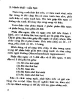
Figure 2–1 Possible configurations of stimuli and responses
in a matching task. In each panel, the lower picture is the sam-
ple stimulus and the upper two pictures are the test stimuli.
The arrow indicates the behavioral response. Although an an-
imal could learn this task by abstracting the rule to choose
the upper picture that matched the lower one, it could equally
learn the task by memorizing the correct response to make to
each of the four possible configurations of stimuli.
26 Rule Representation
received a reward for choosing the arm with the matching test stimulus. Not
only could the bees learn this task, but they also were able to apply the rule
to novel stimuli. Furthermore, they were just as capable of learning to follow
the ‘‘diffe rent’’ rule as they were the ‘‘same’’ rule. This study raises interest-
ing questions. For example, why should the capacity to use an abstract rule be
useful to bees, but not to pigeons? This capacity is not simply the ability to
know that one flower is the ‘‘same’’ as another, a very simple (and useful) be-
havioral adaptation that can be solved through stimulus generalization and
conditioning. Rather, it is using the relationship between two stimuli to gov-
ern behavior in an arbitrary fashion. Quite what use the bee finds for this
ability is a mystery, but it does demonstrate that a remarkably simple ner-
vous system, consisting of a brain of 1mm
3
and fewer than 1 million neurons
(Wittho
¨
ft, 1967) is capable of using abstract information. It remains an open
question whether it can learn a variety of abstract information, as does the
mammalian brain, or whether its abilities are more constrained.
These studies in neuropsychology and comparative psychology thus laid
the groundwork for this exploration of the neuronal mechanisms that might
underlie the use of abstract rules to guide behavior. They suggested a task that
monkeys could perform to demonstrate their grasp of abstract rules and sup-
ported the notion that PFC would be an important brain region for the neu-
ronal representation of such rules.
NEURONAL REPRESENTATION OF ABSTRACT
RULES IN PREFRONTAL CORTEX
Behavioral Paradigm
Although the matching-to-sample task was useful for demonstrating behav-
iorally that monkeys could use abstract rules, this task presented several prob-
lems when it came to exploring the underlying neuronal mechanisms. First,
the task made use of only one rule; to demonstrate neuronal selectivity, we
need at least two rules. To see why this is the case, consider how we would
define a neuron as encoding a face. We would want to show not only that the
neuron responds to faces, but also that it does not respond to non-face stimuli.
Otherwise, the neuron might be encoding any visual stimulus, rather than
faces specifically. In an analogous fashion, to demonstrate that a neuron is
encoding a specific rule, we need to show not only that it responds when the
‘‘same’’ rule is in effect, but also that it does not respond when other rules are
in effect. The matching-to-sample task shows that monkeys can grasp the con-
cept of ‘‘sameness.’’ An obvious second rule to teach the monkey was that of
‘‘difference.’’ Now, the monkey had to choose the test stimulus that did not
match the sample stimulus.
We trained three monkeys to use both of these rules. A sample stimulus
appeared on a computer screen, and we instructed the monkeys to follow ei-
ther the ‘‘same’’ rule or the ‘‘different’’ rule. After a brief delay, one of two test
Neurophysiology of Abstract Rules 27
stimuli appeared. The monkey had to make a given response depending on
which rule was in effect and whether the test stimulus matched or did not
match the sample stimulus. This, of course, raises the following question: How
do you instruct a monkey to follow a given rule? We did this by means of a cue
that we presented simultaneously with the sample stimulus . If the monke y
received a drop of juice, it knew that it should follow the ‘‘same’’ rule, and
if it did not receive juice, it knew that it should follow the ‘‘different’’ rule.
However, this method of cueing the currently relevant rule introduces a po-
tential confounding factor. Any neuron that showed a difference in firing rate
when the ‘‘same’’ or ‘‘different’’ rule was in effect might simply be encoding
the presence or absence of juice. To account for this possibility, we had a sec-
ond set of cues, drawn from a different modality. Thus, a neuron encoding the
abstract rule should be one that shows a difference of activity, irrespective of
the cue that we use to tell the monkey what to do. Figure 2–2 shows the full
task; during the first delay period, the monkey must remember the sample
picture as well as which rule is in effect, to perform the task correctly. Behav-
ioral performance on this task was excellent (the monkeys typically performed
approximately 90% of the trials correctly).
Each day, we used a set of four pictures that the monkey had not previously
seen. We only used four pictures because we wanted to compare the number
of neurons that encoded the sample picture and con trast it with the number of
neurons that encoded the abstract rule. This meant that we needed multiple
trials on which we used the same sample picture to estimate accurately the
neuronal firing rate elicited by a given picture. Unfortunately, this repetition
could conceivably allow the monkeys to learn the task through trial-and-error
configural learning. For example, consider the trial sequence shown in the top
row of Figure 2–2. The monkey might learn that the conjunction of the picture
of a puppy and the cue that indicates the ‘‘same’’ rule (e.g., a drop of juice or a
low t one) indicates that it should release the lever when it sees a picture of a
puppy as a test stimulus. Further analysis of the monkeys’ behavior showed
that this is not how they learned the task (Wallis et al., 2001; Wallis and Miller,
2003a). First, they performed well above chance when applying the rules the
first time they encountered a new picture (i.e., before trial-and-error learning
could have occurred) [70% correct; 4 pictures 55 recording sessions ¼ 220
pictures; p < 10
À8
; binomial test]. Second, in subsequent behavioral tests, the
monkeys performed the task just as easily when new pictures were used on
every trial (performing more than 90% of the trials correctly). Thus, the mon-
keys had to be solving the task by using the abstract rule.
Neurophysiological Results
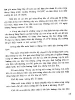
Figure 2–3 shows the activity of a PFC neuron during performance of this task.
This neuron shows a higher firing rate whenever the ‘‘same’’ rule is in effect.
Furthermore, which of the four pictures the monkey is remembering does not
affect the firing rate of the neuron, and neither does the cue that instructs the
28 Rule Representation
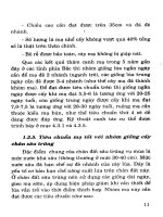
Figure 2–2 Each row (A–D) indicates a sequence of possible
events in the abstract rule task. A trial begins with the animal fix-
ating on a central point on the screen. We then present a sample
picture and a cue simultaneously. We use several cues drawn from
different sensory modalities so that we can disambiguate neuronal
activity to the physical properties of the cue from the abstract rule
that the cue instructs. For our first monkey, we indicate the ‘‘same’’
rule using a drop of juice or a low tone and the ‘‘different’’ rule with
no juice or a high tone. For the second monkey, juice or a blue
border around the sample picture signifies ‘‘same,’’ whereas no
juice or a green border indicates ‘‘different.’’ For the third monkey,
juice or a blue border indicates ‘‘same,’’ whereas no juice or a pink
border indicates ‘‘different.’’ After a short delay, a test picture ap-
pears and the animal must make one of two behavioral responses
(hold or release a lever), depending on the sample picture and the
rule that is currently in effect.
29
rule. In addition, the monkey does not know w hether the test stimulus will or
will not match the sample stimulus; consequently, it does not know whether it
will be holding or releasing the lever. As such, the activity of the neuron during
the delay cannot reflect motor preparation processes. Finally, factors relating
to behavioral performance cannot account for the firing rate, such as differ-
ences in attention, motivation, or reward expectancy. Behavioral performance
was virtually identical in the ‘‘same’’ and ‘‘different’’ trials (0.1% difference in
the per centage of correct trials and 7 ms difference in behavioral reaction time).
The only remaining explanation is that single neurons in PFC are capable of
encoding high-level abstract rules.
We used a three-way analysis of variance (ANOVA) to identify neurons
whose average firing rate during the sample and delay epochs varied signifi-
cantly with trial factors (evaluated at p < 0.01). The factors in the ANOVA
were the modality of the cue, the rule that the cue signified (‘‘same’’ or ‘‘dif-
ferent’’), and which of the four pictures was presented as the sample. We
defined rule-selective neurons as those that showed a significant difference in
firing rate between the two different rules, regardless of either the cue that was
used to instruct the monkey or the picture that was used as the sample stim-
ulus. Likewise, picture-selective neurons were identified as those that showed a
significant difference in firing rates between the four pictures, regardless of ei-
ther the cue or the rule.
We recorded data simultaneously from three major PFC subregions: dor-
solateral PFC, consisting of areas 9 and 46; ventrolateral PFC, consisting of
area 47/12; and orbitofrontal cortex, consisting of areas 11 and 13. The pattern
of neuronal selectivity was similar across the three areas: The most prevalent
selectivity was encoding of the abstract rule, observed in approximately 40% of
Figure 2–3 A prefrontal cortex neuron encoding an abstract rule. Neuronal activity is
consistently higher when the ‘‘same’’ rule is in effect, as opposed to the ‘‘different’’ rule.
We see the same pattern of neuronal activity irrespective of which picture the monkey
is remembering or which cue instructs the rule.
30 Rule Representation
PFC neurons (Table 2–1). There was an even split between neurons encoding
the ‘‘same’’ rule and those encoding the ‘‘different’’ rule. No topographic or-
ganization was evident, and we often recorded the activity of ‘‘same’’ and ‘‘dif-
ferent’’ neurons on the same electrode. The second most prevalent type of
neuronal activity was a CueÂRule interaction (27%). This occurred when a
neuron was most active to a single cue. This may simply reflect the physical
properties of the cue, although , in principle, it could also carry some rule
information. For example, such a neuron might be encoding rule information,
but only from a single modality. In contrast with the extent of rule encoding, a
much smaller proportion encoded which picture appeared in the sample
epoch (13%).
These results suggest that encoding of abstract rules is an important func-
tion of PFC, indeed, more so than the encoding of sensory information.
Having determined this, we wanted to ascertain whether the representation of
abstract rules was a unique property of PFC. We thus recorded from some of
its major inputs and outputs, with the aim of determining whether rule in-
formation arises in PFC.
ENCODING OF ABSTRACT RULES IN REGIONS
CONNECTED TO PREFRONTAL CORTEX
In the next study, we recorded data from three additional areas that are heavily
interconnected with PFC (Muhammad et al., 2006), namely, inferior temporal
cortex (ITC), PMC, and the striatu m (STR). We recorded data from ITC
because it is the major input to PFC for visual information (Barbas, 1988;
Barbas and Pandya, 1991). This was of interest because the rule task requires
the monkey to apply the ‘‘same’’ and ‘‘different’’ rules to complex visual
Table 2–1 Percentage of Neurons Encoding the Various Factors Underlying
Performance of the Abstract Rule Task in Either the Sample
or the Delay Epochs
N
DLPFC
182
VLPFC
396
OFC
150
PFC
728
PMC
258
STR
282
ITC
341
Cue 31% 20% 25% 24% 26% 18% 21%
Rule 42% 41% 38% 41% 48% 27% 12%
Picture 7% 18% 8% 13% 5% 4% 45%
Cue Rule 31% 27% 23% 27% 50% 20% 9%
Rule Picture 2% 1% 0% 1% 0% 1% 6%
Cue Picture 2% 5% 3% 4% 3% 1% 1%
Percentages exceed 100% because neurons could show different types of selectivity in the two epochs.
DLPFC, dorsolateral prefrontal cortex; VLPFC, ventrolateral prefrontal cortex; OFC, orbitofrontal
cortex; PFC, prefrontal cortex; PMC, premotor cortex; STR, striatum; ITC, inferior temporal cortex.
Neurophysiology of Abstract Rules 31
pictures and ITC plays a major role in the recognition of such stimuli (De-
simone et al., 1984; Tanaka, 1996). Furthermore, interactions between PFC and
ITC are necessary for the normal learni ng of stimulus-response associations
(Bussey et al., 2002). We also recorded data from PMC and STR because these
are two of the major outputs of PFC. Within PMC, we recorded data from
the arm area because the monkeys needed to make an arm movement to in-
dicate their response. Within STR, we recorded data from the head and body
of the caudate nucleus, a region known to contain many neurons involved in
the learning of stimulus-response associations (Pasupathy and Miller, 2005;
see Chapter 18).
To compare selectivity across the four brain regions, we performed a re-
ceiver operating characteristic (ROC) analysis. This analysis measures the de-
gree of overlap between two response distributions. It is particularly useful
for comparing neuronal responses in different areas of the brain because it is
independent of the neuron’s firing rate, and so it is easier to compare neurons
with different baseline firing rates and dynamic ranges. It is also nonparametric
and does not require the distributions to be Gaussian.
For each selective neuron, we determined which of the two rules drove its
activity the most. We then compared the distribution of neuronal activity
when the neuron’s preferred rule was in effect and when its unpreferred rule
was in effect. We refer to these two distributions as P and U, respectively. We
then generated an ROC curve by taking each observed firing rate of the neu-
ron (i.e., the unique values from the combined distribution of P and U) and
plotting the proportion of P that exceeded the value of that observation against
the proportion of U that exceeded the value of that observation. The area
under the ROC curve was then calculated. A value of 0.5 would indicate that
the two distributions completely overlap (be cause the proportion of U and P
exceeding that value is equal), and as such, would indicate that the neuron is
not selective. A value of 1.0, on the other hand, would indicate that the two
distributions are completely separate (i.e., every value of U is exceeded by the
entirety of P, whereas none of the values of P is exceeded by any of the values
of U), and so the neuron is very selective. An intuitive way to think about the
ROC value is that it measures the probability that you could correctly identify
which rule was in effect if you knew the neuron’s firing rate.
We used the ROC measure to determi ne the time course of neuronal se-
lectivity and to estimate each neuron’s selectivity latency. We computed the
ROC by averaging activity over a 200-ms window that we slid in 10-ms steps
over the course of the trial. To measure latency, we used the point at which the
sliding ROC curve equaled or exceeded 0.6 for three consecutive 10-ms bins.
We chose this criterion because it yielded latency values that compared favor-
ably with values that we determined by visually examining the spike density
histograms. Other measures yielded similar results, such as values reaching
three standard deviations above the baseline ROC values.
As shown in Figure 2–4, the strongest rule selectivity was observed in the
frontal lobe (PFC and PMC), and there was only weak rule selectivity in STR
32 Rule Representation
and ITC. Figure 2–4 illustrates the time course of rule selectivity across the
four neuronal populations from which we recorded. The x-axis refers to the
time from the onset of the sample epoch, and each horizontal line reflects data
from a single neuron. Color-coding reflects the strength of selectivity, as de-
termined by the ROC analysis. We sorted the neurons along the y-axis so that
neurons with the fastest onset of neuronal selectivity are at the bottom of the
graph. The black area at the top of each graph indicates the neurons that did
not reach the criterion for determining their latency.
The analysis using a three-way ANOVA to define rule- selective neurons
confirmed the results displayed in Figure 2–4. There was a significantly greater
incidence of rule selectivity in the PMC (48% of all recorded neurons, or
Figure 2–4 Time course of neuronal selectivity for the rule across the entire popula-
tion of neurons from which we recorded. Each horizontal line consists of the data from
a single neuron, color-coded by its selectivity, as measured by a receiver operating
characteristic. We sorted the neurons according to their latency. The black area at the
top of each figure consists of the data from neurons that did not encode the rule. Rule
selectivity was strong in premotor cortex and prefrontal cortex, weak in striatum, and
virtually absent in inferotemporal cortex.
Neurophysiology of Abstract Rules 33
125/258) than in PFC (41%, or 297/728), a greater incidence in PFC than in
STR (26%, or 89/341), and a greater incidence in STR than in ITC (12%, or 34/
282; chi-square; all comparisons p < 0.01). In all areas, approximately half of
the rule neurons showed higher firing ra tes to the ‘‘same’’ rule, whereas the
other half showed higher firing rates to the ‘‘different’’ rule. There were also
regional differences in terms of when rule selectivity first appeared. Figure 2–5
shows the distribution of latencies for neurons that reached the criterion for
determining latency (ITC neurons are not included here because so few neu-
rons showed a rule effect). On average, rule selectivity appeared significantly
earlier in PMC (median ¼ 280 ms) than in PFC (median ¼ 370 ms; Wilcox-
on’s rank sum test; p < 0.05). STR latencies (median ¼ 350 ms) were not sig-
nificantly different from those of PFC or PMC.
Figure 2–5 Histogram comparing the latency of rule selectivity across
three of the areas from which we recorded. Rule selectivity appeared
earlier in premotor cortex (PMC) [median ¼ 280 ms] than in prefron-
tal cortex (PFC) [median ¼ 370 ms], whereas striatum (STR) latencies
(median ¼ 350 ms) did not differ from those of PFC or PMC.
34 Rule Representation
When we compared the proportion of neurons with picture selectivity
across regions, we saw a pattern that was quite different from that seen for
rule selectivity. Picture selectivity was strongest in ITC (45% of all neurons, or
126/282), followed by PFC (13%, or 94/728), and finally, PMC (5%, or 12/
258) and STR (4%, or 15/341). The incidence of picture selectivity in PMC
and STR was not significantly different, but all other differences were (chi-
square; p < 0.01). We saw a similar pattern of results with the sliding ROC
analysis using the difference in activity between the most and least preferred
pictures (Fig. 2–6). Once again, each line corresponds to one neuron, and
we sorted the traces by their picture selectivity latency. Picture selectivity was
strongest in ITC, followed by PFC, and it was weak in both PMC and STR. We
used the sliding ROC analysis to determine latencies for picture selectivity
after sample onset (Fig. 2–7). The mean latency for picture selectivity was
significantly shorter in ITC (median ¼ 160 ms) than in PFC (median ¼
220 ms; p < 0.01). Too few neurons reached the criterion in PMC and STR
to allow for meaningful statistical comparisons.
Figure 2–6 Time course of neuronal selectivity for the sample picture across the entire
population of neurons from which we recorded. We constructed the figure in the same
way as Figure 2–4. Picture selectivity was strong in inferotemporal cortex, weak in
prefrontal cortex, and virtually absent in the striatum and premotor cortex.
Neurophysiology of Abstract Rules 35
In summary, PFC was the only area from which we recorded data that
encoded all of the task-relevant information, namely, both the picture and the
rule. In contrast, PMC and STR encoded the rule, but not the picture, whereas
ITC encoded the picture, but not the rule. These results fit with the concep-
tualization of ITC, PFC, and PMC as cortical components of a perception-
action arc (Fuster, 2002). Perceptual information was strongest and tended to
appear earliest in ITC, a sensory cortical area long thought to play a central
role in object recognition, and then in PFC, which receives direct projections
from ITC. ITC does not project directly to PMC (Webster et al., 1994), and
perceptual information was weakest in the PMC. By contrast, information
about the rules was strongest and earliest in frontal cortex (PFC and PMC)
and virtually absent in ITC.
One puzzling feature of our results is that PMC encodes rules more strongly
and earlier than PFC, yet it is not a region that has previously been associated
Figure 2–7 Histogram comparing the latency of picture selectivity in prefrontal
cortex (PFC) and inferotemporal cortex (ITC). Picture selectivity appeared earlier
in ITC (median ¼ 160 ms) than in PFC (median ¼ 220 ms).
36 Rule Representation
with the use of abstract information. One possibility is that we observed
stronger PMC rule effects because the rules were highly familiar to the ani-
mals; they had performed this task for more than a year. Evidence suggests
that PFC is more critical for new learning than for familiar routine s. PFC
damage preferentially affects new learning; animals and humans can still en-
gage in complex behaviors as long as they learned them be fore the damage
occurred (Shallice and Evans, 1978; Shallice, 1982; Knight, 1984; Dias et al.,
1997). PFC neurons also show more selectivity during new learning than dur-
ing the performance of familiar cue-response associations (Asaad et al., 1998).
Human imaging studies report greater blood flow to the dorsal PMC than to
PFC when subjects are performing familiar versus novel tasks (Boettiger and
D’Esposito, 2005) and greater PFC activation when subjects are retrieving
newly learned rules versus highly familiar rules (Donohue, 2005). In addition,
with increasing task familiarity, there is a relative shift in blood flow from
areas associated with fo cal attention, such as PFC, to motor regions (Della-
Maggiore and McIntosh, 2005). Therefo re, it may be that STR is primarily
involved in new learning, but with familiarity, rules become more strongly
established in motor system structures.
A second possibility lies in the design of the task. The task we used ensured
that the perceptual requirements were abstract: Monkeys had to make abstract
judgments about the similarity of pictures. However, the motor requirements
of the task were more concrete: The subjects always indicated their response
with a n arm movement. One could envision a version of the task in which the
subject has to respond with an arm movement to one set of trials, as in the
current task, and with an eye movement to other sets of trials. One might
predict that in such a task, rule activity would only occur in PMC during the
arm movement trials, and might occur in another frontal lobe structure, such
as the frontal eye fields, during eye movement trials. In other words, we pre-
dict that rule activity in PFC would be effector-independent, which would not
be the case for rule activity in PMC. PFC would be the only area to represent
the rule in a genuinely abstract fashion, independent of both sensory input and
the motor effector. These predictions should be tested in future research.
COMPARISON OF ABSTRACT RULES AND CONCRETE
STIMULUS-RESPONSE ASSOCIATIONS
In the experiment described earlier, we found only weak rule selectivity in STR
relative to the front al cortex. Recently, very different results have emerged for
the encoding of lower-level rules, such as the stimulus -response associations
that underpin conditional rules. Pasupathy and Miller (2005) recorded data
simultaneously from PFC and STR while monkeys learned stimulus-response
associations (see Chapter 18). In their task, two stimuli (A and B) instruct one
of two behavioral responses (saccade left or right). Both structures encoded
the associations between the stimuli and the responses, but selectivity ap-
peared earlier in learning in STR than in PFC. Despite this early neural correlate
Neurophysiology of Abstract Rules 37
of learning in STR, the monkey’s behavior did not change until PFC encoded
the associations. These results present us with a challenge: Why would the
monkey continue to make errors, despite the fact that STR was encoding the
correct stimulus-response associations? This finding suggests that not only is
overt behavior under the control of PFC more so than unde r that of STR, but
also that PFC will not necessarily use all of the information available to it to
control behavior.
One possibility is that PFC is integrating information from many low-level
learning systems, not just STR, and that some of these systems may not nec-
essarily agree with STR as to the correct response. For example, consider the
brain systems that acquire stimulus-reward associations or action-reward as-
sociations. It is impossible to learn stimulus-response associations using such
stimulus-reward or action-reward associations because each action and each
stimulus are rewarded equally often. However, this does not necessarily mean
that these systems will be silent during the performance of a task depende nt on
stimulus-response associations. For example, perhaps after a reinforced left-
ward saccade, the action-reward system instructs PFC to make another left-
ward response, oblivious to the fact that on the next trial, the stimulus in-
structs a rightward response. PFC would need to learn that such information is
not useful to solve the task, and ignore this system.
Lesion studies support the idea that these different low-level learning sys-
tems can compete with one another. For example, lesions of ant erior cingu-
late cortex impair the learning of stimulus -reward associations (Gabriel et al.,
1991; Bussey et al., 1997), but facilitate the learning of stimulus-response as-
sociations (Bussey et al., 1996). These findings suggest that in the healthy
animal, anterior cingulate is responsible for learning stimulus-reward asso-
ciations, and that removing the capacity to learn such associations can im-
prove the ability to learn stimulus-response associations.
OTHER FORMS OF ABSTRACT ENCODING
IN PREFRONTAL CORTEX
Recent studies have found that PFC neurons encode a variety of different
kinds of abstract information relating to high-level cognition, including at-
tentional sets (Mansouri et al., 2006), perceptual categories (Freedman et al.,
2001; see Chapter 17), numbers (Nieder et al., 2002), and behavioral strategies
(Genovesio et al., 2005; see Chapter 5). We have recently begun to explore
whether abstract information might also have a role in lower-level behavioral
control, to help guide simple decisions and choices. The neurophysiologi-
cal studies discussed earlier used models derived from sensorimoto r psycho-
physics and animal learning theory to make sense of the neuronal data. Over
the last decade, however, there has been a growing realization that to under-
stand the neuronal mechanisms underlying decision-making, it might help to
widen the fields from which we construct our behavioral models (Glimcher,
2003; Glimcher and Rustichini, 2004; Schultz, 2004; Sanfey et al., 2006).
38 Rule Representation
Evolutionary biologists and economists have constructed detailed models
of the parameters that animals and humans use to make everyday decisions.
These models emphasize the consideration of three basic parameters that must
be considered in making a decision: the expected reward or payoff, the cost in
terms of time and energy, and the probability of success (Stephens and Krebs,
1986; Loewenstein and Elster, 1992; Kahneman and Tversky, 2000). Deter-
mining the value of a choice involves calculating the difference between the
payoff and the cost, and discounting this by the probabi lity of success. One
suggestion is that PFC integrates all of these param eters to derive an abstract
measure of the value of a choice outcome (Montague and Berns, 2002).
To test this hypothesis about the representation of value, we examined
whether PFC neurons encode an abstract repre sentation of value by integrating
the major decision variables of payoff, cost, and risk (Kennerley et al., 2005).
We trained monkeys to choose between pictures while we simultaneously re-
corded data from multiple PFC regions. Each picture was associated with a
specific outcome. Some pictures were associated with a fixed amount of juice,
but only on a certain proportion of trials (risk manipulation). Other pictures
were associated with varying amounts of juice (payoff manipulation). Finally,
some pictures were associated with a fixed amount of juice, but the subject had
to earn the juice by pressing a lever a certain number of times (cost manip-
ulation). A large proportion of PFC neurons encoded the value of the choices
under at least one of these manipulations (Table 2–2). Other neurons encoded
the values under two of the manipulations, and still others encoded the value
under all three manipulations, consistent with encoding an abstract repre-
sentation of value. In other words, some PFC neurons encoded the value of the
choice irrespective of how we manipulated its value. The majority of the se-
lective neurons were located in medial PFC, where approximately half en-
coded the value of the choice outcome in some way.
The encoding of the value of a choice in an abstract manner has distinct
computational advantages. When faced with two choices, A and B, we might
imagine that it would be simpler to comp are them directly rather than going
through an addition al step of assigning them an abstract value. The problem
with this approach is that as the number of available choices increases, the
number of direct comparisons increases exponentially. Thus, choosing among
A, B, and C would require three comparisons (AB, AC, and BC), whereas
choosing among A, B, C, and D requires six comparisons (AB, AC, AD, BC,
BD, and CD). The solution quickly suffers from combinatorial explosion as the
number of choices increases. In contrast, valuing each choice along a common
reference scale provides a linear solution to the problem.
An abstract representation provides important additional behavioral ad-
vantages, such as flexibility and a capacity to deal with novelty. For examp le,
suppose that an animal encounters a new type of food. If the animal relies on
direct comparisons, then to determine whether it is worth choosing this new
food over others, it must iteratively compare the new food with all previ-
ously encountered foods. By deriving an abstract value, on the other hand, the
Neurophysiology of Abstract Rules 39
animal has only to perform a single calculation. By assigning the new food
a value on the common reference scale, it knows the value of this foodstuff
relative to all other foods. In addition, often it is not clear how to compare
directly very different outcomes: How does a monkey decide between groom-
ing a compatriot and eating a banana? Valuing the alternatives along a com-
mon reference scale helps with this decision. For example, although I have
never needed to value my car in terms of bananas, I can readily do so because
I can assign each item a dollar value.
CONCLUSIONS AND FUTURE RESEARCH
In conclusion, numerous studies now suggest that using abstract information
to guide behavior is an important and potentially unique function of PFC. In
turn, this capacity might underlie two of the hallmark functions of PFC,
flexibility and the ability to deal with novelty. A key question that remains is
how we learn such information in the first place. The mechanisms that un-
derpin the learning of abstract information remain unclear. Traditionally,
neurophysiologists record data from animals only once they have learned the
task. There are good reasons for so doing. Collecting an adequate sample of
neurons requires multiple recording sessions, and interpreting the data re-
quires behavior to be stable across those sessions. Even in studies that have
incorporated learning into the design, typically, monkeys are trained until
there is a stable, asymptotic rate of learning (Wallis and Miller, 2003b; Pa-
supathy and Miller, 2005). However, this makes for a rather artificial model of
behavior. In real life, behavior is rarely stable, but instead, constantly changes
and adapts to the environment. Furthermore, the immense amount of train-
ing that the animals often require (usually lasting months, or even years) raises
the possibility that the types of neuronal changes that we observe are not
an accurate reflection of more natural learning, or are perhaps only reflec-
tive of the encoding of highly trained skills. Fortunately, recent advances in
Table 2–2 Percentage of Neurons Encoding Variables Underlying Choices
in Different Prefrontal Cortex Subregions
N
Dorsolateral
108
Ventrolateral
52
Orbital
89
Medial
153
Risk 3% 2% 2% 20%
Payoff 2% 2% 6% 9%
Cost 1% 0% 0% 3%
Risk þ Payoff 0% 0% 2% 13%
Risk þ Cost 0% 0% 1% 4%
Payoff þ Cost 0% 0% 0% 4%
All three 0% 0% 0% 15%
40 Rule Representation
neurophysiological studies, such as chronically implanted electrodes, and the
increase in the number of neurons that can be recorded in a single session raise
the possibility of recording during the learning of these tasks. These and other
methodological advances will help us to understand how the brain achieves its
impressive ability to abstract and generalize.
acknowledgments I would like to thank Earl Miller, in whose laboratory I com-
pleted the abstract rule experiments. Funds from NIH DA019028-01 and the Hellman
Family Faculty Fund supported the abstract value experiments.
REFERENCES
Asaad WF, Rainer G, Miller EK (1998) Neural activity in the primate prefrontal cortex
during associative learning. Neuron 21:1399-407.
Barbas H (1988) Anatomic organization of basoventral and mediodorsal visual re-
cipient prefrontal regions in the rhesus monkey. Journal of Comparative Neurology
276:313–342.
Barbas H, Pandya D (1991) Patterns of connections of the prefrontal cortex in the
rhesus monkey associated with cortical architecture. In: Frontal lobe function and
dysfunction (Levin HS, Eisenberg HM, Benton AL, eds.), pp 35–58. New York: Ox-
ford University Press.
Bartlett FC (1932) Remembering: a study in experimental and social psychology.
Cambridge: Cambridge University Press.
Boettiger CA, D’Esposito M (2005) Frontal networks for learning and executing ar-
bitrary stimulus-response associations. Journal of Neuroscience 25:2723–2732.
Bussey TJ, Muir JL, Everitt BJ, Robbins TW (1996) Dissociable effects of anterior and
posterior cingulate cortex lesions on the acquisition of a conditional visual discri-
mination: facilitation of early learning vs. impairment of late learning. Behavioral
Brain Research 82:45–56.
Bussey TJ, Muir JL, Everitt BJ, Robbins TW (1997) Triple dissociation of anterior
cingulate, posterior cingulate, and medial frontal cortices on visual discrimination
tasks using a touchscreen testing procedure for the rat. Behavioral Neuroscience
111:920–936.
Bussey TJ, Wise SP, Murray EA (2002) Interaction of ventral and orbital prefrontal
cortex with inferotemporal cortex in conditional visuomotor learning. Behavioral
Neuroscience 116:703–715.
Della-Maggiore V, McIntosh AR (2005) Time course of changes in brain activity and
functional connectivity associated with long-term adaptation to a rotational trans-
formation. Journal of Neurophysiology 93:2254–2262.
Desimone R, Albright TD, Gross CG, Bruce C (1984) Stimulus-selective properties of
inferior temporal neurons in the macaque. Journal of Neuroscience 4:2051–2062.
Dias R, Robbins TW, Roberts AC (1997) Dissociable forms of inhibitory control within
prefrontal cortex with an analog of the Wisconsin Card Sort Test: restriction to novel
situations and independence from ‘‘on-line’’ processing. Journal of Neuroscience
17:9285–9297.
Donohue SE, Wendelken C, Crone EA, Bunge SA (2005) Retrieving rules for behavior
from long-term memory. Neuroimage 26:1140–1149
Eldridge MA, Barnard PJ, Bekerian DA (1994) Autobiographical memory and daily
schemas at work. Memory 2:51–74.
Neurophysiology of Abstract Rules 41
Emery NJ, Clayton NS (2004) The mentality of crows: convergent evolution of intel-
ligence in corvids and apes. Science 306:1903–1907.
Freedman DJ, Riesenhuber M, Poggio T, Miller EK (2001) Categorical representation
of visual stimuli in the primate prefrontal cortex. Science 291:312–316.
Fuster JM (2002) Cortex and mind. Oxford: Oxford University Press.
Gabriel M, Kubota Y, Sparenborg S, Straube K, Vogt BA (1991) Effects of cingulate
cortical lesions on avoidance learning and training-induced unit activity in rabbits.
Experimental Brain Research 86:585–600.
Genovesio A, Brasted PJ, Mitz AR, Wise SP (2005) Prefrontal cortex activity related to
abstract response strategies. Neuron 47:307–320.
Giurfa M, Zhang S, Jenett A, Menzel R, Srinivasan MV (2001) The concepts of ‘‘same-
ness’’ and ‘‘difference’’ in an insect. Nature 410:930–933.
Glimcher PW (2003) Decisions, uncertainty, and the brain: the science of neuroeco-
nomics. Cambridge: MIT Press.
Glimcher PW, Rustichini A (2004) Neuroeconomics: the consilience of brain and
decision. Science 306:447–452.
Harlow HF (1949) The formation of learning sets. Psychological Review 56:51–65.
Herman LM, Gordon JA (1974) Auditory delayed matching in the bottlenose dolphin.
Journal of the Experimental Analysis of Behavior 21:19–26.
Hunter MW, Kamil AC (1971) Object discrimination learning set and hypothesis be-
havior in the northern blue jay (Cyanocitta cristata). Psychonomic Science 22:271–
273.
Kahneman D, Tversky A (2000) Choices, values and frames. New York: Cambridge
University Press.
Kastak D, Schusterman RJ (1994) Transfer of visual identity matching-to-sample in
two Californian sea lions (Zalophus californianus). Animal Learning and Behavior
22:427–453.
Kennerley SW, Lara AH, Wallis JD (2005) Prefrontal neurons encode an abstract rep-
resentation of value. Society for Neuroscience Abstracts.
Knight RT (1984) Decreased response to novel stimuli after prefrontal lesions in man.
Electroencephalography and Clinical Neurophysiology 59:9–20.
Loewenstein G, Elster J (1992) Choice over time. New York: Russell Sage Foundation.
Mansouri FA, Matsumoto K, Tanaka K (2006) Prefrontal cell activities related to mon-
keys’ success and failure in adapting to rule changes in a Wisconsin Card Sorting
Test analog. Journal of Neuroscience 26:2745–2756.
Milner B (1963) Effects of different brain lesions on card sorting. Archives of Neu-
rology 9:100–110.
Mishkin M, Prockop ES, Rosvold HE (1962) One-trial object discrimination learning
in monkeys with frontal lesions. Journal of Comparative and Physiological Psy-
chology 55:178–181.
Montague PR, Berns GS (2002) Neural economics and the biological substrates of
valuation. Neuron 36:265–284.
Muhammad R, Wallis JD, Miller EK (2006) A comparison of abstract rules in the
prefrontal cortex, premotor cortex, inferior temporal cortex, and striatum. Journal
of Cognitive Neuroscience 18:974–989.
Nieder A, Freedman DJ, Miller EK (2002) Representation of the quantity of visual
items in the primate prefrontal cortex. Science 297:1708–1711.
Nissen HW, Blum JS, Blum RA (1948) Analysis of matching behavior in chimpanzees.
Journal of Comparative and Physiological Psychology 41:62–74.
42 Rule Representation
Oden DL, Thompson RK, Premack D (1988) Spontaneous transfer of matching by
infant chimpanzees (Pan troglodytes). Journal of Experimental Psychology: Animal
and Behavior Processes 14:140–145.
Pandya DN, Yeterian EH (1990) Prefrontal cortex in relation to other cortical areas
in rhesus monkey: architecture and connections. Progress in Brain Research 85:
63–94.
Pasupathy A, Miller EK (2005) Different time courses of learning-related activity in the
prefrontal cortex and striatum. Nature 433:873–876.
Pepperberg IM (1987) Interspecies communication: a tool for assessing conceptual
abilities in the African Grey parrot (Psittacus arithacus). In: Cognition, language and
consciousness: integrative levels (Greenberg G, Tobach E, eds.), pp 31–56. Hillsdale,
NJ: Lawrence Erlbaum Associates Inc.
Rao SC, Rainer G, Miller EK (1997) Integration of what and where in the primate
prefrontal cortex. Science 276:821–824.
Rehkamper G, Zilles K (1991) Parallel evolution in mammalian and avian brains:
comparative cytoarchitectonic and cytochemical analysis. Cell and Tissue Research
263:3–28.
Restle F (1958) Toward a quantitative description of learning set data. Psychological
Review 64:77–91.
Rolls ET, Baylis LL (1994) Gustatory, olfactory, and visual convergence within the pri-
mate orbitofrontal cortex. Journal of Neuroscience 14:5437–5452.
Romanski LM, Goldman-Rakic PS (2002) An auditory domain in primate prefrontal
cortex. Nature Neuroscience 5:15–16.
Romo R, Brody CD, Hernandez A, Lemus L (1999) Neuronal correlates of parametric
working memory in the prefrontal cortex. Nature 399:470–473.
Sanfey AG, Loewenstein G, McClure SM, Cohen JD (2006) Neuroeconomics: cross-
currents in research on decision-making. Trends in Cognitive Sciences 10:108–116.
Schultz W (2004) Neural coding of basic reward terms of animal learning theory, game
theory, microeconomics and behavioral ecology. Current Opinion in Neurobiology
14:139–147.
Semendeferi K, Lu A, Schenker N, Damasio H (2002) Humans and great apes share a
large frontal cortex. Nature Neuroscience 5:272–276.
Shallice T (1982) Specific impairments of planning. Philosophical Transactions of the
Royal Society London B Biological Sciences 298:199–209.
Shallice T, Evans ME (1978) The involvement of the frontal lobes in cognitive esti-
mation. Cortex 14:294–303.
Stephens DW, Krebs JR (1986) Foraging theory. Princeton: Princeton University Press.
Tanaka K (1996) Inferotemporal cortex and object vision. Annual Review of Neuro-
science 19:109–139.
Wallis JD, Anderson KC, Miller EK (2001) Single neurons in prefrontal cortex encode
abstract rules. Nature 411:953–956.
Wallis JD, Miller EK (2003a) From rule to response: neuronal processes in the pre-
motor and prefrontal cortex. Journal of Neurophysiology 90:1790–1806.
Wallis JD, Miller EK (2003b) Neuronal activity in primate dorsolateral and orbital
prefrontal cortex during performance of a reward preference task. European Journal
of Neuroscience 18:2069–2081.
Webster MJ, Bachevalier J, Ungerleider LG (1994) Connections of inferior temporal
areas TEO and TE with parietal and frontal cortex in macaque monkeys. Cerebral
Cortex 4:470–483.
Neurophysiology of Abstract Rules 43
Wilson B, Mackintosh NJ, Boakes RA (1985) Transfer of relational rules in matching
and oddity learning by pigeons and corvids. Quarterly Journal of Experimental
Psychology 37B:313–332.
Wittho
¨
ft W (1967) Absolute anzahl und Verteilung der zellen im hirn der honigbiene.
Zeitschrift fur Morphologie der Tiere 61:160–184.
44 Rule Representation
3
Neural Representations Used
to Specify Action
Silvia A. Bunge and Michael J. Souza
To understand how we use rules to guide our behavior, it is critical to learn
more about how we select responses on the basis of associations retrieved from
long-term memory and held online in working memory. Rules, or prescribed
guide(s) for conduct or action (Merriam-Webster Dictionary, 1974), are a par-
ticularly interesting class of associations because they link memory and action.
We previously reviewed the cognitive neuroscience of rule representations
elsewhere (Bunge, 2004; Bunge et al., 2005). In this chapter, we focus mainly on
recent functional brain imaging studies from our laboratory exploring the neu-
ral substrates of rule storage, retrieval, and maintenance. We present evidence
that goal-relevant knowledge associated with visual cues is stored in the pos-
terior middle temporal lobe. We further show that ventrolateral prefrontal
cortex (VLPFC) is engaged in the effortful retrieval of rule meanings from long-
term memory as well as in the selection between active rule meanings. Finally,
we provide evidence that different brain structures are recruited, depending on
the type of rule being represented, although VLPFC plays a general role in rule
representation. Although this chapter focuses primarily on the roles of lateral
prefrontal and temporal cortices in rule representation, findings in parietal and
premotor cortices will also be discussed.
LONG-TERM STORAGE OF RULE KNOWLEDGE
Posterior Middle Temporal Gyrus Is Implicated
in Rule Representation
In a previous functional magnetic resonance imaging (fMRI ) study focusing
on rule retri eval and maintenance, we observed activation of left posterior
middle temporal gyrus (postMTG) [BA 21], as well as left VLPFC (BA 44/45/
47), when subjects viewed instructional cues that were associated with specific
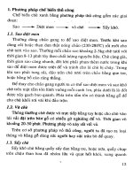
rules (Bunge et al., 2003) [Fig. 3–1]. Although both postMTG and VLPFC were
sensitive to rule complexity during the cue period, only VLPFC was sensitive
to rule complexity during the delay.
45
On the basis of evidence that semantic memories are stored in lateral tem-
poral cortex and that VLPFC assists in memory retrieval (e.g., Gabrieli et al.,
1998; Wagner et al., 2001), we proposed that left postMTG might store rule
knowledge over the long term, and that VLPFC might be important for re-
trieving and using this knowledge (Bunge et al., 2003). However, it is clear that
postMTG is not specifically involved in storing explicit rules for behavior;
rather, the literature on tool use and action representation suggests that this
region more generally represents action-related knowledge associated with
stimuli in the environment (see Donohue et al., 2005).
In ongoing research, we aim to reconcile the disparate views of postMTG
function emerging from the semantic memory literature (i.e., a general role in
semantic memory) and the action representation literature (i.e., a more spe-
cific role in action-related semantic representation). A recent study from our
Figure 3–1 Brain activation related to the retrieval and maintenance of rules uncovered
by functional magnetic resonance imaging (Bunge et al., 2003). Both left ventrolat-
eral prefrontal cortex (L VLPFC) [BA 44/47] and left posterior middle temporal gyrus
(L postMTG) [BA 21] were modulated by rule complexity during the Cue period, but
only the left VLPFC continued this pattern into the Delay period. **p < .01; *p < .05.
(Adapted from Bunge et al., 2003, Journal of Neurophysiology, 90:3419–3428, with per-
mission from the American Physiological Society).
46 Rule Representation
laboratory is consistent with the latter view, although a definitive answer awaits
further experiments.
Intriguingly, our focus in left postMTG was close to a region that is be-
lieved to represent knowledge about actions associated with manipulable ob-
jects (Chao et al., 1999; Martin and Chao, 2001). A large body of research has
shown that this region is active when subjects prepare to use a tool, mentally
conceptualize the physical gestures associated with tool use, make judgments
about the manipul ability of objects, generate action verbs, or read verbs as op-
posed to nouns (for reviews , see Johnson-Frey, 2004; Lewis, 2006).
Although most of these studies involved visual stimuli (images or words),
one group of researchers found that postMTG was engaged by meaningful
relative to meaningless environmental sounds (Lewis et al., 2004), and for tools
relative to animals (Lewis et al., 2005). Thus, the role of postMTG in storing
mechanical or action-related knowledge about stimuli extends to the realm of
auditory information; it is unclear whether it also extends to other modalities.
Given that we likely acquire most of our action-related knowledge through
vision and audition, one might expect that a region that specifically represents
action-related knowledge w ould not be modulated by other modalities. How-
ever, the possibility that postMTG is engaged by other stimulus modalities
remains an open issue, and we know of no functional brain imaging studies or
studies of anatomical connectivity that speak to this issue.
In our rule study, unlike the action knowledge studies mentioned earlier,
participants used recently learned arbitrary mappings between abstract cues
(nonsense shapes or words) and task rules. This finding suggests that left
postMTG plays a broader role in action knowledge than previously assumed.
Rather than specifically representing actions that are non-arbitrarily associ-
ated with real-world objects, left postMTG also represents high-level rules that
we learn to associate with otherwise meaningless symbols.
Explicitly Testing for Involvement of Left
PostMTG in Rule Representation
We sought to further test the hypothesis that left postMTG represents rule
knowledge in an fMRI study in which subjects viewed a series of road signs
from around the world, and considered their meanings (Donohue et al., 2005).
We had two reasons for selecting road signs as experimental stimuli: (1) they
are associated with specific actions or with guidelines that can be used to select
specific actions ; and (2) they allow us to examine the retrieval of rule knowl-
edge acquired long ago. As such, these stimuli enabled us to ask whether pre-
frontal cortex (PFC) [in particular, VLPFC] would be recruited during passive
retrieval of action knowledge asso ciated with well-learned symbols.
The road sign study involved ‘‘Old’’ signs that subjects had used while driv-
ing for at least 4 years, and ‘‘New’’ signs from other countries that they were
unlikely to have been exposed to previously (Fig. 3–2A). Of these New signs,
half were ‘‘Trained’’ (i.e., subjects were told their meaning before scanning, but
Neural Representations Used to Specify Action 47
had had no experience using them to guide their actions). The other half of the
new signs were ‘‘Untrained’’—in other words, subjects had viewed them before
scanning, but were not given their meaning. We predicted that left postMTG
would be active when subjects successfully accessed the meaning of Old and
Trained signs, but not when subjects viewed signs whose meaning they did not
know (‘‘Incorrect’’ trials, of which the majority would be Untrained).
Just as predicted, left postMTG was more active when subjects passively
viewed signs for which they knew the meaning than for signs that were familiar,
but not meaningful to them (Fig. 3–2B). This contrast also identified several
other regions, and all were located in the lateral temporal lobes. However, the
largest and most significant focus was in the predicted region of left postMTG.
Notably, unlike regions in lateral PFC, this region was insensitive to level of ex-
perience with the signs—it was engaged equally strongly for correctly performed
Old and Trained signs (Fig. 3–2B, inset). Thus, it appears that left postMTG
stores the meanings of arbitrary visual cues that specify rules for action, regard-
less of when these cues were originally learned or how much experience one has
had with them. This pattern of activation suggests two points: (1) activation of
the correct representation in temporal cortex contributes to remembering the
sign’s meaning; and (2) these temporal cortex representations can be acti-
vated either through effortful, top-down processes involving VLPFC or through
Figure 3–2 Retrieving well-known and recently learned behavioral rules from long-
term memory (Donohue et al., 2005). A. Domestic, well-known (‘‘Old’’) and foreign,
generally unknown (‘‘New,’’ ‘‘Learned’’) signs were used in the study. B. Activation in
left posterior middle temporal gyrus (L postMTG) [BA 21; circled] was identified in a
group contrast comparing all correct trials relative to fixation. Inset. Activation in this
region was specifically modulated by whether participants knew the meaning of the
sign, not by when the participant learned the meaning of the sign. (Adapted from
Donohue et al., 2005, Neuroimage, 26, 1140–1149, with permission from Elsevier).
48 Rule Representation
automatic, bottom-up means (controlled retrieval of rule-knowledge by VLPFC
is discussed later).
PostMTG: Action Knowledge, Function Knowledge, or Both?
Although left postMTG has been implicated in tasks that promote retrieval of
action knowledge, it has been noted that left postMTG is located near the
posterior extent of the superior temporal sulcus, a region associated with rep-
resentation of biological motion (Chao et al., 1999; Martin and Chao, 2001).
Furthermore, this region is enga ged when subjects think about how living
entities move (Tyler et al., 2003). These observations raise the following ques-
tion: Does left postMTG represent knowledge about specific movements or
actions associated with a visual stimulus, or does it represent semantic mem-
ories associated with an object, such as—in the case of manipulable objects—
knowledge about its function?
To address this question, we designed an fMRI study to investigate wheth er
the left postMTG is sensitive to an object’s function (functional knowledge) or
how the object moves when one uses it (action knowledge) [Souza and Bunge,
under review]. Participants viewed photographs of common household ob-
jects, such as a pair of scissors. The task was a 2 Â 2 factorial design, manip-
ulating whether or not one had to retrieve knowledge about a specific type of
object, as well as the domain of cognitive processing required: verbal or visual-
spatial (Fig. 3–3A).
Based on an instruction that they received on each trial, participants were
asked to do one of the following: (1) imagine themselves using the object in
a typical way (Imagery); (2) consider how they would describe the purpose of
the object to another person (Function); (3) imagine themselves rotating the
object 180 degrees along the surface (Rotate); or (4) identify and verbally re-
hearse the most promine nt color of the object (Rehearse). The Function task
required participants to retrieve information stored in long-term memory
about the use of an object, whereas the Imagery task required participants to
retrieve information about how to handle the object. The Rotate condition
was devised as a control for the visual-spatial and movement-related demands
of the Imagery task, and the Rehearse condition was devised as a control for
the verbal demands of the Function task.
We posited that if left postMTG represents functions associated with ob-
jects, this region should be most active for the Function condition. In contrast,
if this region represents action information, it should be most active for the
Imagery condition. In fact, we found that left postMTG was engaged specifically
when participants were asked to access function knowledge (Fig. 3–3B). These
data indicate that postMTG represents semantic information about the func-
tion of an object, rather than how one interacts with it or how it typically
moves when one uses it. In contrast to left postMTG, left inferior parietal
lobule (IPL) [BA 40] (Fig. 3–3C) and dorsal premotor cortex (PMd) [BA 6]
Neural Representations Used to Specify Action 49
(Fig. 3–3D) were engaged more strongly in the Imagery than in the Function
condition. Unlike PMd, ventral premotor cortex (PMv) [BA 6] was equally
active across all four conditions. The roles of these regions in action repre-
sentation are discussed further later.
Imagery and Semantic Retrieval: Two Routes
to Retrieval of Object Knowledge
In this object knowledge study, we made an effort to direct participants to re-
trieve specific types of information associated with common household ob-
jects. Indeed, the fact that a number of brain regions were modulated by con-
dition (and in opposite ways from other brain regions, in some cases) suggests
that parti cipants did tend to treat the conditions differently. In the real world,
however, we most likely retrieve several typ es of information in parallel when
we perceive a familiar object. Additionally, some individuals may tend to ac-
cess one type of information more readily than another. In this study, we found
that participants with better self-reported imagery ability—as measured by the
Figure 3–3 Brain regions associated with action representation with objects
(Souza and Bunge, under review). A. The object study manipulated whether the action-
knowledge was required and whether the task was primarily verbal or visual-spatial. B.
A 6-mm spherical region-of-interest (ROI) was drawn, centered in the coordinates in
left posterior middle temporal gyrus (postMTG; À56 À40 2) from Donohue et al.
(2005). This ROI was specifically activated by the Function condition. C. Left inferior
parietal (BA 40) activation was modulated by the task (visual-spatial > verbal) and in
fact was greatest for Rotate. D. A similar pattern to that in left inferior parietal region
was also found in left dorsal premotor cortex [BA 6]. E. Activation in left postMTG
(BA 21) positively correlated with imagery ability as assessed by the Vividness of Visual
Imagery Questionnaire (VVIQ) [Marks, 1973]. Note that VVIQ scores are reversed
from the original scale such that higher scores reflect better visual imagery ability.
50 Rule Representation