Báo cáo vật lý: "Resveratrol Derivatives from Stem Bark of Hopea and Their Biological Activity Test" docx
Bạn đang xem bản rút gọn của tài liệu. Xem và tải ngay bản đầy đủ của tài liệu tại đây (213.17 KB, 15 trang )
Journal of Physical Science, Vol. 19(2), 7–21, 2008 7
Resveratrol Derivatives from Stem Bark of Hopea and Their
Biological Activity Test
Sri Atun
1*
, Nurfina Aznam
1
, Retno Arianingrum
1
, Y. Takaya
2
and Niwa Masatake
2
1
Department Chemistry Education, Universitas Negeri Yogyakarta, Karangmalang,
Depok, Sleman, Yogyakarta, 55281, Indonesia
2
Faculty of Pharmacy, Meijo University, Tempaku, Nagoya, Japan
*Corresponding author:
Abstract: From the stem bark of Hopea odorata, H. mengarawan and H. nigra, seven
known resveratrol derivatives, named balanocarpol (1), heimiol A (2), vaticanol G (3),
vaticanol B (4), hopeaphenol (5), ampelopsin H (6), and hemlesyanol C (7) were isolated.
The structure was elucidated by NMR spectroscopy, including 1D and 2D NMR. Some
compounds showed antioxidant activity and cytotoxicity againt HeLa-S3 and Raji cell.
Keywords: resveratrol derivatives, Hopea odorata, H. mengarawan, H. nigra,
antioxidant, cytotoxicity
1. INTRODUCTION
Hopea is one of the main genus of Dipterocarpaceae, consisting of
approximately 100 species and widely distributed in Indonesia specially in
Kalimantan
1,2
and until now only few species have been investigated. This family
of plant is known to produce a variety of resveratrol oligomers.
3–18
These
structures are very interesting and showed interesting biological activity,
such as antibacterial, anticancer, antihepatotoxic and anti-HIV.
3–18
Thus
Dipterocarpaceae plants are very promising for chemical research in natural
product and pharmaceutical industry. In our continuing phytochemical study of
the Dipterocarpaceae family occuring in Indonesia, we have examined resveratrol
oligomer constituents from some species of Hopea odorata, H. mengarawan and
H. nigra. Hopea is widely distributed in tropical rain forest of Sumatra, Malaysia
and up to the Andaman islands, and it is locally known as merawan hitam or
pengarawan
3
This paper reports first investigation of seven resveratrol
derivatives from the stem bark of these species. The structures of these
compounds were derived based on the analysis of the UV, IR, MS and NMR,
including 1D and 2D NMR (
1
H-
1
H COSY, HMQC, HMBC and NOESY)
spectra.
Resveratrol Derivatives from Hopea Stem Bark 8
2. EXPERIMENTAL
2.1 General Experimental Procedure
UV and IR spectra were measured with Varian Cary 100 Conc and
Shimadzu 8300 FTIR, respectively.
1
H and
13
C NMR spectra were recorded with
Jeol JNM A-5000 spectrometers, operating at 600.0 MHz (
1
H) and 150.0 MHz
(
13
C) using residual and deuterated solvent peaks as internal standards. MS
spectra were obtained with a JMS-AM 20 spectrometer, using the mode FAB.
Vacuum liquid chromatography (VLC) was carried out using Si-gel Merck 60
GF
254
(230–400 mesh), column chromatography using Si-gel Merck 60 (200–400
mesh) and TLC analysis on precoated Si gel plates Si-gel Merck Kieselgel 60
F
254
0.25 mm, 20 x 20 cm.
2.2 Plant Material
Samples of the stem bark of H. mengarawan, H. odorata and H. nigra
were collected in December 2003 from the Experimental Garden in Carita,
Banten, Indonesia. The plant was identified by the staff at the Herbarium
Bogoriense, Kebun Raya Bogor, Bogor, and a voucher specimen had been
deposited at the Herbarium.
2.3 Extraction and Isolation
The milled dried stem bark of H. mengarawan (5 kg) was extracted
exhaustively with acetone. The acetone extract on removal of the solvent under
reduced pressure gave a brown residue (400 g). A portion (40 g) of the total
acetone extract was fractionated by VLC and purified by repeated column
chromatography on silica gel eluted with various solvent systems. From this
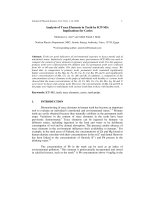
method, we obtained four oligostilbenes, namely balanocarpol (1) (300 mg),
heimiol A (2) (200 mg), vaticanol G (3) (70 mg) and vaticanol B (4) (200 mg).
The structures of these compounds (1–4) were established on the basis of their
spectral data, including UV, IR and NMR spectra in comparison with the
previously reported data
3–18
and by direct comparison with the authentic samples.
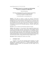
From the dried and milled stem bark of H. odorata (3.8 kg) was isolated four
componds, namely balanocarpol (1) (300 mg), hopeaphenol (5) (1500 mg),
ampelopsin H (6) (250 mg) and hemlesyanol C (7) (120 mg), whereas from the
dried and milled stem bark of H. nigra (4.6 kg) to give vaticanol G (3) (200 mg)
(Fig. 1).
Journal of Physical Science, Vol. 19(2), 7–21, 2008 9
Figure 1: Structure some compounds isolated from Hopea.
3. RESULTS AND DISCUSSION
Balanocarpol (1) was obtained as a pale yellow powder, m.p. 230
o
C, UV
(MeOH) λ
max
(log ε) : 227 (5.6); 283 (3.76) nm, IR (KBr) υ
max
: 3384; 1608; 1405;
1350; 1240; 1132; 1037; 995; 833 cm
–1
,
1
H and
13
C NMR (Me
2
CO-d
6
, 600.0 and
150 MHz) see Table 1. FABMS m/z 470 [M
+
] (C
28
H
22
O
7
).
O
OH
H
H
HO
OH
HO
OH
OH
H
H
O
HO
OH
OH
OH
HO
OH
H
H
H
H
HO
HO
HO
HO
OH
OH
OH
HO
HO
H
H
H
H
H
H
O
O
HO
HO
HO
HO
HO
HO
OH
OH
OH
H
H
H
H
H
H
H
H
OH
(1) (2) (3)
O
HO
HO
HO
OH
OH
H
H
H
O
OH
OH
HO
HO
HO
H
H
H
H
H
O
O
HO
HO
OH
H
H
OH OH
HH
HO
HO
OH
OH
OH
H
H
H
H
A1
A2
B1
B2
1a
4a
7a
8a
10a
12a
7b
8b
1b
4b
12b
14b
(5)
(4)
O
O
H
H
HO
OH
HO
OH
OH
H
H
H
H
H
H
OH
HO
OH
OH
OH
A1
A2
B2
B1
C1
C2
D1
D2
1a
4a
7a
8a
10a
12a
7b
8b
4b
12b
7c
8c
12c
7d
8d
4d
12d
4c
(7)
(6)
Resveratrol Derivatives from Hopea Stem Bark 10
Heimiol A (2) was obtained as a pale yellow powder, m.p. 240
o
C, UV
(MeOH) λ
max
(log ε) : 225 (6.01); 230 (sh 4.83); 282 (3.65) nm, IR (KBr) υ
max
:
3352; 1606; 1512; 1450; 1234; 1141; 1068; 954; 835 cm
–1
,
1
H and
13
C NMR
(Me
2
CO-d
6
, 600.0 and 150 MHz) see Table 1. FABMS m/z 471 [M+H]
+
(C
28
H
22
O
7
).
Vaticanol G (3) was obtained as a brown powder, m.p. 240
o
C, UV
(MeOH) λ
max
(log ε) : 208 (5.95); 234 (sh) (5.72); 280 (5.16)nm, IR (KBr) υ
max
:
3296; 1609; 1510; 1445; 1243; 1142; 1012; 833 cm
–1
,
1
H and
13
C NMR (Me
2
CO-
d
6
, 600.0 and 150 MHz) see Table 1. FABMS m/z 680 [M
+
] (C
42
H
32
O
9
).
Ampelopsin H (6) was obtained as a pale yellow powder, m.p. 240
o
C,
UV (MeOH) λ
max
(log ε) : 225 (6.01); 230 (sh 4.83); 282 (3.65) nm, IR (KBr)
υ
max
: 3352; 1606; 1512; 1450; 1234; 1141; 1068; 954; 835 cm
–1
,
1
H and
13
C
NMR (Me
2
CO-d
6
, 600.0 and 150 MHz) see Table 2. FABMS m/z 906 [M+H]
+
(C
56
H
42
O
12
).
Hemlesyanol C (7) was obtained as white brown powder, UV (MeOH)
λ
max
(log ε): 203 (5.31); 283 (4.33)nm, IR (KBr) υ
max
:
3200, 1612–1454, and 833
cm,
–1 1
H and
13
C NMR (Me
2
CO-d
6
, 600.0 and 150 MHz) see Table 2. FABMS
m/z 906 [M
+
] (C
56
H
42
O
12
).
Vaticanol B (4) and hopeaphenol (5) were identified with UV, IR and
TLC compared with authentic sample.
Table 1:
1
H and
13
C NMR data of compounds (1, 2 and 3)* in acetone-d
6
.
Balanocarpol (1) Heimiol (2) Vaticanol G (3) No
δ H
(m, J in Hz)
δ C δ H
(m, J in Hz)
δ C
δ H
(m, J in Hz)
δ C
1a - 133.7 - 136.8 - 139.8
2a,6a 7.48 (d, 8.8) 131.5 6.90 (d, 8.4) 127.9 6.45 (br s) 130.1
3a,5a 6.95 (d, 8.8) 116.4 6.69 (d, 8.4) 115.3 6.46 (br s) 114.6
4a - 159.2 - 157.2 7.89 (br s) 155.4
7a 5.70 (d, 9.5) 93.5 5.57 (br s) 81.5 4.55 (d, 4.3) 57.1
8a 5.16 (d, 9.5) 52.5 4.24 (br s) 46.9 4.63 (d, 4.3) 50.2
9a - 142.8 - 147.4 - 141.8
10a - 120.5 6.41 (d, 2.6) 107.4 - 125.9
11a - 157.4 - 157.1 8.01 (br s) 153.1
12a 6.09 (d, 2.2) 102.0 6.16 (d, 2.6) 102.0 6.20 (d, 2.8) 101.6
13a - 156.9 - 154.6 7.59 (br s) 155.8
14a 5.96 (d, 2.2) 106.8 - 116.0 5.67 (d, 2.8) 111.4
(continue on next page)
Table 1: (continued)
Balanocarpol (1) Heimiol (2) Vaticanol G (3) No
δ H
(m, J in Hz)
δ C δ H
(m, J in Hz)
δ C
δ H
(m, J in Hz)
δ C
1b - 133.4 - 136.9 - 129.1
2b,6b 6.75 (d, 9.5) 132.0 7.14 (d, 8.4) 130.0 - 141.6
3b,5b 6.42 (d, 9.5) 114.1 6.72 (d, 8.4) 115.5 6.07 (d, 2.6) 119.7
4b - 155.8 - 157.2 7.40 (br s) 154.8
7b 4.89 (br s) 50.2 4.32 (d, 3.3) 50.9 5.77
(dd, 8.4; 2.6)
112.7
8b OH 5.39 (br s) 4.32 (d, 4.4) 73.2 4.97 (d, 3.3) 81.4 6.02 (d, 8.4) 134.9
9b - 140.8 - 142.6 4.89 (d, 3.0) 42.6
10b - 113.9 6.48 (d, 2.2) 104.8 3.85 (dd, 8.9;
3.0)
53.8
11b - 159.2 - 158.1 - 146.9
12b 6.20 (d, 2.2) 95.1 6.21 (d, 2.2) 102.1 - 117.5
13b - 159.7 - 156.2 8.48 (br s) 154.9
14b 6.25 (d, 2.2) 104.5 - 117.0 6.46 (s) 101.8
1c 7.59 (br s) 152.8
2c 5.92 (br s) 127.7
3c 5.98 (br s) 114.6
4c (OH) 7.85 (br s) 156.4
5c 6.67 (br s) 116.2
6c 7.13 (br s) 130.3
7c 3.51 (d, 8.9) 62.9
8c
4.11 56.9
9c - 147.5
10c, 4c 5.96 (d, 2.6) 106.3
11c, 3c
(OH)
7.96 (br s) 158.9
12c 6.12 (t, 2.6; 2.6) 100.9
* measured with acetone-d
6
600.0 MHz (
1
H) and 150.0 MHz (
13
C)
Table 2:
1
H and
13
C NMR data of compounds (6 and 7)* in acetone-d
6
.
Ampelopsin H (6) Hemlesyanol C (7) No
δH ( m, J in Hz) δC δH ( m, J in Hz) δC
1a - 134.8 - 133.2
2a,6a 7.11 (d, 8.4) 127.3 7.58 (d, 8.4) 130.8
3a,5a 6.74 (d, 8.4) 116.1 6.91 (d, 8.4) 115.2
4a - 157.9 - 158.7
7a 5.31 (d, 2.0) 93.8 5.68 (d, 10.6) 94.7
8a 4.33 (d, 2.0) 57.1 5.35 (d, 10.6) 51.8
9a - 148.6 - 138.9
10a 6.29 (br s) 106.6 - 122.8
11a - 160.0 - 157.8
12a 6.32 (t, 2.1; 2.1) 102.2 6.23 (d, 2.2) 102.0
13a - 160.0 - 156.3
14a 6.29 (br s) 106.6 6.05 (d, 2.2) 107.9
1b - 138.8 - 133.4
2b,6b 6.73 (d, 8.4) 129.2 6.11 (d, 8.4) 133.5
3b,5b 6.56 (d, 8.4) 115.5 6.40 (d, 8.4) 114.8
4b - 155.9 - 156.3
7b 4.29 (s) 50.2 4.40 (d, 3.3) 46.2
8b 3.85 (s) 60.5 4.16 (t, 3.3; 3.3) 55.4
9b - 144.6 - 144.3
10b - 126.4 - 115.2
11b - 155.5 - 160.1
12b 6.21 (s) 96.7 6.00 (s) 96.2
13b - 163.2 - 154.7
14b - 116.2 - 122.8
1c - 134.8 - 136.3
2c,6c 7.11 (d, 8.4) 127.3 5.77 (d, 8.8) 129.5
3c,5c 6.74 (d, 8.4) 116.1 6.20 (d, 8.8) 115.1
4c - 157.9 -
156.1
7c 5.31 (d, 2.0) 93.8 3.88 (d, 5.8) 61.2
8c 4.33 (d, 2.0) 57.1 3.19 (d, 5.8) 56.7
9c - 148.6 - 147.5
10c 6.29 (br s) 106.6 - 119.1
11c - 160.0 - 162.8
12c 6.32 (t, 2.1; 2.1) 102.2 6.29 (d, 2.7) 95.4
(continue on next page)
Journal of Physical Science, Vol. 19(2), 7–21, 2008 13
Table 2: (continued)
Ampelopsin H (6) Hemlesyanol C (7) No
δH ( m, J in Hz) δC δH ( m, J in Hz) δC
13c - 160.0 - 160.2
14c 6.29 (br s) 106.6 5.91 (d, 2.7) 106.4
1d - 138.8 - 134.6
2d,6d 6.73 (d, 8.4) 129.2 7.07 (d, 8.4) 127.6
3d,5d 6.56 (d, 8.4) 115.5 6.85 (d, 8.4) 116.0
4d - 155.9 - 157.8
7d 4.29 (s) 50.2 5.08 (d, 3.3) 93.9
8d 3.85 (s) 60.5 3.65 (d, 3.3) 56.2
9d - 144.6 - 148.4
10d - 126.4 5.91 (d, 2.5) 106.6
11d - 155.5 - 160.1
12d 6.21 (s) 96.7 6.11 (d, 2.5) 106.6
13d - 163.2 - 160.1
14d - 116.2 5.91 (d, 2.5) 106.6
* measured with acetone-d
6
600.0 MHz (
1
H) and 150.0 MHz (
13
C)
Balanocarpol (1) was obtained as a pale yellow powder, m.p. 230
o
C. Its
UV spectrum showed absorption maximum at 283 nm suggesting the presence of
unconjugated phenolic chromophore. The IR spectrum exhibited hydroxyl group
(3384 cm
–1
), C=C aromatic (1608; 1405; 1350 cm
–1
), and monosubtituted
benzene (833 cm
–1
), these spectral characteristic absorptions supporting (1) to be
an oligoresveratrol. The positive ion FABMS exhibited an [M]
+
ion at m/z 470
consistent with a molecular formula C
28
H
22
O
7
for a resveratrol dimer and this
suggestion was supported by the NMR data.
13
C NMR spectra showed six signals
for oxyaryl carbon at δ 159.2 (C-4a), 157.4 (C-11a), 156.9 (C-13a), 155.8 (C-4b),
159.2 (C-11b) and 159.7 (C-13b) ppm, characteristics for resveratrol dimer.
Additionally, the
13
C NMR also exhibited one oxyalkyl carbon at δ 73.2 (C-8b),
indicating that C-8b was attached to a hydroxyl functional group. The
1
H NMR
spectrum of (1) in acetone-d
6
exhibited signals for two sets of 4-hydroxybenzene
at δ 7.48 (d, J = 8.8 Hz) and 6.95 (d, J = 8.8 Hz) ppm, each 2H (ring A1) and at
δ 6.75 (d, J = 9.5 Hz) and 6.42 (d, J = 9.5 Hz) ppm, each 2H (ring B1). The
1
H
NMR spectrum also showed two sets of meta-coupled aromatic protons signals at
δ 6.09 (d, J = 2.2 Hz) and 5.96 (d, J = 2.2 Hz) ppm, each 1H (ring A2), and
at δ 6.20 (d, J = 2.2 Hz) and 6.25 (d, J = 2.2 Hz) ppm, each 1H (ring B2).
Additionally, the
1
H NMR spectrum exhibited signals for a set of aliphatics
proton at δ 5.70 (d, J = 9.5 Hz) and 5.16 (d, J = 9.5 Hz), each 1H, characteristic
for trans-2,3-diaryl-dihydrobenzofuran moiety, and signals assignable two
Resveratrol Derivatives from Hopea Stem Bark 14
coupled aliphatic protons at δ 4.89 (br s) and 5.39 (br s) ppm, each 1H. These
spectral data indicated that compound (1) has a dimeric stilbene skeleton as part
of its structure.
Heimiol A (2) was obtained as a pale yellow powder, with of absorption
maxima observed at 225; 230 sh; 282 nm in the UV spectrum attributable to the
phenol rings. The IR spectrum exhibited hydroxyl group (3352 cm
–1
), C=C
aromatic (1606; 1512; 1450 cm
–1
) and monosubstituted benzene (835 cm
–1
). Its
molecular formula of C
28
H
22
O
7
was established by FABMS, showing a [M+H]
+
ion at m/z 471, together with its NMR spectral data, were evidence that (2) was
resveratrol dimer. The
1
H NMR (Table 2) and
1
H-
1
H COSY spectra showed two
sets of AA’BB’ system of aromatic protons assignable to two independent 4-
hydroxyphenyl groups at δ 6.90 (2H, d, J = 8.4 Hz) and 6.69 (2H, d, J = 8.4 Hz)
(ring A1), and δ 7.14 (2H, d, J = 8.4 Hz) and 6.72 (2H, d, J = 8.4 Hz) (ring B2),
two sets of meta-coupled aromatic protons at δ 6.41 (1H, d, J = 2.6 Hz) and δ
6.16 (1H, d, J = 2.6 Hz) (ring A2), 6.48 (1H, d, J = 2.2 Hz) and 6.21 (1H, d, J =
2.2 Hz) (ring B2) assignable to two units 1,2,3,5-tetrasubstituted benzene group.
They also displayed two set of coupled benzyl methine protons at δ 5.57 (1H, br
s) (7a), 4.24 (1H, br s) (8a), 4.32 (1H, d, J = 3.3 Hz) (7b), 4.97 (1H, d, J = 3.3
Hz) (8b). The
13
C NMR spectrum showed that C-7a (δ 81.5 ppm) and C-8b (δ
81.4 ppm) might both be attached to benzylic carbons bearing an oxygen atom.
The connection between protons and their corresponding carbons was established
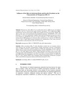
by HMQC. Further support for the structure (2) was obtained form HMBC
measurement (Fig. 2). The HMBC spectrum of (2) showed long-range
correlations between H-2a with C-7a (δ 81.5 ppm), confirming that a
4-hydroxyphenyl group was attached to an oxygen bearing carbon. Long-range
correlations were also observed for the methine proton between H-8b/C-7b
H-7b/C-10b, and H-8a/C-10b, pointing to a fused benzopyran-benzo-oxepane
structure, in the same pattern as those of heimiol A.
18
The relative configuration
of (2) was established on the basis of the NOESY spectrum (Fig. 2). The NOE
correlation showed that the H-8a and H-8b are in a syn configuration, deduced
from the NOE correlations between H-8b/H-7a/H-8a, as well as H-7b which did
not show any correlations. Therefore, it may be concluded that (2) is heimiol A, a
resveratrol dimer.
Journal of Physical Science, Vol. 19(2), 7–21, 2008 15
O
HO
OH
OH
OH
HO
OH
H
H
H
H
A1
A2
B1
B2
1a
4a
7a
8a
14a
12a
1b
4b
7b
8b
9b
10b
12b
14b
O
OH
H
H
HO
OH
HO
OH
OH
H
H
A1
A2
B1
B2
1b
4b
7b
9a
12a
14a
12b
14b
8b
4a
7a
Figure 2: Significant HMBC of (a) balanocarpol (1) and (b) heimiol A (2).
Vaticanol G (3) was obtained as a brown powder, m.p. 240
o
C. Its UV
spectrum showed absorption maximum at 280 nm, suggesting the presence of
unconjugated phenolic chromophore. The IR spectrum exhibited hydroxyl group
(3296 cm
–1
), C=C aromatic (1609; 1510; 1445 cm
–1
) and monosubstituted
benzene (833 cm
–1
). These were characteristic spectral data for supporting (3) to
be an oligostilbene. The positive ion FABMS exhibited an [M]
+
ion at m/z 680,
which together with NMR data, were consistent with a molecular formula
C
42
H
32
O
9
, for a resveratrol trimer. The
1
H NMR spectrum of (3) in acetone-d
6
exhibited signals for two sets of 4-hydroxybenzene at δ 6.45 (br s) and 6.46
(br s), each 2H, at δ 7.13 (br s), 6.67 (br s), 5.98 (br s), and 5.92 (br s), each 1H
(rings of A1 and C1), and one unit of a 1,2,4-trisubstituted benzene at δ 6.07 (1H,
d, J = 2.6 Hz); 6.02 (1H, d, J = 8.4 Hz). Additionally, the
1
H NMR spectrum
exhibited signals for a set of aromatic signals at δ 5.77 (1H, dd, J = 8.4; 2.6 Hz )
(ring B1), one unit of a 1,3,5-trisubstituted benzene at δ 6.12 (1H, t, J = 2.6; 2.6
Hz) and 5.96 (2H, d, J = 2.6 Hz) (ring C2), one unit of a 1,2,3,5-tetrasubstituted
benzene at δ 6.20 (1H, d, J = 2.8 Hz) and 5.67 (1H, d, J = 2.8 Hz) (ring A2), and
one unit of a 1,2,6-trisubstituted-3,5-dihydroxibenzene δ 6.46 (s), (ring B2). The
six substituted benzene rings suggested 24 DBE (double bond equivalents).
Beside that, the
1
H NMR spectrum exhibited two aliphatic proton signals which
correlated at
1
H-
1
H COSY spectrum, characteristic of a unit -CH-CH- [δ 4.63
(1H, d, J = 4.3 Hz) and 4.55 (1H, d, J = 4.3 Hz) (unit D)], and four signals
assignable to two-coupled aliphatic protons characteristic with unit of -CH-CH-
CH-CH- [δ 4.89 (1H, d, J = 3.0 Hz), 3.85 (1H, dd, J = 8.9. 3.0 Hz), 3.51 (1H, d, J
= 8.9 Hz) and 4.11 (1H, s) (unit E)]. The characteristic aliphatic proton signal due
to a trans-2,3-diaryl-dihydrobenzofuran moiety, was not observed, suggesting
that (3) was a trimeric resveratrol with an aliphatic tricyclic skeleton similar to
that of vaticanol G isolated from Vatica rassak.
8
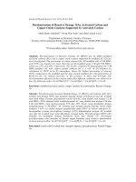
Complete assignment of all
Resveratrol Derivatives from Hopea Stem Bark 16
proton-bearing carbon signals were made possible by analysis of the HMQC
spectrum, and support for structure (3) was obtained from significant cross-peaks
in HMBC measurement (Fig. 3).
Ampelopsin H (6) was obtained as a pale yellow powder, with absorption
maxima observed at 282 nm in the UV spectrum attributable to the phenol rings.
The IR spectrum exhibited hydroxyl group (3352 cm
–1
), C=C aromatic
(1606–1512 cm
–1
), and monosubstituted benzene (835 cm
–1
). Its molecular
formula of C
56
H
42
O
12
was established by FABMS, showing a [M+H]
+
ion at m/z
906, which together with the NMR spectral data, suggested that (5) was a
resveratrol tetramer. The NMR data (
1
H and
13
C), however showed number of
signal corresponding to half the molecular formula, so was suggested that
compound (5) composed of two symmetrical structural units, and each unit was a
resveratrol dimmer (Table 2). The
1
H NMR spectrum of (5) in acetone-d
6
exhibited signals for two sets of 4-hydroxybenzene at δ 7.11 (2H, d, J = 8.4 Hz)
and 6.74 (2H, d, J = 8.4 Hz) ppm, with δ 6.73 (2H, d, J = 8.4 Hz) and 6.56 (2H,
d, J = 8.4 Hz) ppm. The
1
H NMR spectrum also showed two sets of meta-coupled
aromatic protons signals at δ 6.32 (1H, t, J = 2.1; 2.1 Hz) ppm and 6.29 (2H, br s)
ppm indicating the presence of a 3,5-hydroxyphenyl group. Furthermore, the
aromatic proton signal at 6.21 (1H, s) ppm showed existence of a penta-
substituted benzena ring. Two proton signals at δ 5.31 (1H, d, J = 2.0 Hz) ppm
and δ 4.33 (1H, d, J = 2.0 Hz) ppm showed existence of a trans-
dihydrobenzofuran ring. Two proton signals at δ 4.29 (s) ppm and δ 3.85 (s) ppm
indicated that both protons were at different locations.
Figure 3: Significant HMBC (H→C) correlations of vaticanol G (3).
HO
HO
HO
OH
OH
OH
OH
OH
HO
H
H
H
H
H
H
A2
A1
B1
B2
C1
C2
1a
3a
7a
8a
9a
10a
12a
7b
8b
10b
12b
14b
7c
9c
10c
12c
14c
1c
4c
2b
5b
6b
8c
Journal of Physical Science, Vol. 19(2), 7–21, 2008 17
Hemlesyanol C (7), was a brown amorphous powder, with absorption
band (283 nm) in the UV spectrum showing the presence of aromatic rings. The
IR spectrum exhibited hydroxyl group (3200 cm
–1
), C=C aromatic (1612–1454
cm
–1
) and monosubstituted benzene (833 cm
–1
). The [M
+
] ion peak at m/z 906,
corresponded to the molecular formula C
56
H
42
O
12
. The
1
H-NMR spectrum
(Table 2), showed the signals assignable to four 4-hydroxyphenyl groups at δ
7.58 (2H, d, J = 8.4), 6.91 (2H, d, J = 8.4 Hz), 6.11 (2H, d, J = 8.4 Hz), 6.40 (2H,
d, J = 8.4 Hz), δ 5.77 (2H, d, J = 8.8 Hz), 6.20 (2H, d, J = 8.8 Hz), 7.07 (2H, d, J
= 8.4 Hz) and 6.85 (2H, d, J = 8.4 Hz). The presence of a 3,5-dihydroxyphenyl
group at δ 5.91 (2H, d, J = 2.5 Hz) H-10d and 14-d, δ 6.11 (d, J = 2.5 Hz)
H-12d, and two sets of meta-coupled aromatic protons on 1,2,3,5-tetrasubstituted
benzene rings at δ 6.23 (d, J = 2.2 Hz), H-12a; δ 6.05 (d, J = 2.2 Hz), H-14a; δ
6.29 (d, J = 2.7 Hz), H-12c and δ 5.91 (d, J = 2.7 Hz), H-14c were also exhibited.
The spectrum further showed the signals due to an aromatic proton on a
pentasubstituted benzene ring at δ 6.00 (s), H-12b, a sequence of four aliphatic
methine protons coupled successively in the COSY spectrum in the order δ 4.40
(d, J = 3.3 Hz), H-7b; δ 4.16 (t, J = 3.3; 3.3 Hz), H-8b; δ 3.88 (d, J = 5.8 Hz), H-
7c and δ 3.19 (d, J = 5.8 Hz), H-8c, and two sets of mutually coupled aliphatic
protons δ 5.68 (d, J = 10.6 Hz), H-7a and δ 5.35 (d, J = 10.6 Hz), H-8a; δ 5.08
(d, J = 3.3 Hz), H-7d and δ 3.65 (d, J = 3.3 Hz), H-8d, in addition to ten phenolic
hydroxyl groups (δ 6.46–8.57) ppm. These results suggested that compound
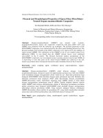
was a stilbene composed of four resveratrol units. Analysis of the HMQC and
HMBC spectra enabled the complete assignments of all protonated carbons
and quarternary carbons corresponding to respective resveratrol units (A–D).
The HMBC spectrum (Fig. 4) showed cross peaks indicating long range
correlations between H-7b/C-14b, C-8c, H-8b/C-14b, and C-9c, H-7c/C-14b; H-
8c/C-14b; and C-8b. Therefore, it may be concluded that the (7) is hemlesyanol
C, a resveratrol tetramer, isolated from Shorea hemsleyana for the first time.
5
O
O
H
H
HO
OH
HO
OH
OH
H
H
H
H
H
H
OH
HO
OH
OH
OH
A1
A2
B2
B1
C1
C2
D1
D2
1a
4a
7a
8a
10a
12a
7b
8b
4b
12b
7c
8c
12c
7d
8d
4d
12d
4c
14b
9c
10b
Figure 4; Significant HMBC (H→C) correlations of hemlesyanol C (7).
Resveratrol Derivatives from Hopea Stem Bark 18
Activity test as antioxidants based on radical scavenger activity using the
Halliwel method,
19
is shown at Table 3. The data IC
50
showed that the activity as
radical hydroxyl scavenger from hopeaphenol (5) was more active than ascorbic
acid, and the IC
50
of oligoresveratrol, balanocarpol (1), heimiol A (2), vaticanol B
(4) and ampelopsin H (6) showed them to be less active. For oligoresveratrol, that
activity as hydroxyl radical scavenger was due to the existence of phenol ring,
stability of molecular structure and existence of double bonds of olefinic unit.
Phenol ring can trap hydroxyl radical by releasing hydrogen radical, by
condensation with hydroxyl radical and form water molecules, whereas radical
phenol will be stabilized by resonance. That is, resveratrol compound is referred
for development as antioxidant. An antioxidant is substance that can prevent or
slow down the reactions of radical oxidation. The role antioxidant in body is to
reduce the amount free radicals, like ROS (reactive oxygen species) that can be
formed in course of metabolism in organism. Antioxidant also can function to
protect low density lipoprotein (LDL) from oxidation reaction, thus preventing
the occurrence of arteriosclerosis.
The in vitro cytotoxicity test was investigated using plate with 96 wells,
with cell density 2 x 10
4
cells per ml. Into each well was added 100 µl cells in
culture medium (87.5% RPMI 10.4 g l
–1
; 2% penstrep; and 10% FBS) which was
then incubated in CO
2
incubator for 12–24 h at 37°C. Each sample was dissolved
in culture medium containing 0.05% DMSO, and 100 μl of each sample in
different concentrations was added into each well in triplicate and was then
incubated in CO
2
incubator for 12–24 h at 37
o
C. MTT solution (10 μl per 100 μl
medium) was added to all wells of an assay, and plates were incubated for 4 h
at 37
o
C in CO
2
incubator. As much as 100 μl formazon (10% SDS and
0.01 M hydrochloric acid) was added into each well and mixed on a shaker for
Table 3: Data of activity test as radical scavengers.
Sample IC
50
(µg ml
–1
)
Observation
Balanocarpol (1) 1802.3 Less active
Heimiol A (2) 4575.3 Less active
Vaticanol G (3) 683.96 Active
Vaticanol B (4) 2146.6 Less active
Hopheaphenol (5) 61.8 High active
Ampelopsin H (6) 4840.0 Less active
Hemlesyanol C (7) 425.5 Active
Ascorbic acid 83.9 High active
Butylated Hydroxy Toluene (BHT) 1328.1 Less active
Note: IC
50
< 100 μg ml
–1
: highly active; 100–1000 μg ml
–1
: active; and 1000–5000 μg ml
–1
: less
active; > 5000 μg ml
–1
: not active
17
Journal of Physical Science, Vol. 19(2), 7–21, 2008 19
5 min. The wells were incubated in the dark room for 12–24 h at room
temperature. The absorbance was measured using multiwell scanning
spectrophotometers (ELISA reader) at wavelength 595 nm. The absorbance is
directly proportional to the number of living cells. So the dead cell could be
calculated to determine LC
50
. Doxorubicin, a medicine for lymphoma, leukaemia
and acute tumor, was also measured its cytotoxic activity as standard comparison.
The cytotoxic activity of the samples against HeLa-S3 cell measured as LC
50
were provided in Table 4. HeLa-S3, a continuous cell line that lived as adherent
cell, is a cell derivate of ephythell cell of human cervix cancer. Further
investigation of cytotoxic activity of the samples was held against Raji cell
(Table 4), the cell that resembles lymphoblast cell found by R.J.V. Pulvertaft
(1963) from Burkitt’s lymphoma at the left of the upper jaw of an 11 year old
negro boy. Table 4 shows that the highest cytotoxic activity against HeLa-S3 and
Raji cell is ampelopsin H (6). This compound is more active than doxorubicin. In
the other hand, heimiol A (2) and vaticanol G (3) showed the lowest cytotoxic
activity against HeLa-S3 and Raji cell. It is necessary to carry out further
investigation about the relationship between the structure and the activities of
these compounds. Some studies of curcumin that has been known as anticancer
indicated that the existence of hydroxyl group at ortho position and
β
-diketone
gave a big contribution as inducer of enzymes in phase two that their function as
protector from carcinogenesis as epoxy hydrolyse, glutathione S-transferase
(GST) and NAD(P)H quinone reductase (QR).
Table 4: LC
50
of some compounds from stem bark of Hopea against HeLa-S3 and Raji
cell.
No Sample HeLa S3 Raji
LC
50
(µg ml
–1
)
Observation LC
50
(µg ml
–1
)
Observation
1 Balanocarpol (1) 682.16 Less active 235.29 Active
2 Heimiol A (2) Very high Not active Very high Not active
3 Vaticanol G (3) Very high Not active Very high Not active
4 Vaticanol B (4) 92.81 Very active 34.45 Very active
5 Hopeaphenol (5) 1931.52 Less active 781.49 Less active
6 Ampelopsin H (6) 129.72 Active 34.69 Very active
7 Hemsleyanol C (7) 557.44 Less active 292.15 Less active
8 Doxorubisin (positive control) 96.82 Very active 94.38 Very active
Resveratrol Derivatives from Hopea Stem Bark 20
4. CONCLUSION
In this paper, we concluded that resveratrol derivatives isolated from the
stem bark of Hopea consist of dimer, trimer and tetramer resveratrol. Some
compounds have biological activity as antioxidant and cytotoxic effect against
Raji and HeLa-S3 cell lines. Hopeaphenol (5) showed the highest activity as
antioxidant, whereas ampelopsin H (6) and vaticanol B (4) gave the highest
cytotoxic effect against HeLa-S3 and Raji cell.
5. ACKNOWLEDGMENT
This work has been supported by competitive grant (Insentif Riset Dasar,
Ristek-2008), Ministry Research and Technology, Republic of Indonesia and
Fundamental Research from DIKTI (2007; 2008). The authors are grateful to the
experimental Garden in Carita, Pandeglang, Banten, Indonesia and Herbarium
Bogoriensis for the sample gift and the identification of the plant specimen.
6. REFERENCES
1. Cronquist , A. (1981). An integrated system of classification of flowering
plants. New York: Columbia In Press.
2. Newman, M.F. (1999). Pedoman identifikasi pohon-pohon
Dipterocarpaceae: Sumatera. Bogor: Prosea Indonesia.
3. Heyne, K. (1987). Tumbuhan berguna Indonesia, Vol. III. Jakarta: Badan
Litbang Kehutanan, 1390–1443.
4. Dai, J.R., Hallock, Y.F., Cardellina, J.H. & Boyd, M.R. (1998). HIV-
inhihibitory and cytotoxic oligostilbenoids isolated from the leaves of
Hopea malibato. J. Nat. Prod., 61, 351–353.
5. Ito, T., Tanaka, T., Ido, Y., Nakaya, K., linuma, M. & Riswan, S. (2000).
Stilbenoids isolated from stem bark of Shorea hemsleyana. Chem.
Pharm. Bull., 48(7), 1001–1005.
6. Ito, T., Tanaka, T., Ido, Y., Nakaya, K., linuma, M. & Riswan, S. (2000).
Four new stilbene C-glycosides isolated from the stem bark of Shorea
hemsleyana. Chem. Pharm. Bull., 48(12), 1959–1963.
7. Ito, T., Tanaka, T., Nakaya, K., linuma M., Takahashi, Y., Naganawa, H.,
Ohyama, M., Nakanishi, Y., Bastow, K.F. & Lee, K H. (2001). A new
resveratrol octamer, vateriaphenol A, in Vateria indica. Tetrahedron
Letters, 42, 5909–5912.
Journal of Physical Science, Vol. 19(2), 7–21, 2008 21
8. Ito, T., Tanaka, T., Ido, Y., Nakaya, K., linuma, M., Takahashi, Y.,
Naganawa, H., Ohyama, M., Nakanishi, Y., Bastow, K.F. & Lee, K H.
(2001). A novel bridged stilbenoid trimer and four highly condensed
stilbenoid oligomers in Vatica rassak. Tetrahedron, 57, 7309–7314.
9. Jang, M., Cai, L., Udeani, G.O., Slowing, K.V., Thomas, C.F., Beecher,
C.W.W., Fong, H.H.S., Farnsworth, N.R., Kinghorn, A.D., Mehta, R.G.,
Moon, R.C. & Pezzuto, J.M. (1997). Cancer chemopreventive activity of
resveratrol, a natural product derived from grapes. Science, 275, 218–220
10. Pryce, R.J. & Langcake, P. (1977). (-)-α-Viniferin: An antifungal
resveratrol trimer from grapevines. Phytochemistry, 16, 1452–1454.
11. Tanaka, T., Ito, T., Ido, Y., Son, T.K., Nakaya, K., linuma, M., Ohyama,
M. & Chelladurai, V.M. (2000). Stilbenoids in the stem bark of Hopea
parviflora. Phytochemistry, 53(8), 1015–1019.
12. Tanaka, T., Ito, T., Nakaya, K., linuma, M. & Riswan, S. (2000).
Oligostilbenoids in the stem bark of Vatica rassak. Phytochemistry,
54, 63–69.
13. Tanaka, T., Ito, T., Nakaya, K., linuma, M., Takahashi, Y., Naganawa,
H., Matsuura, N. & Ubukata, M. (2000). Vaticanol D, a novel resveratrol
hexamer isolated from Vatica rassak. Tetrahedron Letters, 41,
7929–7932.
14. Tanaka, T., Ito, T., Nakaya, K. linuma, M., Takahashi, Y., Naganawa,
H. & Riswan, S. (2001). Six new heterocyclic stilbene oligomers from
stem bark of Shorea hemsleyana. Heterocycles, 55, 729–741.
15. Sri Atun, Nurfina, A., Retno, A. & Niwa, M. (2005). A trimer stilbenoids
compound from stem bark Hopea nigra (Dipterocarpaceae). Indo. J.
Chem., 5(3), 211–214.
16. Sri Atun, Nurfina, A., Retno, A. & Niwa, M. (2006). Balanocarpol and
heimiol A, two resveratrol dimers from stem bark Hopea mengarawan
(Dipterocarpaceae). Indo. J. Chem., 6(1), 75–78.
17. Sri Atun, Sjamsul, A.A., Niwa, M., Retno, A. & Nurfina, A. (2006).
Oligostilbenoids from Hopea mengarawan (Dipterocarpaceae). Biochem.
System. and Ecol., 34, 642–644.
18. Weber, J.F., Wahab, I.A., Marzuki, A., Thomas, N.F., Kadir, A.A.,
Hamid, A., Hadi, A., Awang, K., Latif, A.A., Richomme, P. & Deaunay
J. (2001). Heimiol A, a new dimeric stilbenoid from Neobalanocarpus
heimii. Tetrahedron Letters, 42, 4895–4897.
19. Halliwel, B., Gutteridge, J.M.C. & Aruoma, O.I. (1987). The
deoxyribose method: A simple test tub assay for determination of rate
constans for reaction of hydroxyl radicals. Anal. Biochem., 165, 215–219.