Báo cáo khoa học: "Overexpression of cyclin D1 and cyclin E in 1,2-dimethylhydrazine dihydrochloride-induced rat colon carcinogenesis" potx
Bạn đang xem bản rút gọn của tài liệu. Xem và tải ngay bản đầy đủ của tài liệu tại đây (184.88 KB, 6 trang )
-2851$/# 2)
9HWHULQDU\#
6FLHQFH
J. Vet. Sci. (2000),1(2), 121–126
Overexpression of cyclin D1 and cyclin E in 1,2-dimethylhydrazine
dihydrochloride-induced rat colon carcinogenesis
Kwon Hur, Jung-Rae Kim, Byung-Il Yoon
1
, Jung-Keun Lee, Jae-Hoon Choi, Goo-Taeg Oh
2
and
Dae-Yong Kim
*
Department of Veterinary Pathology, College of Veterinary Medicine and School of Agricultural Biotechnology, Seoul National
University, Suwon, 441-744, Korea
1
National Institute of Health and Sciences, Tokyo 158-8501 Japan
2
Korea Research Institute of Bioscience and Biotechnology, Taejon, 305-333 Korea
Deregulation of G1 cyclins has been reported in several
human and rodent tumors including colon cancer. To
investigate the expression pattern of G1 cyclins in 1,2-
dimethyl-hydrazine dihydrochloride (DMH)-induced rat
colon carcinogenesis, we studied the expression of cyclin
D1 and cyclin E by quantitative reverse transcription-
polymerase chain reaction (RT-PCR) analysis and
immunohistochemistry (IHC). The mRNA level of cyclin
D1 was increased 1.2-fold in adenocarcinomas but not
significantly in adenomas, when compared with normal
rat colonic mucosa (p<0.05). The cyclin E mRNA level
was increased 2.7-fold in adenomas and 3.3-fold in
adenocarcinomas (p<0.05). The PCNA mRNA level was
also increased 1.9-fold in adenomas and 1.8-fold in
adenocarcinomas (p<0.05). Immunohistochemical staining
revealed exclusive nuclear staining of the neoplastic cells
for cyclin D1, cyclin E and PCNA. Cyclin D1 expression
was detected in 56.3% of the adenomas and in 61.5% of
the adenocarcinomas examined, whereas cyclin E
expression was detected in 87.5% of the adenomas and in
92.3% of the adenocarcinomas. Overall, cyclin D1, cyclin
E and PCNA expression was significantly increased at
both the mRNA and protein levels in normal colonic
mucosa, adenomas and adenocarcinomas, but there was
no significant difference in the degree of expression of
these genes in adenomas and adenocarcinomas. Our
results indicate that the overexpression of cyclin D1 and
cyclin E may play an important role during the multistage
process of rat colon carcinogenesis, at a relatively early
stage, and may disturb cell-cycle control in benign
adenomas, and thereafter, participate in tumor
progression.
Key words:
cell cycle, cyclin D1, cyclin E, colon cancer
Introduction
Colorectal cancer in humans is one of the most common
malignancies in the world [6]. Colorectal carcinogenesis is
characterized by multiple genetic alterations and is
preceded by a series of histopathologically recognizable
precancerous lesions that progress to adenocarcinoma over
a period of a year [6]. As with other tumors, cell
proliferation is central to tumor progression in colorectal
cancer [23] and therefore, it is essential to understand the
mechanism and significance of altered cell cycle regulation.
Progression of the cell cycle is regulated by the
sequential formation and degradation of multiple cyclins
that bind to and stimulate the activities of a series of
cyclin-dependent kinases (CDKs) [10, 29]. For example,
cyclin D1 functionally forms a complex with CDK4 and
CDK6, whereas cyclin E complexes with CDK2 during
the G1/S phase [13, 31]. Recent studies have identified
additional regulators of the cell cycle, such as p21
WAF1/CIP1
,
p27
KIP1
, p16
MTS1
and p15
MTS2
tumor suppressor genes, which
bind to the cyclin-CDK complex and inhibit kinase
activities [11, 28]. Altered expression of cell cycle
regulators and the subsequent deregulation of the cell cycle
may be important steps in carcinogenesis and are the most
consistently found events in human malignancies
including colorectal cancer [5, 8]. Among the G1 cyclins,
cyclin D1 and cyclin E are key regulators during the G1/S
cell cycle transition, and perhaps the most important
checkpoint in the mammalian cell cycle [21]. Increased
expression of cyclin D1 and cyclin E has been reported in
various human tumors [1, 15, 17, 18, 25, 32] and several
carcinogen-induced mouse and rat tumor models
[9, 16, 20, 24, 25, 27, 30, 34].
However, there has been insufficient study of the
expression of cyclin D1 and cyclin E in carcinogen-
induced rat colonic carcinogenesis. The purpose of this
*Corresponding author
Phone: +82-331-290-2749; Fax: +82-331-293-6403
E-mail:
122 Kwon Hur et al.
study was to determine whether 1,2-dimethyl-hydrazine
dihydrochloride (DMH)-induced rat colon tumors display
altered expressions of cyclin D1 and cyclin E and to
discover these alterations are linked to cell proliferative
activity in this model.
Materials and Methods
Animals and treatments
Six-week-old, male, Sprague-Dawley rats were purchased
from Charles River Japan (Kanagawa, Japan) and
maintained in a temperature (21 ± 2
o
C) and humidity (50 ±
3%) controlled environment with a 12 hrs light/dark
illumination cycle. The rats were fed a commercial diet
(Jeil Jedang, Co.) and water ad libitum. After a 2-week
acclimatization period, one group of 50 rats was treated
with DMH (Sigma, USA) by subcutaneous injection of 15
mg/Kg body weight once per week for 20 weeks. To
prevent skin irritation during injection, pH of DMH was
adjusted to 6.5. Twenty rats treated with saline in the same
way served as controls. All animals were sacrificed at
week 40 from the initiation of treatment. After both ends
were ligated, the entire colons were injected with saline,
cut along the longitudinal axis, and the neoplastic nodules
harvested. Approximately half of the tumor tissues
removed were snap frozen in liquid nitrogen and stored at
-70
o
C until analysis. Normal colonic tissues of the control
group were also harvested. For histopathology and
immunohistochemistry (IHC), the remaining tumor tissues
were fixed in 10% neutral phosphate-buffered formalin,
routinely processed and embedded in paraffin. During 40
week exposure 3 rats died. The remaining 47 rats were
examined for tumor development and 35 rats were found
to have tumor (74.5% incidence). The neoplastic nodules
from each mouse were classified as either adenoma or
adenocarcinoma. Sixteen adenoma and 13 adenocarcinoma
were used for RT-PCR analysis of cyclin D1 and cyclin E.
RNA isolation and quantitative RT-PCR analysis
Frozen tissue specimens were ground with liquid nitrogen
and total cellular RNA isolated, based on the method of
Chomczynski et al [4]. Two-step quantitative RT-PCR
analysis was performed as previously described [19]. Two
µ
g of total RNA was reverse transcribed into first strand
cDNA in a volume of 25
µ
l at 37
o
C for 60 min using a first
strand cDNA synthesis kit (Novagen, Madison, WI), and
heated at 95
o
C for 5 min to terminate the reverse
transcription reaction. Cyclin D1, cyclin E, proliferating
cell nuclear antigen (PCNA) and hypoxanthine-guanine
phosphoribosyltransferase (HPRT: housekeeping gene) genes
were amplified from 2
µ
l cDNA mixtures in a final
volume of 20
µ
l PCR reaction mixture containing, 10 mM
Tris-HCl (pH 8.3), 50 mM KCl, 2.5 mM MgCl
2
, 2 mM
each of dNTPs, 0.25 µM each of sense and antisense
primers (Bioneer, Seoul, Korea), 1.25 U Taq DNA
polymerase (Bioneer, Seoul, Korea) and [
α
-
32
P]dCTP
(3000 Ci/mmol, Amersham, Arlington Heights, IL). The
PCR reactions were carried out using a Perkin-Elmer
Thermocycler 9600 (Perkin-Elmer, Norwalk, CT). Reaction
mixtures were first denatured at 95
o
C for 5 min, and
amplification was performed for 35 cycles , at 95
o
C for 45
sec, 60
o
C for cyclin D1 (61
o
C for cyclin E, 58
o
C for PCNA,
and 61.5
o
C for HPRT) for 1 min, and at 72
o
C for 1 min,
followed by an extension for 7 min at 72
o
C. Primer sets for
the PCR amplification of cyclin D1, cyclin E, PCNA and
HPRT genes were selected based on published sequences.
The PCR primer pairs used were as follows:-cyclin D1,
sense, 5’-TGGAGCCCCTGAAGAAGAG-3’ and antisense,
5’-AAGTGCGTTGTGCGGTAGC-3’; cyclin E, sense, 5’-
CTGGCTGAATGTTTATGTCC-3’ and antisense 5’-TC-
TTTGCTTGGGCTTTGTCC-3’; PCNA, sense, 5’-GC-
CCTCAAAGACCTCAT CAA-3’ and antisense, 5’-GC-
TCCCCACTCGCAGAAAAC-3’; and HPRT, sense, 5’-
CGGGGGAC ATAAAAGTTAT-3’ and antisense, 5’-GG-
ACGCAGCAACAGACATT-3’. After running the amplified
PCR products of each gene on a 1.8% agarose gel, the gels
were dried at 80
o
C for 60 min and exposed to a
Phosphoimaging plate (Fuji, Minami-Ashigara) for 3 days.
After autoradiography, the imaging plate was scanned on
an Image Reader BAS-2500 (Fuji, Tokyo). For
quantification of the RT-PCR products, the levels of
incorporated [
α
-
32
P]dCTP in each band were measured
with a liquid scintillation counter (Walac, OY, Finland).
The radioactivity in the cyclin D1, cyclin E and PCNA
band was normalized to the radioactivity of the
corresponding HPRT internal control band.
Immunohistochemical staining
Immunohistochemical staining was performed to detect
the degree of cyclin D1, cyclin E and PCNA expression on
replicate sections of the selected neoplastic tissues used for
RT-PCR analysis. Tissue sections were placed on Probe-
On slides (Fisher scientific, Pittsburgh, PA), deparaffinized
and rehydrated. After inhibiting endogenous peroxidase
activity with methanol containing 3% H
2
O
2
, tissue sections
were heated in 10 mM sodium citrate (pH 6.0) in a
pressure cooker for 6 min for antigen retrieval. After
blocking non-specific binding by treating the slides with
10 % normal goat serum at 37
o
C for 60 min, the slides
were incubated at 4
o
C overnight with commercially
available antibodies to cyclin D1 (mouse monoclonal;
Santa Cruz Biotech., Santa Cruz, CA), cyclin E (rabbit
polyclonal; Santa Cruz Biotech., Santa Cruz, CA) and
PCNA (mouse monoclonal; Novocatra, Newcastle, UK) at
1 : 100, 1 : 100 and 1 : 200 dilutions, respectively. After
washing, the sections were incubated with biotinylated
goat anti-mouse IgG or goat anti-rabbit IgG (Vector Lab,
Burlingame, CA) at 37
o
C for 60 min. Sections were then
Cyclin D1 and E expression in rat colon tumor 123
washed and incubated with Streptavidin (DAKO,
Copenhagen, Denmark) at 37
o
C for 60 min. 3,3-
diaminobenzidine was used as a chromogen to show the
antigen and sections were counterstained with Harris
hematoxylin. Negative control tissues were prepared in the
same manner as that described above, except for the
omission of primary antibodies and the substitution of an
isotype-matched but irrelevant antibody.
Results
Quantitative RT-PCR analysis of cyclin D1, cyclin E
and PCNA mRNA expression
Quantitative RT-PCR analysis of the tissue samples using
primers specific for cyclin D1, cyclin E, PCNA and HPRT
revealed product bands of the expected size. The results of
autoradiography are shown in Figure 1. The width and
intensity of the cyclin D1, cyclin E and PCNA bands were
markedly increased from those normal mucosa (Fig. 1A)
in both adenomas (Fig. 1B) and adenocarcinomas (Fig.
1C). The mRNA levels of cyclin D1, cyclin E and PCNA
in each stage, as quantified by measuring the radioactivity
of each band, are shown in Figure 2. The mRNA levels of
cyclin D1 (Fig. 2A), cyclin E (Fig. 2B) and PCNA (Fig.
2C) were all significantly increased in the tumor tissues
compared with normal colon tissues. The mRNA level of
cyclin D1 was increased 1.2-fold in adenocarcinomas
(p<0.05) but in adenomas it was not significantly
increased. Cyclin E mRNA was increased 2.7-fold in
adenomas and 3.3-fold in adenocarcinomas (p<0.05)
compared to normal mucosa. The proliferative activity of
the tumor cells as determined by PCNA mRNA level was
also increased, 1.9-fold in adenomas and 1.8-fold in
adenocarcinomas (p<0.05), respectively. However, there
were no significant differences in the cyclin D1, cyclin E
and PCNA mRNA expression levels of adenomas and
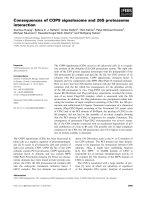
Fig. 1.
RT-PCR analysis of cyclin D1, cyclin E and PCNA
mRNA levels using HPRT as an internal control in the rat colon
carcinogenesis model. (A) Normal colorectal mucosa from the
control group. (B) adenomas harvested from DMH-treated rats.
(C) adenocarcinomas harvested from DMH-treated rats.
Fig. 2.
Quantitation of cyclin D1, cyclin E and PCNA mRNA
expression by quantitative RT-PCR analysis. Bars represent
levels of incorporation of [
α
-
32
P]dCTP in cyclin D1, cyclin E and
PCNA PCR products after normalization to HPRT, by measuring
the radioactivity (c.p.m.) of each band in Figure 1. Results
quoted are the mean ± SE of each group of tissues. mRNA levels
of (A) cyclin D1, (B) cyclin E and (C) PCNA. NS, not
significant. *, P<0.05. N, normal colonic mucosa. A, colonic
adenomas. C, colonic adenocarcinomas.
124 Kwon Hur et al.
adenocarcinomas (p<0.05, Fig. 2).
Immunohistochemical analysis of cyclin D1, cyclin E
and PCNA protein expression
Before IHC, neoplastic nodules were examined micro-
scopically and classified as normal mucosa, adenomas or
adenocarcinomas, respectively. Sixteen adenomas and 13
adenocarcinomas from different rats and 10 normal
colonic mucosa were selected for immunohistochemical
study.
Immunoreactivity for cyclin D1, cyclin E and PCNA
was confined predominantly to the nuclei of the neoplastic
cells (Fig. 3, C-H). Normal colonic mucosa showed only
weak to undetectable staining for cyclin D1 and cyclin E,
whereas positively stained cells for PCNA were primarily
detected in the basal layer of normal colonic mucosa. The
number and distribution of cyclin D1- and cyclin E-
positive cells in both adenomas and adenocarcinomas was
generally variable and heterogeneous, whereas PCNA was
diffusely positive, and irrespective of cyclin D1 and cyclin
E positivity. PCNA protein was expressed in almost all the
tumor cells of the adenomas and adenocarcinomas
examined, but the topological distribution of PCNA-
positive cells was not colocalized with that of cyclin D1-
and cyclin E-positive cells (Fig. 3, C-H). Cyclin D1
immunoreactivity was noted in 9/16 (56.3%) of the
adenomas and in 8/13 (61.5%) of the adenocarcinomas
examined (Fig. 3, E and F). Cyclin E expression was
detected in 14/16 (87.5%) of the adenomas and in 12/13
(92.3%) of the adenocarcinomas (Fig. 3, G and H).
Although the staining intensity of cyclin D1 and cyclin E
was variable among the cases studied, the degree of
immunoreactivity was generally weak in adenomas and
relatively strong in adenocarcinomas.
Discussion
The deregulation of cell cycle regulators is one of the most
common events in tumor development. Numerous studies
have indicated that G1 cyclins are frequently deregulated
in various human malignancies including breast [26], lung
[17], gastric [1], urinary bladder [15] and colorectal
cancers [18, 32]. Similar findings have been reported in
rodent tumor models, such as, mouse and rat mammary
tumors [25, 27], mouse skin carcinogenesis [24, 34], rat
bladder carcinogenesis [16] and rat esophageal
carcinogenesis [30, 33]. Recently, Otori et al. [20] reported
that the overexpression of cyclin D1 occurs early in rat
colon tumor induced by azoxymethane. However, this
work studied cyclin D1 expression only at the protein level
by IHC, not at the mRNA level, and did not investigate the
expression status of other important G1 cell cycle
regulators, such as cyclin E. Thus, in the present study, we
analyzed the expression pattern of cyclin D1 and cyclin E
at the protein and mRNA levels and compared their
expressions with the expression of PCNA.
In the present study, we observed significantly increased
expressions of cyclin D1 and cyclin E mRNA in both
adenomas and adenocarcinomas, as compared with normal
colonic tissues (Fig. 1 and 2). Immunohistochemical
findings also revealed that the expressions of cyclin D1
and cyclin E were increased in both adenomas and
adenocarcinomas, but that it is undetectable in normal
colonic mucosa, indicating that the degree of induction of
these proteins during carcinogenesis may be related to
oncogenic transformation. However, there was no
Fig. 3.
Topologic distributions of PCNA, cyclin D1 and cyclin E
in DMH-induced rat colonic adenoma (A, C, E, G) and
adenocarcinoma (B, D, F, H). (A, B) H&E staining. (C, D) IHC
of PCNA. (E, F) IHC of cyclin D1. (G, H) IHC of cyclin E.
Exclusive nuclear staining of PCNA, cyclin D1 and cyclin E was
observed. PCNA positive nuclei were confined to the highly
proliferative regions, but the topological distribution of PCNA-
positive nuclei often did not colocalized with those of cyclin D1-
and cyclin E-positive nuclei. Magnification: A, B,
50; C-H,
200.
Cyclin D1 and E expression in rat colon tumor 125
significant difference in either the mRNA levels or protein
expressions of cyclin D1 and cyclin E in adenomas and
adenocarcinomas. These results suggest that once the
tumor has been established at the adenoma stage, there is
no need for further expression of these proteins for
malignant transformation. Therefore, the overexpression
of these genes may be involved in the development and
progression of colorectal adenocarcinomas and seems to
be an early event during the multistage carcinogenesis of
rat colon tumor. Similar results were also found in rat
esophageal tumor [30] and in human colorectal
carcinogenesis [2].
One recent study has shown that PCNA, a marker for
cell proliferation, is maximally elevated in the late G1 and
S phases of proliferating cells [14]. Furthermore, it has
also been reported that the degree of PCNA expression
generally correlates well with the mitotic activity of the
neoplastic cells and the grade of tumor [7]. Thus, we
compared cyclin D1 and cyclin E mRNA levels with tissue
PCNA in the same stage. We also compared the topologic
distributions of cyclin D1 and cyclin E with that of PCNA
by IHC. The present study revealed that the topologic
distributions of cyclin D1- and cyclin E-immunoreactive
cells did not correspond to PCNA-immunoreactive cells in
either adenomas or adenocarcinomas (Fig. 3). These
findings suggest that there was no specific association
between the overexpression of cyclin D1 or cyclin E with
PCNA, and that cyclin D1 and cyclin E overexpression
occurred independently of PCNA. These results also
suggest that the overexpression of either cyclin D1 or
cyclin E is not a mere consequence of cellular proliferative
activity, but rather represents a true difference between the
normal and tumorous states. Our findings are consistent
with several previous reports showing that no simple
correlation was observed between cyclin D1 and PCNA
expression, nor was there a correlation between cyclin E
and PCNA expression [3, 12, 16, 25, 33]. Since the
overexpressions of cyclin D1 and cyclin E have been
shown to cause abnormalities in growth control and cell
cycle progression, the increased expression of PCNA in
our study is probably a consequence of these events. In
addition, no association was found between the
overexpressions of cyclin D1 and cyclin E, suggesting that
multiple independent mechanisms of cell cycle deregulation
may be present during colonic carcinogenesis in our
model.
So far, several studies have been performed to
investigate the possiblity of using cyclin D1 and cyclin E
overexpression as a prognostic factor for tumors
[1, 2, 15, 18, 22] but the results have been conflicting. In
gastric, urinary bladder, and breast tumors, cyclin D1 and
cyclin E overexpression correlates highly with tumor
clinical and pathologic parameters [1, 15, 22], whereas
other studies have failed to find any correlation between
cyclin D1 and cyclin E overexpression and the
clinicopathologic factors of colorectal cancer [2, 18].
Further investigation is needed to determine whether the
altered expressions of cyclin D1 and cyclin E can be used
as an independent prognostic markers in an animal
colorectal carcinogenesis model that is relevant to human
colorectal carcinoma.
In conclusion, our findings indicate that the overexpression
of cyclin D1 and cyclin E may play an important role
during the early progression of DMH-induced rat colon
carcinogenesis deregulating cell cycle control at the in
benign adenoma stage, and thereafter, participating in
tumor progression. Furthermore, our results also suggest
that the overexpressions of cyclin D1 and cyclin E occur
independently of PCNA expressions, and therefore, that
the overexpression of these genes is not just a secondary
phenomenon following cell proliferation.
Acknowledgements
This work was supported by the Brain Korea 21 Project.
The authors also wish to acknowledge the financial
support of Research Institute for Veterinary Science from
of the College of Veterinary Medicine, Seoul National
University, Korea.
References
1.
Ahn M. J., Kim B. H., Jang S. J., Hong E.K., Lee W.M.,
Baik H.K., Park H.K, Lee C.B. and Ki M.
Expression of
cyclin D1 and cyclin E in human gastric carcinoma and its
clinicopathologic significance. J. Korean. Med. Sci. 1998,
13
, 513-518.
2.
Arber N., Hibshoosh H., Moss S. F., Sutter T., Zhang Y.,
Begg M., Wang S., Weinstein I.B. and Holt P.R.
Increased
expression of cyclin D1 is an early event in multistage
colorectal carcinogenesis. Gastroenterology 1996,
110
, 669-
674.
3.
Bianchi A. B., Fischer S. M., Robles A. J., Rinchik E.M.
and Conti C.J.
Overexpression of cyclin D1 in mouse skin
carcinogenesis. Oncogene 1993,
8
, 1127-1133.
4.
Chomczynski P., and Sacchi N.
Single-step method of
RNA isolation by acid guanidinium thiocyanate-phenol-
chloroform extraction. Anal. Biochem 1987,
162
, 156-159.
5.
Dutta A., Chandra R., Leiter L. M. and Lester S.
Cyclins
as markers of tumor proliferation: Immunocytochemical
studies in breast cancer. Proc. Natl. Acad. Sci.USA 1995,
92
,
5386-5390.
6.
Fearon E. R. and Vogelstein B. A.
genetic model for
colorectal tumorigenesis. Cell 1990,
61,
759-767.
7.
Hall P. A., Levison D. A. and Woods L. A.
Proliferating
cell nuclear antigen (PCNA) immunolocalization in paraffin
s] ections: an index of cell proliferation with evidence of
deregulated expression in some neoplasms. J. Pathol. 1990,
162
, 285-294.
8.
Hartwell L. H. and Kastan M. B.
Cell cycle control and
126 Kwon Hur et al.
cancer. Science 1994,
266
, 1821-1828.
9.
Hayashi S. M., Mitsumori K., Yasuhara K., Mori, I.,
Imazawa T., Onodera H., Nonoyama T., Takahashi M.
and Hayashi Y.
Significance of cyclin D1 overexpression
and K-ras point mutations in lung tumors induced by N-
methyl-N-nitrosourethane in Hamsters. J. Toxicol. Pathol
1997,
10
, 137-143.
10.
Hunter T. and Pines J.
Cyclins and cancer. II. Cyclin D and
CDK inhibitors come of age. Cell 1994,
79
, 573-582.
11.
Jacks T. and Weinberg R. A.
Cell cycle control and its
watchman. Nature 1996,
381
, 643-644.
12.
Keyomarsi K., OLeary N., Molnar G., Lees E., Fingert
H.J. and Pardee A.B.
Cyclin E, a potential prognostic
marker for breast cancer. Cancer Res. 1994,
54
, 380-385.
13.
Koff A., Giordano A., Desai D., Yamashita K., Harper
B.R. and Roberts J.M.
Formation and activation of a cyclin
E-cdk2 complex during the G1 phase of the human cell
cycle. Science 1992,
257
, 1689-1694.
14.
Kovac D., Rubinic M., Krasevic M., Krizanac S.,
Petrovecki M., Stimac D. and Melato M.
Proliferating cell
nuclear antigen (PCNA) as a prognostic factor for colorectal
cancer. Anticancer Res. 1995,
15
, 2301-2302.
15.
Lee C. C. R., Yamamoto S., Morimura K., Waibunchi H.,
Nishisaka N., Ikemoto S., Nakatani T., Wada S.,
Kishimoto T. and Fukushima S.
Significance of cyclin D1
overexpression in transitional cell adenocarcinomas of the
urinary bladder and its correlation with histopathologic
features. Cancer 1997,
79
, 780-789.
16.
Lee C. C. R., Yamamoto S., Wanibuchi H., Wada S.,
Sugimura K., Kishimoto T. and Fukushima S.
Cyclin D1
overexpression in rat two-stage bladder carcinogenesis and
its relationship with oncogenes, tumor suppressor genes, and
cell proliferation. Cancer Res 1997,
57
, 4765-4776.
17.
Lonardo F., Rusch V., Langenfeld J., Dmitrovsky E. and
Klimstra D.S.
Overexpression of cyclin D1 and E is
frequent in bronchial preneoplasia and precedes squamous
cell carcinoma development. Cancer Res. 1999,
59
, 2470-
2476.
18.
Maeda K., Chung Y. S., Kang S. M., Ogawa M., Onoda
N., Nishiguchi Y., Ikehara T., Nakata B., Okuno M. and
Sowa M.
Cyclin D1 overexpression and prognosis in
colorectal adenocarcinoma. Oncology 1998,
55
, 145-151.
19.
Murphy L. D., Herzog C. E., Rudick J. B., Fojo A. T. and
Bates S. E.
Use of polymerase chain reaction in the
quantitation of mdr-1 gene expression. Biochemistry 1990,
29
, 10351-10356.
20.
Otori K., Sugiyama K., Fukushima S. and Esumi H.
Expression of the cyclin D1 gene in rat colorectal aberrant
crypt foci and tumors induced by azoxymethane. Cancer
Letters 1999,
140
, 99-104.
21.
Pardee A. B.
G1 events and regulation of cell proliferation.
Science 1989,
246
, 603-608.
22.
Porter P. L., Malone K. E., Heagerty P. J., Alexander
G.M., Gatti L.A., Firpo E.J. and Roberts J.M.
Expression
of cell-cycle regulators p27 and cyclin E, alone and in
combination, correlate with survival in young breast cancer
patients. Nat. Med. 1997,
3
, 222-225.
23.
Rew D.A.
Cell proliferation, tumor growth and clinical
outcome: gains and looses in intestinal cancer. Ann. R. Coll.
Surg. Engl. 1993,
75,
397-404.
24.
Robles A. I. and Conti C. J.
Early overexpression of cyclin
D1 protein in mouse skin carcinogenesis. Carcinogenesis
1995,
16
, 781-786.
25.
Said T. K. and Medina D.
Cell cyclins and cyclin-
dependent kinase activities in mouse mammary tumor
development. Carcinogenesis 1995,
16
, 823-830.
26.
Sasano H., Frost A. R., Saitoh R., Taniyama Y., Nagura
H., Matsunaga G., Takehana K., Kimura M. and
Silverberg S.
Immonolocalization of cyclin D and E and
cyclin dependent kinase (cdk) 2 and 4 in human breast
carcinoma. Anticancer Res. 1997,
17
, 3685-3690.
27.
Sgambato A., Han E. K. H., Zhang Y. J., Moon R.C.,
Santella R.M. and Weinstein I.B.
Deregulated expression
of cyclin D1 and other cell cycle-related genes in
carcinogen-induced rat mammary tumors. Carcinogenesis
1995,
16
, 2193-2198.
28.
Sherr C. J.
Cancer cell cycles. Science 1996,
274
, 1672-
1677.
29.
Sherr C. J.
Mammalian G1 cyclins. Cell 1993,
79,
1059-
1065.
30.
Wang Q. S., Sabourin C. L. K., Wang H. and Stoner G.
D.
Overexpression of cyclin D1 and cyclin E in N-
nitrosomethylbenzylamine-induced rat esophageal tumori-
genesis. Carcinogenesis 1996,
17
, 1583-1588.
31.
Xiong Y., Zhang H. and Beach D.
D type cyclins associate
with multiple protein kinases and the DNA replication and
repair factor PCNA. Cell 1992,
71
, 505-514.
32.
Yasui W., Kuniyasu H., Yokozaki H., Semba S.,
Shimamoto F. and Tahara E.
Expression of cyclin E in
colorectal adenomas and adenocarcinomas: correlation with
exrpession of Ki-67 antigen and p53 protein. Virchows
Arch. 1996,
429
, 13-19.
33.
Youssef E. M., Hasuma T., Morishima Y., Takada N.,
Osugi H., Higashino M., Otani S. and Fukushima S.
Overexpression of cyclin D1 in rat esophageal
carcinogenesis model. Jpn. J. Cancer. Res. 1997,
88
, 18-25.
34.
Zhang S. Y., Liu S. C., Goodrow T., Morris R. and Klein-
Szanto A.J.P.
Increased expression of G1 cyclins and
cyclin-dependent kinases during tumor progression of
chemically induced mouse skin neoplasms. Mol.
Carcinogenesis 1997,
18
, 142-152.