Báo cáo khoa học: "Detection of canine distemper virus (CDV) through one step RT-PCR combined with nested PCR" ppt
Bạn đang xem bản rút gọn của tài liệu. Xem và tải ngay bản đầy đủ của tài liệu tại đây (484.82 KB, 5 trang )
9HWHULQDU\
6FLHQFH
J. Vet. Sci. (2001), 2(1), 59–63
Detection of canine distemper virus (CDV) through one step RT-PCR
combined with nested PCR
Yong-Hwan Kim, Kyu-Woan Cho
1
, Hwa-Young Youn*, Han Sang Yoo and Hong-Ryul Han
Department of Internal Medicine and Infectious Diseases, College of Veterinary Medicine and School of Agricultural
Biotechnology, Seoul National University, Seoul 151-742, Korea
1
Department of Internal Medicine, College of Veterinary Medicine, Chungnam National University, Taejon 305-764, Korea
A one step reverse transcription PCR (RT-PCR) com-
bined nested PCR was set up to increase efficiency in the
diagnosis of canine distemper virus (CDV) infection after
developement of nested PCR. Two PCR primer sets were
designed based on the sequence of nucleocapsid gene of
CDV Onderstepoort strain. One-step RT-PCR with the
outer primer pair was revealed to detect 10
2
PFU/m
l.
The
sensitivity was increased hundredfold using the one-step
RT-PCR combined with the nested PCR. Specificity of the
PCR was also confirmed using other related canine virus
and peripheral blood mononuclear cells (PBMC) and
body secretes of healthy dogs. Of the 51 blood samples
from dogs clinically suspected of CD, 45 samples were
revealed as positive by one-step RT-PCR combined with
nested PCR. However, only 15 samples were identified as
positive with a single one step RT-PCR. Therefore approx-
imately 60% increase in the efficiency of the diagnosis was
observed by the combined method. These results sug-
gested that one step RT-PCR combined with nested PCR
could be a sensitive, specific, and practical method for
diagnosis of CDV infection.
Key words:
CDV, RT-PCR, nested PCR
Introduction
Canine distemper (CD) is a worldwide, highly conta-
gious disease in young dogs, particularly in 3 to 6 months
of age, with high morbidity and mortality. It is manifested
by a diphasic fever curve and acute rhinitis, and later by
bronchitis, catarrhal pneumonia, severe gastroenteritis, and
nervous signs [17]. The disease spread mainly in the winter
since canine distemper virus (CDV), a member of the
genus
Morbillivirus
of family
Paramyxoviridae,
could sur-
vive for a longer period of time under cold condition [11].
It is comparatively rare in many developed countries, being
well-controlled through vaccination using the attenuated
live virus [6]. However, in areas with unvaccinated popula-
tions, CD occurs whereever dogs are raised. Recently,
many cases of CD have been reported regardless of the
seasons in Korea.
Diagnosis of CD in acute or subacute form had been
done usually based on clinical signs and history in unvacci-
nated puppies. But it was difficult to differentiate CD from
other diseases such as kennel cough in the early stage.
Serologic diagnosis might be accomplished through detec-
tion of anti-CDV IgM antibody [4, 9], but it still pose as a
problem in vaccinated dogs due to a measurable IgM anti-
body titer to CDV within 3 weeks after vaccination [9].
Definitive diagnosis could be made through isolation of the
virus or detection of CDV in epithelial cells after fluores-
cent antibody (FA) staining [6]. However, the virus isola-
tion takes several days to weeks and is frequently not
effective in acute stage of the infection [1, 15]. FA test was
successful only during the first few days of acute signs of
distemper [3, 7].
After a technique of
in vitro
DNA amplifi-
cation with a thermostable DNA polymerase was intro-
duced [13], it has been widely applied to diagnosis of
several types of diseases including viral infection.
Recently, infections of CDV [14] and other morbilliviruses
[8, 10, 16] were also determined through reverse transcrip-
tion PCR (RT-PCR). But detection of CDV with the RT-
PCR was not satisfactory during the first and end stages of
the infection. Therefore, in this study, to increase efficiency
in the diagnosis of CD, a one-step RT-PCR combined with
nested PCR was developed after the establishment of
nested PCR.
Materials and Methods
Dogs
Five healthy, vaccinated dogs with an attenuated live
canine distemper vaccine (DaeSung Microbiology co.,
Korea) and 61 affected dogs clinically suspected of CD
*Corresponding author
Phone: +82-2-880-8685; Fax: +82-2-880-8682
E-mail:
60 Yong-Hwan Kim et al.
were used. The clinically suspected dogs, prepared from
Veterinary Medical Teaching Hospital of Seoul National
University and local veterinary hospitals in Seoul area,
Korea, revealed typical clinical signs of CD such as con-
junctivitis, bronchitis, catarrhal pneumonia, gastroenteritis,
and neurological disturbances.
Preparation of samples
Blood, ocular discharge, nasal discharge, saliva, and
feces were collected from the vaccined dogs at 0, 2, 7, 14
days after vaccination. Blood samples from healthy and
suspected dogs were also collected. Peripheral blood
mononuclear cells (PBMC) were isolated from 2 m
l
of
whole blood treated with anticoagulant (CPD-A1
®
, Green
Cross co., Korea) by centrifugation over Ficoll-sodium dia-
trizoate solution.
c
Ocular and nasal discharges, saliva, and
feces were swabbed and then eluted with 0.5 m
l
phos-
phated buffered saline (PBS). Supernatants of urine col-
lected through cystocentesis were obtained by
centrifugation for 10 min at 12,000 rpm.
Other common canine viruses (parainfluenzavirus 2,
canine coronavirus, infectious canine hepatitis virus, and
canine parvovirus) were also prepared (DaeSung Microbi-
ology co., Korea). To determine the sensitivity of one-step
RT-PCR and nested PCR, CDV vaccine strain was diluted
tenfold from 10
3
to 10
0
PFU/m
l
and used for isolation of
RNA.
Isolation of RNA
Total RNA was prepared using acids guanidium thiocy-
anate-phenol-chloroform extraction method
d
following the
manufacture's instruction (Total RNA Isolation Reagent,
Advanced Biotechnologies Ltd., Epsom, UK) and then
washed with 75% ethanol.
Amplification of CDV Nucleocapsid (NP) gene
Primers specific to nucleocapsid gene of CDV Onder-
stepoort strain were designed (Fig. 1)
14
and synthesized
with a DNA synthsizer. The synthesis of first strand cDNA
was carried out in a 20
µ
l reaction mixture containing 13.3
µ
l of the annealed RNA-primer mixture (50 pmol outer
primer set and 12.3
µ
l RNA extract), 50 mM Tris-HCl (pH
8.3), 100 mM KCl, 4 mM DTT, 10 mM MgCl
2
, 1 mM
dNTP mix, 1 U/
µ
l RNase inhibitor (Takara, Japan), and
0.32 U/
µ
l AMV (avian myeloblastosis virus) reverse tran-
scriptase (Takara, Japan). cDNA was synthesized through
incubation at 42
o
C
for 50 min, and the enzyme was dena-
tured by heating at 69
o
C
for 5 min. The cDNA was used as
a template in the 50
µ
l PCR reaction with 10 mM Tris-HCl
(pH 8.3), 50 mM KCl, 1.5 mM MgCl
2
, 0.2 mM dNTP mix,
50 pmol 1st forward and reverse primers, and 0.05 U/
µ
l
Taq polymerase (Takara, Japan). PCR amplification of
CDV was carried out in 30 sequential cycles at 94
o
C
for 30
sec, 54
o
C
for 30 sec, and 72
o
C
for 1 min.
One-step RT-PCR was performed in the 50
µ
l reaction
volume with 20
µ
l RNA extract, 20 mM Tris-HCl (pH
8.3), 100 mM KCl, 3 mM MgCl
2
, 0.4 mM dNTP mix, 50
pmol first forward and reverse primers, 0.13 U/
µ
l AMV
reverse transcriptase, 0.8 U/
µ
l RNase inhibitor, and 0.05
U/
µ
l Taq polymerase. One-step RT-PCR amplification of
CDV was carried out in 30 sequential cycles at 94
o
C
for 30
sec, 54
o
C
for 30 sec, and 72
o
C
for 1 min after incubation at
42
o
C
for 50 min for the synthesis of cDNA.
In nested PCR, 1
µ
l
(the detected case in first RT-PCR)
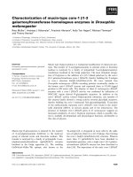
Fig. 1. Diagramatic representation of CDV NP gene to be amplified by RT-PCR and nested PCR and position of the outer and inne
r
primer pairs. Primers were designed from CDV nucleocapsid gene of Onderstepoort strain. The 549 and 419 bps fragments o
f
nucleocapsid gene were amplified by RT-PCR and nested PCR with the outer (primers 1 and 2) and inner primer pairs (primers 3 and 4),
respectively. Primer 1 = outer forward primer positioned at 675 to 692, primer 2 = outer reverse primer positioned at 1206 to 1223,
primer 3 = inner forwared primer positioned at 768 to 785, primer 4 = inner reverse primer positioned at 1169 to 1186. Amplified
products were identified by restrionion endonuclease, Ava I (restriction site: 1063). L = large virus-specific RNA-directed RNA
polymerase protein, H = hemagglutinin protein, F = fusion protein, M = matrix protein, P = phosphoprotein, N = nucleocapsid.
Detection of canine distemper virus through PCR 61
or 5
µ
l
(non-detected case) of the PCR products amplified
with the outer primer pairs was used as a template and
amplified with the inner primer pairs using the same proce-
dure described in the first PCR after cDNA synthesis.
Amplified PCR products were visualized under UV illumi-
nator after Etbr staining.
Analysis of PCR product
The PCR products were analyzed by 1.5% agarose gel
electrophoresis after digestion with restriction endonu-
clease,
AvaI
.
e
The DNA fragment of the first PCR was
cloned into plasmid using a TA-cloning method (TOPO
TM
TA Cloning
®
, Invitrogen, USA).
f
Extracting plasmid
DNAs (GENOMED plasmid kit
®
, Genomed, Germany),
both strands of plasmid inserts were sequenced using the
dideoxy chain termination method (Dye Terminator Ampl-
iTaq kit
®
, PE Applied Biosystems, USA). The similarity of
nucleotide sequence of PCR product obtained through
sequencing analyzer was calculated (BLAST program,
/>Serologic test
Serum neutralization test (SN) was performed using the
Chalmers and Baxendale's method
6
with a minor modifica-
tion. Sera were collected from five vaccinated dogs weekly
for three weeks after vaccination. Heat inactivated and
serially diluted sera were mixed with equal volume of
CDV suspensions containing 200 TCID
50
/m
l
. After incu-
bation for 1 h at 37
o
C
, 0.1 m
l
of the mixtures were inocu-
lated on to monolayered Vero cell and incubated at 37
o
C
for four days in 5% CO
2
. SN titer was determined by cal-
culating the 100% inhibition dilution dose of cytopathic
effect.
Results
Amplification of CDV NP gene by a RT-PCR and
nested PCR
Five-hundred and forty-nine bp fragment of NP gene
was successfully amplified from tissue culture fluid con-
taining CDV vaccine strain (Lederle; 10
3
PFU/m
l
) by a RT-
PCR with the outer primer pair (Fig. 2). From 10 cases of
clinically suspected dogs for CD, only 8 dogs were found
positive by a previous RT-PCR. However, the gene was not
detected from 2 cases of the positive dogs after 9 days with
the same RT-PCR.
With inner primer pair for nested PCR to increase the
sensitivity and specificity, 419 bp fragment was success-
fully amplified from 1
µ
l of the first PCR product (Fig. 2).
With the nested PCR, the positive band of 419 bp was suc-
cessfully amplified from all samples clinically suspected
of CD including 2 negative products in the reexamination
of the positives (data not shown).
Amplification of CDV NP gene by one-step RT-PCR
with nested PCR
Five-hundred and forty-nine and 419 bp fragments of
NP gene were successfully amplified by one-step RT-PCR
and nested PCR with the outer and inner primer pairs,
respectively (Fig. 3). In the digestion of the PCR products
with
Ava I
, the products with outer and inner primer sets
were 389 and 160 bp fragments and 296 and 123 bp frag-
ments, respectively (Fig. 3). The nucleotide sequence of
the one-step RT-PCR product showed a 98% identity with
the sequence of CDV NP gene from a previous report.
14
In
the sensitivity of one-step RT-PCR and nested PCR, the
Fig. 2. Amplification of CDV NP gene by previous RT-PCR and
nested PCR. Five-hundred and forty-nine bp (panel A) and 419
bp (panel B) fragments were successfully amplified from tissue
culture fluid containing CDV vaccine strain (Lederle; 10
3
PFU
/
ml) and visualized by ethidium bromide staining.
Fig. 3. Detection of CDV NP gene by one-step RT-PCR
combined with nested PCR. One-step RT-PCR and nested PCR
products were visualized by ethidium bromide staining and
treated with restriction endonuclease, Ava I. Lane 1: one-step RT-
PCR product (549 bp), lane 2: digestion product of one-step RT-
PCR product (389 & 160 bp), lane 3: nested PCR product (419
bp), and lane 4: digestion product of nested PCR product (296 &
123 bp band).
62 Yong-Hwan Kim et al.
detection limits were 10
2
and 10
0
, respectively (Fig. 4).
PBMC and normal body secretes (ocular discharge, nasal
discharge, saliva, feces, and urine) of healthy dogs and
other common canine viruses (parainfluenzavirus 2, canine
coronavirus, infectious canine hepatitis virus, and canine
parvovirus) were also tested with the same primers. No
detectable bands were produced by one-step RT-PCR and
nested PCR (data not shown).
Detection of CDV in vaccinated dogs
CDV NP gene from 5 vaccinated dogs with one-step RT-
PCR and nested PCR was detected at 2 days after vaccina-
tion, but not at 7 days, in PBMC only by one-step RT-PCR.
However, combined with nested PCR, 4 of the 5 samples
were positive at 7 days. It was also detected with combined
nested PCR at 2 and 7 days in other samples (ocular dis-
charge, nasal discharge, saliva, feces, and urine) with vari-
ous ratios. However, no amplified band was observed after
14 days (Table 1). SN titer was >250 at 1-2 weeks and 128
at 3 weeks after vaccination in all vaccinated dogs.
Detection of CDV in clinically affected dogs
Of the 51 PBMC samples from dogs with the typical
clinical signs of CD, the amplified NP gene was detected
in 45 dogs by one-step RT-PCR combined with nested
PCR. Of the 45 positive samples, however, only 15 sam-
ples were revealed as positive through single one-step RT-
PCR. The last 6 cases shown local myoclonus of temporal
muscle or thoracic and pelvic limb were also suspected of
being infected with CDV even though the gene was not
detected by one-step RT-PCR combined with nested PCR.
Discussion
CD is the most important viral, contagious disease
found in dogs, particularly 3 to 6 months of age, with high
morbidity and mortality. Diagnosis of CD in acute or sub-
acute forms had been done usually based on clinical signs
such as conjunctivitis, bronchitis, catarrhal pneumonia,
gastroenteritis, and neurological disturbances. However,
some problems arose in the differentiation with other dis-
eases such as kennel cough or other clinical forms such as
delayed-onset and chronic distemper encephalitis, among
others. Although detection of anti-CDV IgM antibody, FA,
and virus isolation had been used, these methods also had
several problems such as time-consuming, time-limitation,
and cross-reaction in vaccinated dogs in the diagnosis of
CD. Therefore, development of a sensitive, specific, and
practical method was required. With growing knowledge
in molecular biology, a RT-PCR was developed to detect
CDV. However, this method still had problems in sensitiv-
ity and specificity due to contamination error since the
reaction was carried out in separate tubes for RT and PCR.
We established a one-step RT-PCR. Moreover, a nested
PCR was developed from the product of one-step RT-PCR.
Sensitivity of the one-step RT-PCR combined with nested
PCR increased hundredfold than the previous PCR using
culture supernatant containing CDV vaccine strain. The
Fig. 4.
Sensitivity of one-step RT-PCR and nested PCR. Lanes 1-
4 of panel A: amplified products with one-step RT-PCR using
virus titier 10
3
to 10
0
PFU/ml and lanes 1-4 of panel B: amplifie
d
products with nested PCR using PCR products of one-step RT-
PCR for virus titer 10
3
to 10
0
PFU/ml.
Table 1.
Detection of canine distemper virus in vaccinated dogs by one step RT-PCR combined with nested PCR
Samples
Days after vaccination
02714
1st 2nd 1st 2nd 1st 2nd 1st 2nd
PBMC 0/5* 0/5 5/5 5/5 0/5 4/5 0/5 0/5
Conjunctival swab 0/4 0/4 0/4 4/4 0/4 2/4 0/4 0/4
Nasal discharge 0/4 0/4 0/4 4/4 0/4 1/4 0/4 0/4
Saliva 0/4 0/4 0/4 3/4 0/4 2/4 0/4 0/4
Feces 0/4 0/4 0/4 3/4 0/4 2/4 0/4 0/4
Urine 0/4 0/4 0/4 2/4 0/4 0/4 0/4 0/4
*No. of positive/No. of tested;
1st = one-step RT-PCR, 2nd = nested PCR.
Detection of canine distemper virus through PCR 63
sensitivity was confirmed using blood samples of dogs
clinically suspected of CDV infection. Specificity of the
PCR was confirmed by PBMC and body secretes of
healthy dogs and other viruses which could infect dogs and
showed similar clinical signs with CD (parainfluenzavirus
2, canine coronavirus, infectious canine hepatitis virus, and
canine parvovirus). Identity of the PCR products was con-
firmed by digestion with
AvaI
and nucleotide sequencing
of the PCR products.
Within 6 days after infection, all lymphatic tissues are
infected, and viremia is developed. Dogs without antibody
against CDV die approximately 3 weeks after exposure,
showing widespread distribution of virus in lymphatic tis-
sue, epithelium, and brain, with signs of illness. But viral
antigens disappear within 2 weeks if infected dogs obtain
high serum antibody titer.
2
These phenomena were con-
firmed in this experiment through detection of CDV from
PBMC and body secretes of vaccinated dogs. These results
from vaccinated dogs suggested the importance of deter-
mining the time period for the effective application of the
PCR method. In the comparision of the diagnostic effi-
cency of the two PCR methods with 51 PBMC samples
suspected of CD, the efficiency of one-step RT-PCR com-
bined with nested PCR was increased up to 60%. Although
the one-step PCR combined with nested PCR was found to
be the most sensitive method to detect CDV from the spec-
imen, further studies to find the proper time to take sam-
ples from dogs should be performed.
References
1. Appel, M. and Robson, D. S. A microneutralization test for
canine destemper virus. Am. J. Vet. Res. 1973, 34, 1459-
1463.
2. Appel, M. J. G. Pathogenesis of canine distemper. Am. J.
Vet. Res. 1969, 30, 1167-1182.
3.
Blixenkrone-M
φ
ller, M.
Detection of intracellular canine
distemper virus antigen in mink inoculated with an attenu-
ated or a virulent strain of canine distemper virus. Am. J.
Vet. Res. 1989,
50
, 1616-1620.
4.
Blixenkrone-M
φ
ller, M., Pedersen, I. R., Appel, M. J. and
Griot, C.
Detection of IgM antibodies against canine dis-
temper virus in dogs and mink sera employing enzyme-
linked immunosorbent assay(ELISA). J. Vet. Diag. Invest.
1991,
3
, 3-9.
5. Chalmers, W. S. K. and Baxendale, W. A comparison of
canine distemper vaccine and measles vaccine for the pre-
vention of canine distemper in young puppies. Vet. Rec.
1994, 135, 349-353.
6. Ettinger, S. J. and Feldman, E. C. Textbook of veterinary
internal medicine. pp.400-402. 4th ed. B. W. Saunders, Phil-
adelpia, 1995.
7. Fairchild, G. A., Wyman, M. and Donovan, E.F. Fluores-
cent antibody technique as a diagnostic test for canine dis-
temper infection: detection of viral antigen in epithelial
tissues of experimentally infected dogs. Am. J. Vet. Res.
1967, 28, 761-768.
8. Godec, M. S., Asher, D. M., Swoveland, P. T., Eldadah, Z.
A., Feinstone, S. M., Goldfarb, L. G., Gibbs, C. J. and
Gajdusek, D. C. Detection of measles virus genomic
sequences in SSPE brain tissue by the polymerase chain
reaction. J. Med. Virol. 1990, 30, 237-244.
9. Guy, J. S. Diagnosis of canine viral infections. Vet. Clin.
Nor. Am.: Small Ani. Prac. 1986, 16, 1145-1156.
10. Haas, L., Subbarao, S. M., Harder, T., Liess, B. and Bar-
rett, T. Detection of phocid distemper virus RNA in seal tis-
sues using slot hybridization and the polymerase chain
reaction amplification assay: genetic evidence that the virus
is distinct from canine distemper virus. J. Gen. Virol. 1991,
72, 825-832.
11. Ho, C. K. and Babiuk, L. A. A new plaque system for
canine distemper: characteristic of the green strain of canine
distemper virus. Can. J. Microbiol. 1979, 25, 680-685.
12. Rozenblatt, S., Eizenberg, O., Ben-Levy, R., Lavie, V. and
Belli ,W. J. Sequence homology within the morbilliviruses.
J. Virol. 1985, 53, 684-690.
13. Saiki, R. K., Gelfand, D. H., Stoffel, S., Scharf, S. T.,
Higuchi, R., Horn, G. T., Mullis, K. B. and Erlich, H. A.
Primer-directed enzymatic amplification of DNA with a
thermostable DNA polymerase. Science 1988, 239, 487-491.
14. Shin, Y. S., Mori, T., Okita, M., Gemma, T., Kai, C. and
Mikami T. Detection of canine distemper virus nucleo-
capsid protein gene in canine peripheral blood mononuclear
cells by RT-PCR. J. Vet. Med. Sci. 1995, 57, 439-445.
15. Stephensen, C. B., Welter, J., Thaker, S. R., Taylor, J.,
Tartaglia, J. and Paoletti, E. Canine distemper virus (CDV)
infection of ferrets as a model for testing Morbillivirus vac-
cine strategies: NYVAC- and ALVAC- based CDV recombi-
nants protect against symptomatic infection. J. Virol. 1997,
71, 1506-1513.
16.
Visser, I. K. G., Marie-Fran
ç
oise Van Bressem, Rik L de
Swart, van de Bildt M. W., Vos H. W., van der Heijden R.
W., SalikiJ, T., Orvell, C., Kitching, P. and Kuiken, T.
Characterization of morbilliviruses isolated from dolphins
and porpoises in Europe. J. Gen. Virol. 1993,
74
, 631-641.
17. Winters, W. D. Time dependent decreases of maternal
canine virus antibodies in newborn pups. Vet. Rec. 1981,
108, 295-299.