Báo cáo khoa học: "Immunohistochemical Localization of Bcl-2 in the Spinal Cords of Rats with Experimental Autoimmune Encephalomyelitis" docx
Bạn đang xem bản rút gọn của tài liệu. Xem và tải ngay bản đầy đủ của tài liệu tại đây (209.78 KB, 5 trang )
J O U R N A L O F
Veterinary
Science
J. Vet. Sci. (2002), 3(4), 279-283
Abstract
5)
We examined the localization of the anti-apoptotic
molecule Bcl-2 in the spinal cords of Lew is rats with
experimental autoim mune encephalomyelitis (EAE).
Western blot analysis show ed that Bcl-2 w as
constitutively expressed in normal spinal cords, and
w eakly increased in response to complete Freund's
adjuvant(CFA) immunization. In EAE, with infiltration
of inflammatory cells into spinal cords, Bcl-2 declined
during the peak stage and further decreased during
the recovery stage. Immunohistochemically, some
neurons and glial cells constitutively expressed Bcl-2
in normal rat spinal cords. In the spinal cords of rats
w ith EAE, Bcl-2 w as also im munoreacted in some
perivascular inflammatory cells while some brain
cells, such as neurons and GFAP (+) astrocytes
showed less Bcl-2 immunoreaction.
These findings suggest that in EAE, Bcl-2 expression
in the CNS host cells decreases with CNS inflammation,
possibly progressing to cell death in some cases,
w hile the survival of host cells, including neurons,
astrocytes, and some inflammatory cells, is associated
w ith activation of the anti-apoptotic m olecule Bcl-2.
Taking all into considerations, its is postulated that
Bcl-2 either beneficially or detrimentally functions in
some host cells depe nding on the activation stage of
each cell type.
Key words :
apoptosis, autoimmune encephalomyelitis,
Bcl-2
Introduction
Experimental autoimmune encephalomyelitis (EAE) is an
autoimmune disease of the central nervous system (CNS)
*
Corresponding author: Tae-Kyun Shin
Department of Veterinary Medicine, Cheju National University,
Jeju 690-756, Korea
Tel : +82-64-754-3363, Fax : +82-64-756-3354
E-mail :
that is used to study human demyelinating diseases such as
multiple sclerosis [2, 12]. The clinical course of EAE is
characterized by weight loss, ascending progressive paralysis,
and finally spontaneous recovery. These steps are matched
by the inflammatory response in the CNS, which is charac-
terized by the infiltration of T cells and macrophages, and
the activation of microglia and astrocytes at the peak stage
[13, 16]. Apoptosis is one possible mechanism for the recovery
in EAE, because invading cells are eliminated through
apoptosis during the peak stage [1, 5, 10,15]. Apoptotic cells
are found mainly in the parenchyma, where many apoptosis-
related molecules are found, including p53 and Bax, while
they are rarely found in perivascular EAE lesions [8]. Brain
cells can survive in the CNS of EAE rats, despite the
increased infiltration of inflammatory cells and the resulting
secretion of many cytokines and cyto-toxic molecules [6, 11].
Bcl-2 is an anti-apoptotic molecule that is normally
expressed in neurons and cancer cells [14, 17]. Although
Bcl-2 is expressed in multiple sclerosis lesions [19] and its
animal model EAE [3, 18], little is known about the
localization of Bcl-2 in rat EAE in relation to escape from
apoptosis in host and some inflammatory cells.
In this study, we examined the distribution of the
anti-apoptotic molecule Bcl-2 in EAE lesions of the spinal
cord in Lewis rats, and studied the relationship between the
distribution of this molecule and apoptosis.
Material and Methods
Animals
Lewis rats of both sexes (7-12 weeks old) were obtained
from the Korea Research Institute of Bioscience and
Biotechnology, KIST (Daejeon, Korea) and bred in our
animal facility.
EAE induction
EAE was induced in Lewis rats with a slight modification
of a previously described method [16]. Briefly, each rat was
subcutaneously injected in the hind footpads bilaterally with
an emulsion containing equal parts of fresh rat spinal cord
homogenates in phosphate buffer (mg/ml) and complete
Freund's adjuvant (CFA; Mycobacterium tuberculosis H37Ra,
Immunohistochemical Localization of Bcl-2 in the Spinal Cords of Rats with
Experimental Autoimmune Encephalomyelitis
Chang-Jong Moon, Yong-Duk Lee and Tae-Kyun Shin*
Department of Veterinary Medicine, Cheju National University, Jeju 690-756, Korea
Received May 3, 2002 / Accepted November 14, 2002
280 Chang-Jong Moon, Yong-Duk Lee and Tae-Kyun Shin
mg/ml)(Difco, Detroit, MI, U.S.A.). On the day of immunization,
the rats were injected with 2 g of pertussis toxin intraperi-
toneally (Sigma, St. Louis, MO, U.S.A.). Control animals
received either CFA or pertussis toxin only. Immunized rats
were observed daily for clinical signs of EAE. Clinically, EAE
was separated into five stages (grade 0, no signs; grade 1, floppy
tail; grade 2, mild paraparesis; grade 3, severe paraparesis;
grade 4, tetraparesis or moribund condition [10, 16].
Tissue sampling
Tissue samples were taken on days 10-14 and 21-25
post-immunization (PI), during the peak and recovery stages
of EAE, respectively. Experimental rats (n=3) in each group
were sacrificed under ether anesthesia, and the spinal cords
were removed and frozen in a deep freezer (-70 C) for
protein analysis. Pieces of the spinal cords were processed
for paraffin embedding after fixation in 4% paraformaldehyde
in phosphate-buffered saline (PBS), pH 7.4.
Western blot analysis
Frozen nervous tissue was thawed at room temperature,
minced, lysed in a buffer consisting of 40 mM Tris-HCl, pH
7.4, 120 mM NaCl, and 0.1% Nonidet P-40 (polyoxyethylene
(9) p-t-octyl phenol) containing the protease inhibitors
leupeptin (0.5
㎍
/ml), PMSF (1 mM), and aprotinin (5
㎍
/ml),
and homogenized. Equal amounts of protein (200 g/20 l)
were loaded in each lane, and electrophoresed under denaturing
conditions in sodium dodecyl sulfate-polyacrylamide gels
(SDS-PAGE). After electrophoresis, the proteins were electro-
transferred onto nitrocellulose transfer membranes (Schleicher
and Schuell, Keene, NH). Blotting with rabbit anti-Bcl-2
(1:200 dilution, Santa Cruz, CA) was performed as described
in a previous paper [8]. Visualization was achieved using
Amersham ECL reagents (Amersham Life Science, Little
Chalfont, Buckinghamshire, UK). The results were quantified
with a densitometer (M GS-700 Imaging Densitometer,
Bio-Rad Laboratories, Hercules, CA).
Terminal deoxynucleotidyl transferase (TdT)-mediated
dUTP nick end-labeling (TUNEL)
DNA fragmentation was detected by in situ nick end-
labeling, as described in the manufacturer’s instructions
(Intergen, Purchase, NY). In brief, the paraffin sections
were deparaffinized, rehydrated, and washed in PBS. The
sections were treated with 0.005% pronase (Dako, glostrup,
Denmark) for 20 minutes at 37
℃
and subsequently incubated
with TdT buffer solution (140 mM sodium cacodylate, 1 mM
CoCl, 30 mM Tris-HCl, pH 7.2) containing 0.15 U/
μ
L TdT
and 0.004 nmol/L digoxigenin-dUTP for 60 minutes at 37
℃
,
and then in TB buffer (300 mM NaCl, 30 mM sodium
citrate) for 15 minutes. They were then reacted with peroxidase-
labeled anti-digoxigenine antibody for 60 minutes. Positive
cells were visualized by using a diaminobenzidine substrate
kit and counterstained with hematoxylin.
Immunohistochemistry
Staining followed the labeled-streptavidin-biotin (LAB-SA)
method (Histostain®-Plus Kits, Zymed Laboratories Inc, San
Francisco, CA) according to the manufacturer’s instructions.
In brief, 5-m-thick sections of paraffin-embedded spinal
cords were deparaffinized and treated with 0.3% H2O2 in
methyl alcohol for 20 minutes to block endogenous peroxidase.
The sections were exposed to normal goat serum, and then
incubated in optimally diluted primary antisera including
rabbit anti-Bcl-2 (1:200 dilution, Santa Cruz Biotechnology,
Santa Cruz, CA) for 1 hour at room temperature. To
distinguish cell types in the CNS, either rabbit anti-GFAP
serum (1:800, Dako) specific for astrocytes or ED1 for
macrophages was applied to adjacent sections. The peroxidase
was developed with diaminobenzidine- H2O2 solution (0.001%
3,3
′
-diaminobenzidine [Sigma] and 0.01% H2O2 in 0.05 M
Tris-buffered saline [TBS, pH 7.4]). The sections were
counterstained with hematoxylin before mounting.
Results
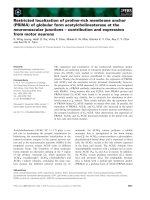
Western blot analysis of Bcl-2 in EAE
Bcl-2 was constitutively expressed in the normal rat
spinal cord (Fig. 1, lane 1), and expression increased in
response to immunization with CFA (Fig. 1, lane 2). The
degree of Bcl-2 expression in the early stage of EAE (G1,
day 9 PI) (Fig. 1, lanes 3 and 4) was the same as in the
Bc l-2
Norma l
CFA Ea rly Pea k Re c o ve ry
Fig. 1.
A representative photograph of Western blot analysis of Bcl-2 in the spinal cord in normal, CFA-immunized, and
EAE rats: lane 1; normal, lane 2; CFA immunized (day 9 PI), lanes 3 and 4; EAE (G1, day 9 PI), lanes 5 and 6; EAE (G3,
day 12 PI), lanes 7 and 8; EAE (R0, day 21 PI).
Immunohistochemical Localization of Bcl-2 in the Spinal Cords of Rats with Experimental Autoimmune Encephalomyelitis
281
CFA-immunized group on the same day (day 9) (Fig. 1, lane
2). Surprisingly, Bcl-2 expression declined at the peak stage
(day 12 PI, G3) (Fig. 1, lanes 5 and 6), and further decreased
during the recovery stage (day 21 PI, R0) (Fig. 1, lanes 7 and
8). This suggests that Bcl-2 is constitutively expressed in
normal adult CNS tissues, and its expression may increase
in response to peripheral stimulation, such as immunization
with CFA. However, relative amount of Bcl-2 expression
decreases in the EAE affected spinal cords, suggesting that
some cells exhibit less Bcl-2.
Distribution of Bcl-2 immunoreactivity and TUNEL
reaction in EAE lesions
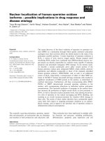
Bcl-2 was expressed in some neurons and glial cells in
the normal rat spinal cord (Fig. 2A). In EAE lesions, Bcl-2
immunoreactivity was found in some inflammatory cells in
the perivascular cuffing, rather than in those in the
parenchyma, as well as in some neurons and glial cells (Fig.
2B). Table 1 summarizes the expression of Bcl-2 by cell
phenotype. The intensity of Bcl-2 staining in neurons in EAE
was weaker than in neurons in the normal and CFA-
immunized groups. It suggests that the decreased expression
of Bcl-2 in EAE by western blot might come from the less
immunoreactivity of Bcl-2 in host cells, or the amount of
Bcl-2 expression in normal spinal cords overwhelm those of
both host cells and inflammatory cells in EAE lesions.
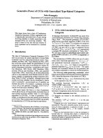
The localization of the TUNEL reaction (Fig. 3) was
inversely related to Bcl-2 immunoreactivity (Fig. 2B) in
EAE lesions. In EAE, TUNEL (+) apoptotic cells were
scattered throughout the spinal cord parenchyma, but were
rarely found in perivascular lesions (Fig. 3), as previously
shown [8]. Moreover, the TUNEL reaction was barely seen
in neurons and glial cells (Fig. 3), suggesting that host cells
escape death in autoimmune CNS inflammation.
Discussion
Bcl-2 is a survival molecule that allows neuronal cells to
survive in vitro [7], and is an anti-apoptotic factor in
primary carcinoid tumors [20]. Consistent with previous
findings, multiple sclerosis lesions were found to contain
Bcl-2-expressing T lymphocytes, which may continuously
injure brain tissues [19]. Previously, it was reported that in
EAE, astrocytes and oligodendroglial cells expressing Bcl-2
do not undergo apoptosis [2]. Furthermore, the lack of
apoptosis in perivascular cuffing in EAE is caused by the
generation of superoxide in invading macrophages, at least
in part [4].
Our results suggest that the apoptosis of inflammatory
cells in EAE parenchyma cells lacking anti-apoptotic Bcl-2
requires additional molecules from the apoptosis cascade. In
this study, in EAE spinal cords, the TUNEL reaction was
barely seen in neurons and glial cells that showed Bcl-2
immunoreactivity, suggesting that the host cells escape
death in autoimmune CNS inflammation. This is one
possible reason why brain cells, including neurons and glial
cells, survive autoimmune injury. Our finding that some
inflammatory cells do not undergo apoptosis in perivascular
lesions suggests that in these cells, during the peak stage of
EAE, anti-apoptotic Bcl-2 predominates, rather than the
cytotoxic effectors. Similar findings are consistently seen in
cancer cells [17] and multiple sclerosis lesions [3]. Our
findings are further supported by the observation that
effector cells, such as oligodendroglial cells expressing many
death signals, including Fas, do not undergo apoptosis in
the murine EAE model, while homing inflammatory cells
are selectively vulnerable to the death signals associated
with apoptosis [2]. Moreover, Offen et al. [9] reported that,
in MOG-induced EAE, Bcl-2 reduces axonal damage and
attenuates the clinical severity.
In conclusion, we hypothesize that the anti-apoptotic molecule
Bcl-2 allows the survival of host cells and perivascular
inflammatory cells in autoimmune CNS inflammation, while
inflammatory cells in the parenchyma undergo apoptosis
because they lack survival genes.
Table 1.
Bcl-2 immunoreactivity in cell phenotypes of the spinal cords of normal and EAE rats
Normala CFA EAE(G3, day 12PI)a EAE(R0, day 21 PI)a
Neurons
Astrocytes
Ependymal cells
Macrophages/activated microglia
T-cells
+++
++
+
-
-
+++
++
+
-
-
+
+
+
++
++
+
+
+
+
+
a Three to five animals were examined in each group.
b Normal and EAE spinal cord sections were analyzed using an apoptosis detection kit and immunohistochemistry was
examined using antibodies to detect specific markers. Stained sections were scored according to the number of cells per
field that were positive. The number of positive cells in an average of five randomly chosen 100 fields was scored as:
-
, no positive cells;
+
, <10 cells per field;
++
, <30 cells;
+++
, 30 cells.
282 Chang-Jong Moon, Yong-Duk Lee and Tae-Kyun Shin
Acknowledgments
This study was supported by a grant from the Korean
Health 21 R & D Project, The Ministry of Health & Welfare,
Republic of Korea (02-PJ1-PG10-21305-0003).
References
1.
Bauer, J., Bradl, M., Hickley, W.F., Forss-Petter,
S., Breitschopf, H., Linington, C., Wekerle, H.,
Lassmann, H.
T-cell apoptosis in inflammatory brain
lesions: destruction of T cells does not depend on
antigen recognition. Am. J. Pathol. 1998,
153
, 715-724.
2.
Bonetti, B., Pohl, J., Gao, Y.L., Raine
, C.S. Cell
death during autoimmune demyelination: effector but
not target cells are eliminated by apoptosis. J. Immunol.
1997,
159
, 5733-5741.
3.
Bonetti, B., Raine, C.S.
Multiple sclerosis: oligoden-
drocytes display cell death related molecules in situ but
do not undergo apoptosis. Ann. Neurol. 1997,
42
, 74-84.
4.
Brune, B., Gotz, C., Messmer, U.K., Sandau, K.,
Hirvonen, M.R., Lapetina, E.G.
Superoxide formation
and macrophage resistance to nitric oxide-mediated
apoptosis. J. Biol. Chem. 1997,
272
, 7253-7258.
5.
Kohji, T., Tanuma, N., Aikawa, Y., Kawazoe, Y.,
Suzuki, Y., Kohyama, K., Matsumoto, Y.
Interaction
between apoptotic cells and reactive brain cells in the
central nervous system of rats with autoimmune
encephalomyelitis. J . Neuroimmunol. 1998,
82
, 168-74.
6.
Korner, H., Sedgwick, J.D.
Tumour necrosis factor
and lymphotoxin: molecular aspects and role in tissue-
specific autoimmunity. Immunol. Cell. Biol. 1996,
74(5)
,
465-472.
7.
Melkova, Z., Lee, S.B., Rodriguez, D., Esteban, M.
Bcl-2 prevents nitric oxide-mediated apoptosis and
poly(ADP-ribose) polymerase cleavage. F.E.B.S. Lett.
1997,
403
, 273-278.
8.
Moon, C., Kim, S., Wie, M., Kim, H., Cheong, J.,
Park, J., Jee, Y., Tanuma, N., Matsumoto, Y., Shin,
T.
Increased expression of p53 and Bax in the spinal
cords of rats with experimental autoimmune encepha-
lomyelitis. Neurosci. Lett. 2000,
289(1)
, 41-44.
9.
Offen, D., Kaye, J.F., Bernard, O., Merims, D.,
Coire, C.I., Panet, H., Melamed, E., Ben-Nun, A.
Fig. 3.
TUNEL reaction in EAE lesions. TUNEL (+) Apoptotic
cells were scattered throughout the parenchyma of the
spinal cords of rats with EAE. The TUNEL reaction was
commonly present around neurons and some GFAP (+)
processes identical to astrocytes (A). Some apoptotic cells
were colocalized with ED1 (+) cells (B). TUNEL and ABC-
alkaline phosphatase reaction. A and B: TUNEL and either
GFAP (A) or ED1 (B). Scale = 30
㎛
. B; EAE (G3, day 12 PI).
Fig. 2.
Immunostaining for Bcl-2 (A, B) in EAE. Intense
Bcl-2 immunoreactivity is found in neurons and glial cells in
the spinal cords of normal rats (A). In EAE, intense Bcl-2
immunoreactivity is present in perivascular clusters (arrowhead)
of inflammatory cells and weak immunoreactivity is seen in
some neurons in the parenchyma during the peak stage (B).
Arrows indicate blood vessels. Scale =30
㎛
. B; EAE (G3, day
12 PI).
Immunohistochemical Localization of Bcl-2 in the Spinal Cords of Rats with Experimental Autoimmune Encephalomyelitis
283
Mice overexpressing Bcl-2 in their neurons are resistant
to myelin oligodendrocyte glycoprotein (MOG)-induced
experimental autoimmune encephalomyelitis (EAE). J.
Mol. Neurosci. 2000,
15(3)
, 167-176.
10.
Ohmori, K., Hong, Y., Fujiwara, M., Matsumoto, Y.
In situ demonstration of proliferating cells in the rat
central nervous system during experimental autoimmune
encephalomyelitis. Evidence suggesting that most infil-
trating T cells do not proliferate in the target organ.
Lab. Invest. 1992,
66
, 54-62.
11.
Okuda, Y., Sakoda, S., Fujimura, H., Yanagihara,
T.
Nitric oxide via an inducible isoform of nitric oxide
synthase is a possible factor to eliminate inflammatory
cells from the central nervous system of mice with
experimental allergic encephalomyelitis. J. Neuroimmunol.
1997,
73
, 107-116.
12.
Raine, C.S.
The Dale E. McFarlin Memorial Lecture:
the immunology of the multiple sclerosis lesion. Ann.
Neurol. 1994,
36
, S61-72.
13.
Raine, C.S., Cannella, B., Duijvestijn, A.M., Cross,
A.H.
Homing to central nervous system vasculature by
antigen-specific lymphocytes. II. Lymphocyte/endothelial
cell adhesion during the initial stages of autoimmune
demyelination. Lab. Invest. 1990,
63
, 476-489.
14.
Reed, J.C., Jurgensmeier, J.M., Matsuyama, S.
Bcl-2 family proteins and mitochondria. Biochem. Biophys.
Acta. 1998,
1366
, 127-137.
15.
Schmied, M., Breitschopf, H., Gold, R., Zischler,
H., Rothe, G., Wekerle, H., Lassmann, H.
Apoptosis
of T lymphocytes in experimental autoimmune encepha-
lomyelitis. Evidence for programmed cell death as a
mechanism to control inflammation in the brain. Am. J.
Pathol. 1993,
143
, 446-452.
16.
Shin, T., Kojima, T., Tanuma, N., Ishihara, Y.,
Matsumoto, Y.
The subarachnoid space as a site for
precursor T cell proliferation and effector T cell selection
in experimental autoimmune encephalomyelitis. J .
Neuroimmunol. 1995,
56
, 171-178.
17.
Waggoner, S.E., Baunoch, D.A., Anderson, S.A.,
Leigh, F., Zagaja, V.G.
Bcl-2 protein expression associated
with resistance to apoptosis in clear cell adenocarcinoma
of the vagina and cervix expressing wild-type p53. Ann.
Surg. Oncol. 1998,
5
, 544-547.
18.
White, C.A., McCombe, P.A., Pender, M.P.
The roles
of Fas, Fas ligand and Bcl-2 in T cell apoptosis in the
central nervous system in experimental autoimmune
encephalomyelitis. J . Neuroimmunol. 1998,
82
, 47-55.
19.
Zettl, U.K., Kuhlmann, T., Bruck, W.
Bcl-2 expressing
T lymphocytes in multiple sclerosis lesions. Neuropathol.
Appl. Neurobiol. 1998,
24
, 202-208.
20.
Zirbes, T.K., Lorenzen, J., Baldus, S.E., Moenig,
S.P., Wolters, U., Ottlik, A., Thiele, J., Holscher,
A.H., Dienes, H.P.
Apoptosis and expression of Bcl-2
are inverse factors influencing tumor cell turnover in
primary carcinoid tumors of the lung. Histopathology
1998,
33
, 123-128.