Báo cáo khoa học: "Identification and epidemiological characterization of Streptococcus uberis isolated from bovine mastitis using conventional and molecular methods" pdf
Bạn đang xem bản rút gọn của tài liệu. Xem và tải ngay bản đầy đủ của tài liệu tại đây (1.61 MB, 11 trang )
-2851$/ 2)
9H W H U L Q D U \
6FLHQFH
J. Vet. Sci.
(2003),
/
4
(3), 213–223
Identification and epidemiological characterization of
Streptococcus uberis
isolated from bovine mastitis
using conventional and molecular methods
I. U. Khan
1
, A. A. Hassan
2
, A. Abdulmawjood
2
C. Lämmler
3,
*, W. Wolter
4
and M. Zschöck
4
Department of Environmental Health, Toxicology Division, 3223 Eden Ave, University of Cincinnati, Medical Center.
Cincinnati OH, 45267-0056, USA
1
Institut für Tierärztliche Nahrungsmittelkunde, Professur für Milchwissenschaften, Justus-Liebig-Universität Gießen,
Ludwig Str. 21, 35390 Gießen, Germany
2
Institut für Pharmakologie und Toxikologie, Justus-Liebig-Universität Gießen, Frankfurter Str. 107, 35392 Gießen, Germany
3
Staatliches Untersuchungsamt Hessen, Marburger Str. 54, 35396, Gießen, Germany
In the present study 130
S. uberis
strains and one
S.
parauberis
strain isolated from bovine milk samples of 58
different farms of various locations in Hesse, Germany, as
well as two reference strains of each species were
comparatively investigated for cultural, biochemical,
serological and molecular properties. All
S. uberis
strains
produced the enzyme
β
-D-glucuronidase, while the
S. parauberis
strains were negative. The
S. uberis
and
S.
parauberis
16S rRNA genes were amplified by polymerase
chain reaction and subsequently digested with the
restriction enzymes
Rsa
I and
Ava
II yielding species-
specific restriction patterns. Both species were
additionally identified by amplifying species-specific parts
of the genes encoding the 16S rRNA, the 23S rRNA and
the 16S-23S rDNA intergenic spacer region, respectively.
The CAMP factor gene
cfu
, a potential virulence factor of
S. uberis
, was amplified, corresponding to a
phenotypically positive CAMP-reaction, using
cfu
-specific
oligonucleotide primers. In addition the streptokinase/
plasminogen activator encoding genes
skc
/
pau
A, a second
potential virulence factor, could be amplified for 126 of
the 130
S. uberis
but not for
S. parauberis
. A DNA
fingerprinting of
S. uberis
strains, performed by
macrorestriction analysis of their chromosomal DNA by
pulsed-field gel electrophoresis, revealed that most of the
isolates were not related to each other. However, identical
DNA patterns were noted for some of the isolates within
different quarters of an individual cow and also for
different cows within the same farm. The generally
unrelated DNA patterns indicated that
S. uberis
is a
pathogen with multiple environmental habitats and that
infections are caused by a great variety of strains.
Key words:
Streptococcus uberis
,
Streptococcus parauberis
,
16S rDNA, 23S rDNA, 16S-23S rDNA intergenic spacer
region, CAMP factor gene
cfu
,
skc
/
pauA
genes
Introduction
Streptococcus uberis
is world wide known as an
environmental pathogen responsible for a high proportion
of cases of clinical, mostly subclinical mastitis in lactating
cows and is also the predominant organism isolated from
mammary glands during the nonlactating period [37].
S. uberis
differs from other mastitis-causing streptococci
in that it can also be isolated from the udder surface, from
other sites on the body of cows and also from the cows
environment. The most important reservoirs for infections
of the mammary gland parenchyma appears to be the skin
and the udder surface [35,52].
S. uberis
can also be isolated
from numerous sites including belly, lips, teats, urogenital
tract, tonsils, rectum, rumen, nostrils, eye, poll, chest,
sacrum, caudal folds and feces [15,18,42,44,57,63]. In
addition,
S. uberis
had been isolated in large numbers from
the straw bedding of housed cattle usually during the
winter housing period and from the pasture grazed by
infected cattle [10].
According to Sherman [58] and Slot [61]
S. uberis
showed some similarities to bacteria of genus
Enterococcus
. However, the studies summarized by
Schleifer and Kilpper-Bälz [53] and Lämmler and Hahn
Part of the results were presented as poster presentation at the 41.
Arbeitstagung des Arbeitsgebietes “Lebensmittelhygiene” der
Deutschen Veterinärmedizinischen Gesellschaft. 25-28. September 2000
in Garmisch-Partenkirchen, Germany.
*Corresponding author
Phone: 0049641-38406; Fax: 0049641-38409
E-mail:
214 I. U. Khan
et al.
[37] revealed that
S. uberis
seems to be more related to the
pyogenic group of genus
Streptococcus
. On the basis of
chromosomal DNA hybridizations Garvie and Bramley
[23] and Collins
et al
. [14] suggested the existence of two
distinct
S. uberis
genotypes, designated as
S. uberis
type I
and II. According to a proposal of Williams and Collins
[68] type II
S. uberis
were classified as
S. parauberis
.
In the present study
S. uberis
and
S. parauberis
strains
isolated during routine diagnostics from bovine milk
samples of one region in Germany were investigated
together with reference strains of both species for cultural,
biochemical, serological and molecular properties. The
latter included the detection of various genes by
polymerase chain reaction and the determination of
epidemiological relationships by pulsed-field gel
electrophoresis (PFGE).
Materials and Methods
Collection and cultivation
For the present study 342 bovine milk samples from 342
quarters of 269 cows from 93 different farms were initially
collected within three months from January to March 1999
at different locations in Hesse, Germany. Approximately
0.1 ml milk obtained from clinical as well as subclinical
milk samples were initially plated on sheep blood agar
(Oxoid, Wesel, Germany), while subclinical samples were
subjected to total somatic cell count (SCC) in order to
confirm the subclinical status of the collected samples. The
determination of cell count was performed with the
Fossomatic system (360 N. Foss Electronic A/S, Hamburg,
Germany).
All bacteria suspected to belong to genus
Streptococcus
were subsequently cultivated on Columbia esculin blood
agar (Merck, Darmstadt, Germany) to determine their
culturing ability. The esculin-hydrolyzing cultures were
further cultivated on five different selective growth media
specific for enterococci. This included Citrate azide tween
carbonate agar (CATC, Merck), Kanamycin esculin azide
agar (KAA, Merck), Esculin bile agar (Oxoid),
Chromocult enterococci agar (Merck), and Slanetz-Bartley
media (Oxoid). All media were prepared, used and the
results interpreted according to the manufacturers
instructions. An
Enterococcus faecalis
strain, obtained
from the institutes strain collection (Institute of Milk
Science, Giessen University, Giessen, Germany), was used
as positive control.
On the basis of the above mentioned cultural ability and
growth patterns 131 isolates from 112 cows of 58 different
farms affected with subclinical and clinical mastitis were
further processed. The isolates were investigated together
with the
S. uberis
reference strains NCDO 2038 and
NCDO 2086, the
S. parauberis
reference strain NCDO
2020 and the
S. parauberis
strain 94/16. The latter,
originally isolated from a diseased turbot, was kindly
obtained from J. F. Fernández-Garayzábal (Faculty of
Veterinary Medicine, Complutense University, Madrid,
Spain) [19].
Biochemical characterization
Carbohydrate fermentation tests were determined by
using phenol-red broth (Merck) containing 1% arabinose,
fructose, glucose, inulin, lactose, maltose, mannitol,
raffinose, ribose, saccharose, salicin, sorbitol and
trehalose, respectively. Esculin hydrolysis was carried out,
using Brain Heart Infusion (BHI, Merck) containing 0.1%
esculin and 0.05% iron (III) citrate. For determination of
sodium-hippurate hydrolysis the method described by
Hwang and Ederer [29] was used. For arginine hydrolysis
commercial diagnostic test tablets (Rosco, Hiss
Diagnostics, Freiburg, Germany) were used as substrate.
The tests were carried out as described by the
manufacturer. Commercial diagnostic test tablets (Rosco,
Hiss Diagnostics) were also used as substrates for
determination of â-D-glucuronidase, and pyrrolidonyl
aminopeptidase
enzyme activities. In addition,
hyaluronidase enzyme activities were investigated by
cultivation of the bacteria in close proximity of a mucoid
growing
S. equi
subsp.
zooepidemicus
strain, obtained
from the institutes strain collection, as described by Winkle
[70].
Serogrouping
Serological grouping of the cultures was performed with
autoclaved extracts [47] and specific antisera of Lancefield
groups A, B, C, E, G, P, U and V. The antisera were
obtained from the institutes collection [36].
Other phenotypic characteristics
Synergistic CAMP-like hemolytic activities were
determined together with a
β
-toxin producing
S. aureus
on
sheep blood agar plates [37], lectin agglutination reactions
with the lectin from Helix pomatia (Sigma, Deisenhofen,
Germany), on microscopic slides [43]. Self-agglutinating
bacterial cultures were pretreated with 5
µ
l trypsin (1 mg
trypsin/ml PBS) for 1 hr at 37
o
C, washed, resuspended in
PBS and subsequently used for lectin agglutination as
described [43].
Genotypic characterization
The extraction of the DNA of the isolates was performed
as described [28]. The gene encoding the 16S rRNA was
amplified using the oligonucleotide primer ARI with the
sequence 5' GAGAGTTTGATCCTGGCTCAGGA 3' [8]
and the primer AmII with the sequence 5' CGGGTGTTAC
AAACTCTCGTGGT 3' [3]. The oligonucleotide primers
were synthesized by MWG-Biotech (Ebersberg,
Germany). Restriction fragment length polymorphism
Identification and epidemiological characterization of
Streptococcus uberis
isolated from bovine mastitis 215
analysis (RFLP) of the amplified 16S rRNA gene was
performed as recommended by Jayarao
et al
. [30] and
Lämmler
et al.
[38]. The amplicon was digested for 1 hr at
37
o
C in a water bath in 30
µ
l volumes with 1µl (10 U/µl)
Rsa
I and
Ava
II (New England Biolabs, Frankfurt,
Germany), respectively.
A molecular identification was additionally performed
by using species-specific oligonucleotide primers for the
genes encoding the 16S rRNA and 23S rRNA as well as
the 16S-23S rDNA intergenic spacer region with
oligonucleotide primers described previously [28]. In
addition, phenotypically CAMP positive and selected
CAMP-negative
S. uberis
and
S. parauberis
were
investigated for CAMP factor gene
cfu
. For amplification
of
S. uberis
CAMP factor gene
cfu
the oligonucleotide
primers were designed according to the
cfu
sequence of
S.
uberis
described by Jiang
et al
. [33]; (accession no.
U34322) by using computer program OLIGO 4.0. The
primer 1 had the sequence cfu-I 5' CTTTATTTTCCCCAA
3' and primer 2 the sequence cfu-II 5' ATTTCTTGGTCAA
CTTGT 3'. The PCR temperature program of 30 cycles
was: 92
o
C for 60 sec, 45
o
C for 1.5 min, 72
o
C for 1.5 min.
The final cycle was followed by an extension at 72
o
C for 5
min.
The amplification of another potential virulence factor
gene of
S. uberis
known as streptokinase/plasminogen
activator gene
skc
/
pauA
was performed as described by
Rosey et al. [51]; (accession no. AJ012549) and Johnsen
et
al.
[34]; (accession no. AJ131604), respectively.
Amplification of the gene
skc
was conducted using the
oligonucleotide primer SKC-I as primer 1 with the
sequence 5' CTCCTCTCCAACAAAGAGG 3' and SKC-
II as primer 2 with the sequence 5' GAAGGCCTTCCCCT
TTGAAA 3' according to Rosey
et al
. [51]. The PCR
temperature program consisted of 30 cycles: 94
o
C for 60
sec, 52
o
C for 60 sec and 72
o
C for 90 sec. The amplification
of
pauA
gene was performed with the oligonucleotide
primer 1 P38 5' AATAACCGGT TATGATTCCGACTAC
3' and primer 2 P39 5' AAAATTTACTCGAGACTTCCTT
TAAGG 3' described by Johnsen
et al
. [34]. The thermal
cycler program consisted of 30 cycles: 94
o
C for 60 sec,
54
o
C for 60 sec and 72
o
C for 90 sec. The final cycle was
followed by an extension incubation at 72
o
C for 5 min,
respectively. The PCR products was determined by
electrophoresis of 12
µ
l of the reaction product in a 2% (w/
v) agarosegel (Sigma) with Tris acetate-electrophoresis
buffer (TAE) (4.0 mmol/l Tris, 1 mmol/l EDTA, (pH 7.8)
and a 100 bp DNA ladder (Gibco BRL, Eggenstein,
Germany) as molecular marker.
Finally a macrorestriction analysis of the chromosomal
DNA of the cultures was performed according to
Soedarmanto
et al
. [62]. The DNA-fingerprinting was
carried out by preparation of whole bacterial DNA of the
isolates in agarose gel plugs and subsequent digestion of
the bacterial DNA with the restriction enzyme
Sma
I and
separation of the fragments by PFGE using the pulse time
described by Baseggio
et al
. [7]. The interpretation of the
restriction patterns was performed as described by Tenover
et al
. [64].
Results
All 131 streptococci investigated in the present study
were Gram positive chain forming cocci. The somatic cell
count analysis of the corresponding milk samples revealed
the subclinical status of mastitis for 126 of the 131 selected
samples. However, five of the samples exhibited a clinical
status of mastitis. By cultivation on sheep blood agar 126
isolates were
α
-hemolytic while the remaining five strains
were non-hemolytic. After cultivation on Columbia esculin
blood agar, all 131 isolates degraded esculin. None of the
131 isolates grew on CATC and KAA media. However,
nine, seven and four isolates showed a weak growth on
Esculin bile agar, Chromocult enterococci agar and
Slanetz-Bartley agar, respectively. The reference strains of
both species could not be cultivated on all five media
specific for enterococci.
All 131 isolates and the four reference strains exhibited
degradation of fructose, glucose, maltose, mannitol,
saccharose, salicin, sorbitol and trehalose and hydrolyzed
esculin and sodium hippurate, while all isolates did not
ferment arabinose. All 135 isolates except one fermented
lactose, ribose and hydrolysed arginine, respectively.
Among the 135 streptococci investigated 94 and 4 of the
strains fermented inulin and raffinose, respectively.
Additionally, 130 of the investigated strains, and the
S.
uberis
reference strains NCDO 2038 and NCDO 2086
showed
β
-D-glucuronidase enzyme activity whereas
isolate 138/80 and the
S. parauberis
strains NCDO 2020
and 94/16 were negative in this enzyme. Investigating
pyrrolidonyl aminopeptidase enzyme activities 120
isolates yielded a positive reaction. Hyaluronidase enzyme
activity, demonstrated by forming non-mucoid colonies of
the mucoid growing
S. equi
subsp.
zooepidemicus
indicator strain, could be observed for 47 of the 135
isolates. The remaining strains, also including the
reference strains of both species, appeared to be
hyaluronidase negative.
Among the 131 strains investigated, 130 strains were
classified as
S. uberis
and strain 138/80 as
S. parauberis
.
The serological investigations revealed that 42 of the 130
S. uberis
strains,
S. uberis
reference strain NCDO 2038
and
S. parauberis
reference strain NCDO 2020, reacted
with group E specific antiserum, whereas 11 and 9 of the
130
S. uberis
strains reacted with group P and group U
specific antiserum, respectively. One of the
S. uberis
isolates reacted with group A,
S. uberis
reference strain
NCDO 2086 with group G, one
S. uberis
strain
216 I. U. Khan
et al.
simultaneously with group E and group P and one
S. uberis
with group E and group U specific antisera, respectively.
The remaining 65
S. uberis
strains,
S. parauberis
138/80
and
S. parauberis
94/16 were categorized as non-
groupable.
A synergistic hemolytic CAMP-like reaction on sheep
blood agar within the zone of staphylococcal
α
-toxin could
be observed for five of the 130
S. uberis
strains. In lectin
agglutination reactions, 43 of 130
S. uberis
strains
exhibited agglutination reactions with the lectin of Helix
pomatia. A self-agglutination reaction was observed for
two
S. uberis
strains even after trypsin pretreatment of the
bacteria. None of the remaining
S. uberis
and
S. parauberis
strains also including the reference strains of both species
showed a comparable reaction with the lectin investigated.
A molecular characterization of the bacteria was
performed by RFLP of the 16S rRNA gene. All 131 strains
investigated and the four reference strains of both species
displayed an amplicon size of the 16S rRNA gene of 1430
bp. The amplified 16S rRNA gene was digested with the
restriction endonucleases
Rsa
I and
Ava
II, respectively. The
restriction profiles confirmed the classification of 130 strains
as
S. uberis
and strain 138/80 as
S. parauberis
. Identical
restriction profiles could be observed for the reference
strains of both species, respectively.
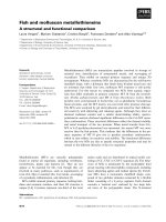
Rsa
I restriction of the
S.
uberis
16S rRNA gene revealed four fragments with sizes of
approximately 140, 190, 220 and 700 bp and for the
S.
parauberis
16S rRNA gene four fragments with sizes of
approximately 140, 190, 380 and 700 bp.
Ava
II restriction
revealed three different fragments with sizes of 230, 310 and
900 bp for
S. uberis
and fragment sizes of 230 and 1,200 bp
for
S. parauberis
(Fig. 1).
Using oligonucleotide primers amplifying
S. uberis
specific parts of the 16S rRNA gene, the 23S rRNA gene
and the 16S-23S rDNA intergenic spacer region revealed
amplicons with sizes of 440, 450 and 340 bp, respectively.
This could be observed for all 130
S. uberis
and both
S.
uberis
reference strains but not for
S. parauberis
. Typical
F
ig. 1.
Typical fragments of the PCR amplified 16S rRNA gene of
S. uberis
(1, 2, and 3) and
S. parauberis
(4, 5, and 6) after digesti
on
w
ith the restriction enzymes
Rsa
I and
Ava
II, respectively. M = a 100 bp ladder size marker.
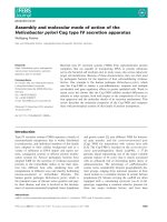
F
ig. 2.
Amplicons of
S. uberis
(1, 2, 3, 4) with a size of 340
bp
u
sing the
S. uberis
16S-23S rDNA intergenic spacer regi
on
s
pecific oligonucleotide primers;
S. parauberis
(5) served
as
n
egative control. M = see Fig. 1.
Identification and epidemiological characterization of
Streptococcus uberis
isolated from bovine mastitis 217
amplicons using
S. uberis
16S-23S rDNA intergenic
spacer region specific oligonucleotide primers are shown
in Fig. 2. For
S. parauberis
species specific parts of the
16S rRNA gene, the 23S rRNA gene and the 16S-23S
rDNA intergenic spacer region with sizes of 880, 480 and
200 bp, respectively, could be observed (data not shown).
Using oligonucleotide primers specific for CAMP factor
gene
cfu
an amplicon with a size of 680 bp could be
observed for five
S. uberis
strains. All five strains positive
for gene
cfu
were also phenotypically CAMP positive.
Selected phenotypically CAMP negative
S. uberis
(n = 31)
and all three phenotypically CAMP negative
S. parauberis
strains were also genotypically negative. Investigating the
130
S. uberis
,
S. parauberis
138/80 and the four reference
strains of both species for gene
skc
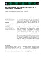
a specific amplicon
with a size of 1130 bp could be observed for 126 of the
investigated
S. uberis
and both
S. uberis
reference strains
(Fig. 3). The remaining four
S. uberis
and the three
S.
parauberis
strains were negative. Investigating the strains
for gene
pauA
a specific amplicon with a size of 800 bp
could be observed for all 128
skc
positive
S. uberis
strains.
No amplicon could be observed for the remaining strains.
Some phenotypical and genotypical characteristics are
summarized in Table 1.
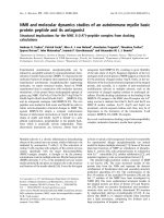
For DNA fingerprinting, a macrorestriction analysis of
the chromosomal DNA of the bacteria was determined by
PFGE. This was performed with 69 arbitrarily selected
strains obtained from 57 cows of 26 different farms.
Digestion of the chromosomal DNA of the isolates was
performed with the endonuclease
Sma
I. The 69 selected
strains displayed 55 different DNA patterns. Identical
PFGE patterns could be observed for some of the isolates
within different quarters of an individual cow and between
different cows within the same farm. The PFGE patterns of
9 isolates from five different cows of farm 2 are shown in
Fig. 4.
Discussion
S. uberis
is important to the veterinary domain because
of its increasing association with bovine mastitis. The
S.
uberis
mastitis causes a tremendous economic loss in milk
production and has become the major environmental
mastitis agent [66].
The present results strongly support the findings
described by Lerondelle [42] that a
S. uberis
infection
rarely gives rise to clinical mastitis. The infection remains
subclinical during long periods of time. In the absence of
treatment, this causes serious losses in milk production.
Also corresponding to the present work, Bramley [9] and
Jayarao
et al
. [32] described a high prevalence of
subclinical forms of
S. uberis
intramammary infections in
dairy cows. According to these authors, a
S. uberis
subclinical mastitis frequently occurs before parturition
and near drying-off period, whereas a clinical mastitis with
S. uberis
could be observed more frequently in the first five
weeks of lactation.
The esculin positive bacteria of the present investigation
were further cultivated on five different media selective for
enterococci. The growth patterns of the cultures were
clearly different to a comparatively cultivated
E. faecalis
strain indicating that all five media could be used to
differentiate between esculin degrading enterococci and
S.
uberis
.
According to hemolysis on blood agar plate and the
carbohydrate fermentation tests, all strains displayed,
comparable to various authors [16,17,19,23,56,68], the
typical properties of
S. uberis
and
S. parauberis.
However, according to Williams and Collins [69] and
Doménech
et al
. [19] and the results of the present study
the enzyme
β
-D-glucuronidase seems to be the only
criterion allowing a differentiation of
S. uberis
and
S.
parauberis
.
Serogrouping of the bacteria revealed that 42 isolates
and one
S. uberis
and
S. parauberis
reference strain were
positive with group E specific antisera, some strains were
positive with group A, G, P and U specific antisera alone
or in combination. Comparable to the present studies
Lämmler [36] and Roguinsky [49,50] also reported that
S.
uberis
strains could serologically be classified into
Lancefield group E, P, G and U. Some of the strains
investigated by Roguinsky [49,50] simultaneously reacted
with group E and group P, group P and group U, group P
and group G specific antiserum, respectively; some strains
were serologically non-groupable. A reaction of some
S.
F
ig. 3. Amplicons of
S. uberis
(1, 2, 3) with a size of 1130
bp
u
sing the
S. uberis
skc
gene specific primers SKC-I and SKC-
II;
s
kc
gene negative
S. uberis
and
S. parauberis
are shown in lane
4
a
nd 5. M = see Fig. 1.
218 I. U. Khan
et al.
uberis
with group P, G, U and B specific antisera had also
been reported by other authors [12,23,26,54,55]. Among
the three
S. parauberis
strains reference strain NCDO
2020 reacted with group E specific antiserum whereas the
remaining two strains were non-groupable.
Lectin agglutination reactions were conducted with the
lectin from Helix pomatia. Corresponding to Niewerth
et
al.
[43], Lämmler [36], Christ and Lämmler [13] and
Abdulmawjood
et al.
[1] some
S. uberis
of the present
study specifically reacted with the lectin from Helix
pomatia indicating the usefulness of lectin agglutination
reactions to phenotypically characterize bacteria of this
species.
A molecular identification of both species could be
performed by RFLP analysis of the 16S rRNA gene.
Corresponding to Jayarao
et al
. [31], as well as Lämmler
et
al
. [38] and Hassan
et al.
[27] all
S. uberis
and
S.
parauberis
strains of the present investigation showed a
specific restriction profile using the restriction enzymes
Rsa
I and
Ava
II. RFLP analysis of the 16S rRNA gene had
Table 1.
Some pheno- and genotypic characteristics of 132
S. uberis
and 3
S. parauberis
S. uberis
*
(n=132)
S. parauberis
**
(n=3)
Growth on
CATC
no growth
KAA
Esculin bile agar 9
1
no growth
Chromocult enterococci agar 7
1
Slanetz-Bartly agar 4
1
Haemolysis
alpha 126
2
-
non 6 3
Carbohydrate
fermentation
arabinose - -
fructose, glucose, maltose, mannitol,
saccharose, salicin, sorbitol, trehalose
132 3
lactose, ribose 131 3
Inulin 92 2
raffinose 2 2
Hydrolysis of
esculin 132 3
hippurate 132 3
arginine 131 3
Enzyme activities
β
-D-glucuronidase 132 -
Pyrrolidonyl aminopeptidase 117 3
Hyaluronidase 47 -
Serogrouping
E431
P11-
U9-
A1-
G1-
E and P 1 -
E and U 1 -
non-groupable 65 2
Lectin agglutination Helix pomatia 43 -
CAMP-like factor sheep blood agar plates 5 -
Specific
PCR reaction
cfu
gene 5 -
skc
gene 128 -
pauA
gene 128 -
*including
S. uberis
reference strains NCDO 2038 and NCDO 2086
**including
S. parauberis
reference strain NCDO 2020, strain 94/16 and strain 138/80
1
weak growth
2
number of strains showing a positive reaction
-negative reaction
Identification and epidemiological characterization of
Streptococcus uberis
isolated from bovine mastitis 219
already been used for characterization of
S. agalactiae
and
S. porcinus
[2,39]. Comparable to the present results, these
authors also found no intraspecies variations for the 16S
rRNA genes of
S. agalactiae
and the serologically
heterogenous species
S. porcinus
. However, an intraspecies
variation in the sequence of the 16S rRNA gene was
observed for
S. suis
[11] and for
S. equi
subsp.
zooepidemicus
[4].
In further studies, a PCR-based identification with
specific oligonucleotide primers targeted to species-
specific regions of the gene encoding the 16S rRNA, the
gene encoding the 23S rRNA and the 16-23S rDNA
intergenic spacer region of
S. uberis
and
S. parauberis
respectively, was performed. All three target genes could
successfully be used to identify and differentiate both
species. Comparable investigations were carried out by
Forsman
et al.
[22] investigating
S. uberis
specific parts of
the 16-23S rDNA intergenic spacer region and by Hassan
et al
. [28] using
S. uberis
and
S. parauberis
specific
regions of the genes encoding the 16S rRNA and the 23S
rRNA, and
S. parauberis
specific regions of the 16S-23S
rDNA intergenic spacer region. In addition, Tilsala-
Timisjärvi
et al
. [65] used species-specific oligonucleotide
primers targeted to the 16-23S rDNA intergenic spacer
region for differentiation of
S. uberis
and other pathogenic
streptococcal and staphylococcal species. Moreover,
Phuektes
et al.
[45] used a 16-23S rDNA intergenic spacer
region based multiplex PCR assay for identification and
differentiation of
S. uberis
and other mastitis pathogens.
Similarly, Riffon et al. [48] described species-specific parts
of the 23S rRNA gene and the 16-23S rDNA intergenic
spacer region of
S. uberis
as well as species-specific parts
of the 23S rRNA gene of
S. parauberis
.
An additional potential virulence factor investigated in
the present study was the CAMP factor and the CAMP
factor encoding gene
cfu
. The importance of “uberis
factor” for the virulence of
S. uberis
has been pointed out
by Skalka and Smola [60]. These authors parenterally
administered an “uberis factor” containing exosubstance of
S. uberis
to rabbits and white mice causing the death of the
animals. In 1979, Skalka
et al
. [59] reported that 58 of 81
investigated
S. uberis
strains produced a hemolytically
active exosubstance showing an identical effect as the
CAMP factor of
S. agalactiae
. Similarly, Christ
et al
. [12]
and Lämmler [36] found 10% and 28% CAMP positive
S.
uberis
strains, respectively. A positive CAMP-reaction and
the detection of gene
cfu
could be observed for five
S.
uberis
of the present investigation. The latter could be
demonstrated, with oligonucleotide primers designed in
the present study. In 1996 Jiang
et al
. [33] cloned and
sequenced the CAMP factor gene
cfu
. According to these
authors the CAMP factor gene cfu of
S. uberis
and the
deduced amino acid sequence appeared to be highly
homologous to the
cfb
gene and amino acid sequence of
S.
agalactiae
. Similarly, Gase
et al
. [24] described a sequence
homology of CAMP factor gene
cfa
of group A
streptococci,
cfb
of group B streptococci and
cfu
of
S.
uberis
. These authors also suggested that CAMP factor
and CAMP factor-like genes are fairly widespread among
streptococci, at least in serogroups A, B, C, G, M, P, R and
U. In addition, Hassan
et al
. [27] found a close relation of
the CAMP gene
cfa
of
S. pyogenes
,
cfb
of
S. agalactiae
,
cfu
of
S. uberis
and
cfg
of
S. canis
.
An additional potential virulence gene investigated in the
present study was the gene pauA/skc encoding a
plasminogen activator. According to previous
investigations, bovine plasminogen activated by
streptokinase seemed to be a virulence factor of
S. uberis
during early stages of infection. This activation might
cause a rapid growth of the bacteria in the lactating bovine
mammary gland [40]. For
S. uberis
the plasminogen
activator gene pauA and the plasminogen activator gene
designated as streptokinase gene skc was cloned and
sequenced by Rosey
et al.
[51] and Johnsen
et al
. [34],
respectively. According to Leigh [41] the gene pauA was
produced by the majority of the
S. uberis
strains isolated
from clinical cases of bovine mastitis. Comparable to these
findings most of the
S. uberis
of the present investigation
were pauA and skc positive. However, comparing the
sequence of both genes revealed their complete sequence
identity (data not shown).
To determine the possibly existing epidemiological
relationship of the collected
S. uberis
strains of the present
*
number of cows
F
ig. 4.
Pulsed-field gel electrophoretic restriction patterns
of
c
hromosomal DNA of 9
S. uberis
isolated from 5 cows of
a
s
ingle farm using the restriction enzyme
Sma
I. Six differe
nt
D
NA restriction patterns were observed; pattern I (lane 1
),
p
attern II (lane 2, 4, 7), pattern III (lane 3), pattern IV (lane 5
),
p
attern V (lane 6, 8) and pattern VI (lane 9).
220 I. U. Khan
et al.
investigation, a macrorestriction analysis of the
chromosomal DNA of arbitrarily selected
S. uberis
was
performed by PFGE. DNA macrorestriction analysis by
PFGE is an essential tool for epidemiological
investigations to identify specific strains of a causative
bacterial species as well as for the comparison of strains
between cows and farms, and has already successfully
been used to investigate restriction patterns among strains
of
S. uberis
[7]. Gordillo
et al
. [25] used PFGE for typing
group B streptococci and described that PFGE patterns
could easily be discerned, interpreted and potentially
utilized for epidemiological investigations.
The isolates of the present study were collected from
bovine milk of a defined area within a time period of three
months. This collection corresponded to the criteria of
epidemiological isolates proposed by Tenover
et al
. [64].
These authors additionally defined a set of guidelines for
interpreting DNA restriction patterns generated by PFGE
and for using these results as epidemiologically useful
information.
The PFGE restriction patterns obtained from 69 selected
S. uberis
strains were comparatively investigated after
digestion with the endonuclease
Sma
I. The results of the
present study revealed mostly nonidentical PFGE patterns.
However, for some strains identical PFGE patterns could
be observed for isolates within different quarters of an
individual cow and different cows within the same farm.
Among 69
S. uberis
strains isolated from 57 cows from 26
different farms 55 different DNA restriction patterns were
observed, indicating that a wide variety of
S. uberis
strains
might infect and cause mammary gland infection due to
the contamination of the gland from the environment. This
high degree of heterogeneity supports the epidemiological
studies by Baseggio
et al
. [7], also suggesting a limited
transmission of infection from cow-to-cow during milking
process. These authors examined and differentiated
S.
uberis
,
S. agalactiae
and
S. dysgalactiae
isolates by PFGE
also after digestion with the restriction enzyme
Sma
I. The
S. uberis
isolates investigated in these studies displayed
diverse restriction patterns. However, the investigated
S.
dysgalactiae
had most diverse and complex restriction
patterns. In contrast to the latter the species
S. agalactiae
had identical restriction patterns within the herds but
distinct between herds. The studies of Douglas
et al
. [20]
additionally supported the results of the macrorestriction
analysis of the
S. uberis
isolates of the present
investigation. According to these authors 330 different
PFGE patterns could be observed from 343 isolates. In
addition Wang
et al
. [67] reported that S. uberis had most
diverse PFGE patterns as compared to
S. agalactiae
and
S.
dysgalactiae
. According to these authors, 74 distinct PFGE
patterns could be observed among 130
S. uberis
strains
collected from 73 different cows of 3 farms. In contrast to
S. uberis
, the
S. agalactiae
isolates examined by Wang
et
al.
[67] exhibited, corresponding to the results of Baseggio
et al.
[7], identical patterns within the same farm but
different patterns between various farms. The latter
indicated that a single clone was transmitted between
cows. Fink
et al.
[21] also analysed and compared
macrorestriction patterns of
S. agalactiae
isolated from
bovine mastitis. According to these authors, also a single
clone seemed to be responsible for the mastitis situation
within a herd. The clones differed between herds.
Moreover, Annemüller
et al
. [6] analysed PFGE patterns
of
Staphylococcus aureus
strains isolated from cows with
mastitis. These studies revealed that isolates from a single
farm generally had identical restriction patterns. This could
also be observed for isolates of different herds. Akineden
et al
. [5] also described that
S. aureus
had identical PFGE
restriction patterns within the same farm but different
patterns between the farms investigated.
Despite the degree of heterogeneity in DNA restriction
patterns, some
S. uberis
strains of the present study
isolated from a single cow as well as from different cows
of the same farm displayed identical PFGE patterns,
indicating that some
S. uberis
strains might be transmitted
from quarter to quarter and cow to cow of a single farm.
This also corresponded to the findings of Phuektes
et al.
[46]. These authors investigated the epidemiological status
of
S. uberis
mastitis in dairy cows and detected
nonidentical and also identical PFGE patterns.
According to the results of the present study, a
macrorestriction analysis of the
S. uberis
isolates by PFGE
appears to be a useful and reliable method to study the
epidemiological relationship of the investigated strains.
The conventional and molecular methods used in the
present study allowed a reliable identification and further
characterization of
S. uberis
and
S. parauberis
and might
help investigate the importance of both species as causative
agents of bovine mastitis. However, according to the
present results, the occurrence of
S. parauberis
as mastitis
causing pathogen seems to be rare.
References
1. Abdulmawjood, A., Soedarmanto, I. and Lämmler, C.
Identification of streptococci of serological group C using
fluorescein and peroxidase conjugated lectins. Med. Sci. Res.
1996, 24, 375-376.
2. Abdulmawjood, A., Weiß, R. and Lämmler, C. Species
identification of
Streptococcus porcinus
by restriction
fragment length polymorphism analysis of 16S ribosomal
DNA. Res. Vet. Sci. 1998, 65, 85-86.
3. Abdulmawjood, A. and Lämmler, C. Amplification of 16S
ribosomal RNA gene sequences for the identification of
streptococci of Lancefield group B. Res. Vet. Sci. 1999, 67,
159-162.
4. Abdulmawjood, A. and Lämmler, C. 2000. Determination
of intraspecies variations of the V2 region of the 16S rDNA
Identification and epidemiological characterization of
Streptococcus uberis
isolated from bovine mastitis 221
gene of
Streptococcus equi
subsp.
zooepidemicus
. Res. Vet.
Sci. 2000,
68
, 33-39.
5.
Akineden, Ö., Annemüller, C., Hassan, A. A., Lämmler,
C., Wolter, W. and Zschöck, M.
Toxin genes and other
characteristics of
Staphylococcus
aureus
isolates from milk
of cows with mastitis. Clin. Diagn. Lab. Immunol. 2001,
8
,
959-964.
6.
Annemüller, C., Lämmler, C. and Zschöck, M.
Genotyping of
Staphylococcus aureus
isolated from bovine
mastitis. Vet. Microbiol. 1999,
69
, 217-224.
7.
Baseggio, N., Mansell, P. D., Browning, J. W. and
Browning, G. F.
Strain differentiation of isolates of
streptococci from bovine mastitis by pulsed-field gel
electrophoresis. Mol. Cell. Probes. 1997,
11
, 349-354.
8.
Bentley, R. W. and Leigh, J. A.
Development of the PCR-
based hybridization protocol for identification of
streptococcal species. J. Clin. Microbiol. 1995,
33
, 1296-
1301.
9.
Bramley, A. J.
Streptococcus uberis
udder infection-a major
barrier to reducing mastitis incidence. Beecham Mastitis
Series, Shinfield, Reading RG.1980,
29AT
, 328-335.
10.
Bramley, A. J.
Source of
Streptococcus uberis
in the dairy
herd. Isolation from bovine feces and from straw bedding of
cattle. J. Dairy Res. 1982,
49
, 369-373.
11.
Chatellier, S., Harel, J., Zhang, Y., Gottschalk, M.,
Higgins, R., Devriese, L. A. and Brousseau, R.
Phylogenetic diversity of
Streptococcus suis
strains of
various serotypes as revealed by 16S rDNA gene sequence
comparison. Int. J. Syst. Bacteriol. 1998,
48
, 581-589.
12.
Christ D., Schwarz, S. and Lämmler, C.
DNA-
fingerprinting of
Streptococcus uberis
. Med. Sci. Res. 1988,
16
, 1297-1298.
13.
Christ, D. and Lämmler, C.
Characterization of
Streptococcus uberis
with special consideration of supposed
pathogenicity factors. Berl. Münch. Tierärztl. Wschr. 1992,
105
, 224-230.
14.
Collins, M. D., Farrow, J. A. E., Katic, V. and Kandler, O.
Taxonomic studies on streptococci of serological group E, P,
U and V: Description of
Streptococcus porcinus
sp. nov. Syst.
Appl. Microbiol. 1984,
5
, 402-413.
15.
Cullen, G. A.
The ecology of
Streptococcus uberis
. Br. Vet.
J. 1966,
122
, 333-339.
16.
Cullen, G. A.
Streptococcus uberis
and its relationship to
bovine mastitis. Univ. of London. Ph.D. Thesis. 1968.
17.
Cullen, G. A.
Isolation of
S. uberis
from lactating and non-
lactating cows. Br. Vet. J. 1969,
125
, 145-148.
18.
Daleel, E. E. and Frost, A. J.
Some observations on the
bacterial flora of the bovine tonsil. Br. Vet. J. 1967,
123
, 232-
236.
19.
Doménech, A., Fernández-Garayzábal, J. F., Pascual, C.,
Garcia, J. A., Cutuli, M.T., Moreno, M. A., Collins, M. D.
and Dominguez, L.
Streptococcosis in cultured turbot,
Scophthalmus maximus
(L.), associated with
Streptococcus
parauberis
. J. Fish Dis. 1996,
19
, 33-38.
20.
Douglas, V. L., Fenwick, S. G., Pfeiffer, D. U., Williamson,
N. B. and Holmes, C. W.
Genomic typing of
Streptococcus
uberis
isolates from cases of mastitis, in New Zealand dairy
cows, using pulsed-field gel electrophoresis. Vet. Microbiol.
2000,
75
, 27-41.
21.
Fink, K., Abdulmawjood, A., Lämmler, C. and Zschöck,
M.
Genotypisierung von
Streptococcus agalactiae
-Kulturen,
isoliert von subklinischen Rindermastitiden. 41
Arbeitstagung des Arbeitsgebietes “Lebensmittelhygiene”
der Deutschen Veterinärmedizinischen Gesellschaft in
Garmisch-Partenkirchen. Tagungsbericht. pp. 177-182, 2000.
22.
Forsman, P., Tilsala-Timisjärvi, A. and Alatossava, T.
Identification of staphylococcal and streptococcal causes of
bovine mastitis using 16S-23S rDNA spacer regions.
Microbiol. 1997,
143
, 3491-3500.
23.
Garvie, E. I. and Bramley, A. J.
Streptococcus uberis
: an
approach to its classification. J. Appl. Bacteriol. 1979,
46
,
295-304.
24.
Gase, K., Ferretti, J. J., Primeaux, C., McShan, M. W.,
Identification, cloning, and expression of the CAMP factor
gene (
cfa
) of group A streptococci. Infect. Immun. 1999,
67
,
4725-4731.
25.
Gordillo, M. E., Singh, K. V., Baker, C. J. and Murray, B.
E.
Typing of group B streptococci: comparison of pulsed-
field gel electrophoresis and conventional electrophoresis. J.
Clin. Microbiol. 1993,
31
, 1430-1434.
26.
Hahn, G.
Ergebnisse aus der Streptokokken-Zentrale in Kiel
von 1965-1978: Mastitis-Streptokokken. Zbl. Bakt. Hyg. I.
Abt. Orig. 1981,
A249
, 223-340.
27.
Hassan, A. A., Abdulmawjood, A., Yildirim, A. Ö., Fink,
K., Lämmler, C. and Schlenstedt, R.
Identification of
streptococci isolated from various sources by determination
of
cfb
gene and other CAMP-factor genes. Can. J. Microbiol.
2000,
46
, 946-951.
28.
Hassan, A. A., Khan, I. U., Abdulmawjood, A. and
Lämmler, C.
Evaluation of PCR methods for the rapid
identification and differentiation of
Streptococcus uberis
and
Streptococcus parauberis
. J. Clin. Microbiol. 2001,
39
, 1618-
1621.
29.
Hwang, M. N. and Ederer, G. M.
Rapid hippurate
hydrolysis for presumptive identification of group B-
streptococci. J. Clin. Microbiol. 1975,
30
, 2235-2240.
30.
Jayarao, B. M., Doré, Jr. J. J. E., Baumbach, G. A.,
Matthews, K. R. and Oliver, S. P.
Differentiation of
Streptococcus uberis
from
Streptococcus parauberis
by
polymerase chain reaction and restriction fragment length
polymorphism analysis of the 16S ribosomal DNA. J. Clin.
Microbiol. 1991,
29
, 2774-2778.
31.
Jayarao, B. M., Doré, Jr. J. J. E., and Oliver, S. P.
Restriction fragment length polymorphism analysis of 16S
ribosomal DNA of
Streptococcus
and
Enterococcus
species
of bovine origin. J. Clin. Microbiol. 1992,
30
, 2235-2240.
32.
Jayarao, B. M., Gillespie, B. E., Lewis, M. J., Dowlen, H.
H. and Oliver, S. P.
Epidemiology of
Streptococcus uberis
intramammary infections in a dairy herd. J. Vet. Med. 1999,
46
, 433-442.
33.
Jiang, M., Babiuk, L. A. and Potter. A. A.
Cloning,
sequencing and expression of the CAMP factor gene of
Streptococcus uberis
. Microb. Pathol. 1996,
20
, 297-307.
34.
Johnsen, L. B., Poulsen, K., Kilian, M. and Peterson, T. E.
Purification and cloning of a streptokinase from
Streptococcus uberis
. Infect. Immun. 1999,
67
, 1072-1078.
222 I. U. Khan
et al.
35.
King, J. S.
Streptococcus uberis
: a review of its role as a
causative organism of bovine mastitis. II. Control of
infection. Br. Vet. J. 1981,
137
, 160-165.
36.
Lämmler, C.
Biochemical and serological properties of
Streptococcus uberis
. J. Vet. Med. 1991,
38
, 737-742
.
37.
Lämmler, C. and Hahn, G. Streptokokken In: Blobel, H.
und Schließer, T.
(eds): Handbuch der bakteriellen
Infektionen bei Tieren. Band II/2: Streptokokken-Infektionen
und Rotlauf. 2. Auflage. Gustav Fischer Verlag, Jena/
Stuttgart. 1994.
38.
Lämmler, C., Abdulmawjood, A., Danic, G., Vaillant, S.
and Weiß, R.
Differentiation of
Streptococcus uberis
and
Streptococcus parauberis
by restriction fragment length
polymorphism analysis of the 16S ribosomal RNA gene and
further studies on serological properties. Med. Sci. Res.
1998,
26
, 177-179.
39.
Lämmler, C., Abdulmawjood, A. and Weiß, R.
Properties
of serological group B streptococci of dog, cat and monkey
origin. J. Vet. Med. 1998,
45
, 561-566.
40.
Leigh, J. A.
Activation of bovine plasminogen by
Streptococcus uberis
. FEMS Microbiol. Lett. 1993,
114
, 67-
72.
41.
Leigh, J. A.
Purification of a plasminogen activator from
Streptococcus uberis
. FEMS Microbiol. Lett. 1994,
118
, 153-
158.
42.
Lerondelle, C.
Les mammites a
Streptococcus uberis
. Res.
Méd. Vét. Ec. Alfort. 1985,
161
, 539-744.
43.
Niewerth, B.
Wechselwirkung zwischen Gram-positiven
Bakterien, Lektinen und Körperzellen. Vet. Med. Diss.,
Justus-Liebig-Universität Giessen. Ph.D. Thesis. 1987.
44.
Obiger, G.
Untersuchungen über die in Rindertonsillen
vorkommenden Streptokokken. Kieler Milchwirtschaftl.
Forschungsber. 1954,
6
, 595-603.
45.
Phuektes, P., Mansell, P. D. and Browning, G. F.
Multiplex
polymerase chain reaction assay for simultaneous detection
of
Staphylococcus aureus
and streptococcal causes of bovine
mastitis. J. Dairy Sci. 2001,
84
, 1140-1148.
46.
Phuektes, P., Mansell, P. D., Dyson, R. S., Hooper, N. D.,
Dick, J. S. and Browning, G. F.
Molecular epidemiology of
Streptococcus uberis
isolates from dairy cows with mastitis.
J. Clin. Microbiol. 2001,
39
, 1460-1406
.
47.
Rantz, L. A. and Randall, E.
Use of autoclaved extracts of
hemolytic streptococci for serological grouping. Stanford
Med. Bull. 1955,
13
, 290-291.
48.
Riffon, R., Sayasith, K., Khalil, H., Dubreuil, P., Drolet,
M. and Lagace, J.
Development of a rapid and sensitive test
for identification of major pathogens in bovine mastitis by
PCR. J. Clin. Microbiol. 2001,
39
, 2584-2589
.
49.
Roguinsky, M.
Reactions de
Streptococcus uberis
avec les
serums G et P. Ann. Inst. Pasture. 1969,
117
, 529-532.
50.
Roguinsky, M.
Caracteres biochimiques et serologiques de
Streptococcus uberis
. Ann. Inst. Pasture. 1971,
126
, 529-532.
51.
Rosey, E. L., Lincoln, R. A., Ward, P. N., Yancey, Jr. R. J.
and Leigh, J. A.
PauA: a novel plasminogen activator from
Streptococcus uberis
. FEMS Microbiol. Lett. 1999,
178
, 27-
33.
52.
Schalm, O. W., Carroll, E. J. and Jain, N. C.
In bovine
mastitis. Lee and Febiger. Philadelphia. PA. USA. 1971.
53.
Schleifer, K. H. and Kilpper-Bälz, R.
Molecular and
chemotaxonomic approaches to the classification of
streptococci, enterococci and lactococci: A review. Syst.
Appl. Microbiol. 1987,
10
, 1-19.
54.
Schuman, R. D., Nord, N., Brown, R. W., Wessman, G. E.
and Wessman, G. E.
Biochemical and serological
characteristics of Lancefield groups E, P, and U streptococci
and
Streptococcus uberis
. Cornell Vet. 1972,
62
, 540-562.
55.
Schuman, R. D., Wood, R. L. and Nord, N.
Swine
abscesses caused by Lancefields group E streptococci. X.
Specificity of the precipitin test for their detection with
relation to the streptococcal antigens of groups R and T,
Streptococcus uberis
and
Staphylococcus aureus
. Cornell
Vet. 1972,
62
, 61-73.
56.
Seeley, H. W.
The physiology and nutrition of
Streptococcus
uberis
. J. Bacteriol. 1951,
62
, 107-115.
57.
Sharma, R. M. and Parker, R. A.
Occurrence and ecologic
features of
Streptococcus uberis
in the dairy cows. Am. J.
Vet. Res. 1970,
31
, 1197-1202.
58.
Sherman, J. M.
The streptococci. Bacteriol. Rev. 1937,
1
, 3-
97.
59.
Skalka, B., Smola, J. and Pilich, J.
Diagnostic availability
of the hemolytically active exosubstance of
Corynebacterium
pseudotuberculosis
for isolation and identification of
Streptococcus agalactiae
and its comparison with the beta-
toxin of
Staphylococcus aureus
. Zbl. Vet. Med. 1979,
B26
,
679-687.
60.
Skalka, B. and Smola, J.
Lethal effect of CAMP-factor and
Uberis-factor- a new finding about diffusible exosubstances
of
Streptococcus agalactiae
and
Streptococcus uberis
. Zbl.
Bakt. Hyg. I. Abt. Orig. 1981,
A249
, 190-194.
61.
Slot, P. A.
Classification, systemics and occurrence. Nord.
Vet. Med. 1958,
10
, 143-152.
62.
Soedarmanto, I., Pasaribu, F. H., Wibawan, I. W. and
Lämmler C.
Identification and molecular characterization of
serological group C streptococci isolated from diseased pigs
and monkeys in Indonesia. J. Clin. Microbiol. 1996,
34
,
2201-2204
.
63.
Sweeney, E. J.
Observation on the epidemiology of mastitis
due to
Streptococcus uberis
in a laboratory herd during a
complete lactation. Res. Vet. J. 1964,
5
, 483
-
487.
64.
Tenover, F. C., Arbeit, R. D., Goering, R. V., Mickelsen, P.
A., Murray, B. E., Persing, D. H. and Swaminathan, B.
Interpreting chromosomal DNA restriction patterns produced
by pulsed-field gel electrophoresis: criteria for bacterial strain
typing. J. Clin. Microbiol. 1995,
33
, 2233-2239.
65.
Tilsala-Timisjärvi, A., Forsman, P. and Alatossava, T.
Bovine mastitis diagnosis from milk by a polymerase chain
reaction-based reaction. J. Nutri. Res. Food Sci. 2000,
55
,
488-492.
66.
Todhunter, D. A., Smith, K. L. and Hogan, J. S.
Environmental streptococcal mastitis. In: Proc. pp. 92-95,
Natl. Mast. Counc., Arlington. 1994.
67.
Wang, S. M., Deighton, M. A., Capstick, J. A. and
Gerraty, N.
Epidemiological typing of bovine streptococci
by pulsed-field gel electrophoresis. Epidemiol. Infect.
1999,
123
, 317-324.
68.
Williams, A. M. and Collins, M. D.
Molecular taxonomic
Identification and epidemiological characterization of
Streptococcus uberis
isolated from bovine mastitis 223
studies on
Streptococcus uberis
types I and II. Description of
Streptococcus parauberis
sp. nov. J. Appl. Bacteriol. 1990,
68
, 485-490.
69.
Williams, A. M. and Collins, M. D.
DNA fingerprinting of
Streptococcus uberis
based on polymorphism of DNA
encoding rRNA. Lett. Appl. Microbiol. 1991,
12
, 23-28.
70.
Winkle, S.
Mikrobiologische und Serologische Diagnostik.
3. Auflage, Gustav Fischer Verlag, Stuttgart/New York. 1979
.