Báo cáo khoa học: "Glucocorticoid-induced laminitis with hepatopathy in a Thoroughbred filly" ppt
Bạn đang xem bản rút gọn của tài liệu. Xem và tải ngay bản đầy đủ của tài liệu tại đây (1.14 MB, 4 trang )
-2851$/ 2)
9H W H U L Q D U \
6FLHQFH
J. Vet. Sci.
(2004),
/
5
(3), 271–274
Glucocorticoid-induced laminitis with hepatopathy in a Thoroughbred filly
Seung-ho Ryu
1
, Byung-sun Kim
1
, Chang-woo Lee
2,
*, Junghee Yoon
3
, Yonghoon Lyon Lee
4
1
Equine Hospital, Korea Racing Association, Kwachon 427-070, Korea
2
Department of Clinical Pathology, College of Veterinary Medicine, Seoul National University, Seoul 151-742, Korea
3
Department of Radiology, College of Veterinary Medicine, Seoul National University, Seoul 151-742, Korea
4
Department of Anesthesia, Pain Management and Perioperative Medicine, Boren Veterinary Medical Teaching Hospital and
College of Veterinary Medicine, Oklahoma State University, Stillwater, OK 74074, USA
A 3-year-old Thoroughbred filly was referred to the
Equine Hospital, Korea Racing Association for evaluation
of hematuria, inappetite, weight loss and depression.
From 25 days prior to admission, the horse was treated
for right carpal lameness with 20 mg intramuscular
administration of triamcinolone acetonide per day for
consecutive 10 days by a local veterinarian. Clinical and
laboratory findings included vaginal hyperemia, flare in
bladder wall, neutrophilia, lymphopenia, polyuria,
polydipsia and laminitis in the end. High activities of
aspartate transaminase and gamma glutamyltransferase
and high concentration of total bilirubin indicated
hepatopathy. Further hematology, serum biochemistry
and urinalysis did not reveal any abnormalities. Medical
history, physical and clinicopathologic findings suggest
that the laminitis and hepatopathy in this horse were most
likely induced by repeated administration of exogenous
corticosteroid. However, guarded prognosis of treating
laminitis undermined the benefit of improvement of
hematuria following electroacupuncture stimulation. The
combined stimulation of kidney related acupoints (Shen
Peng, Shen Shu), lumber related acupoints (Yao Qian, Yao
Zhong) and associate acupoints (Guan Yuan Shu, Bai
Hui) at 5Hz, 1-2V, for 40 minutes was of value in the
treatment of hematuria. This case shows that horses
under steroids may exhibit laminitis and steroid
hepatopathy. Early recognition and good management of
laminitis are important in the limitation of complications.
Key words:
hematuria, hepatopathy, laminitis, Thorough-
bred, triamcinolone acetonide
Laminitis is recognized as a potentially crippling
condition in the horse that frequently progressed to
euthanasia for humane reasons. It is believed that
inflammatory mediators and other unknown local factors
associated with these systemic diseases alter the
hemodynamics within the digit and this alteration leads to
laminitis [13].
There is no data reporting clinical case of laminitis in
horses in Korea. The purpose of this report is to describe
the first case of glucocorticoid-induced laminitis with
hepatopathy and hematuria in a Thoroughbred filly in
Korea.
Clinical findings and clinical pathology:
A 3-year-old
Thoroughbred filly was referred to the Equine Hospital,
Korea Racing Association for evaluation of hematuria,
inappetite, weight loss and depression. From 25 days prior
to admission, the horse was treated for right carpal lameness
with 20 mg (10 ml) intramuscular administration of
triamcinolone acetonide (Retardoesterode, Laboratorios
Calier, Barcelona, Spain) per day for consecutive 10 days
(total amount: 200 mg) by a local veterinarian.
When admitted, the horses rectal temperature, heart rate
and respiratory rate were 39.2
o
C
,
60 beats/min and
36 breaths/min, respectively. The mucous membranes were
congested and slightly cyanotic. There was severe
thrombophlebitis on both jugular veins. Decreased intestinal
sounds were auscultated in all 4 abdominal quadrants. There
was vaginal hyperemia in speculum examination. Flare in
bladder wall (Fig. 1) and hematuria in bladder (Fig. 2) were
observed in endoscopic examination. No cystic calculi or
neoplasia was observed in ultrasound examination.
Radiograph of thorax was normal.
The horse had mild neutrophilia (14,852 neutrophils/
µ
l
)
and lymphopenia (948 lymphocytes/
µ
l). Abnormal serum
biochemical values were high activities of aspartate
transaminase (558 IU/L), gamma glutamyltransferase (39
IU/L), creatine phosphokinase (493 IU/L), lactic
dehydrogenase (814 IU/L) and high concentration of total
bilirubin (3.8 mg/dl). Urinary specific gravity and RBC
*Corresponding author
Phone: 82-2-880-1273; Fax: 82-2-880-8662
E-mail:
Case Report
272 Seung-ho Ryu
et al.
counts were 1.023 and 190,000/
µ
l
,
respectively. No
abnormality in renal function was indicated by urinary
specific gravity and within normal range concentrations of
BUN (2 weeks after admisson 11 mg/dl: on admission 9 mg/
dl) and creatinine (2 weeks after admisson 1.3 mg/dl: on
admission 1.4 mg/dl) and no glucose and ketone in urine.
Polydipsia was observed. Polyuria was presumed on the
basis of wetter bedding in the horses stall.
Therapy and course of condition:
There was a difficulty
in medical treatment because of severe thrombophlebitis on
both jugular veins, and therefore a decision to treat the filly
with electroacupuncture therapy was made. Kidney related
acupoints including Shen Peng (kidney shelf) and Shen Shu
(kidney association point), and associate acupoints including
Guan Yuan Shu (association point of enclosed original
energy, BL-26), Bai Hui (hundred meetings, GV-20), Yao
Qian (cranial lumber) and Yao Zhong (Middle lumber) were
selected for the treatment of hematuria and stimulated at 1-
2V and 5Hz, for 40 minutes. Color of urine changed
gradually into almost normal yellow color and RBC counts
in urine gradually decreased (from
190,000/
µ
l to 8,000/
µ
l
)
by the 2 weeks of electroacupuncture
therapy.
On the 8th day of hospitalization, the horse became lame
and showed signs of laminitis in the front feet. Lateral
radiography of the front feet revealed 17 degrees of ventral
deviation of the third phalanges of both front feet (Fig. 3).
Extra-deep bedding was placed in the stall. Medical
treatment with mineral oil, flunixin meglumine and
antihypertensive such as acepromazine and both front Qian
Ti Tou (toe of the hoof) and Qian Jiu (central bulb)
hemoacupuncture for 4 days failed to alleviate clinical signs.
Serum chemical values were evaluated daily; serum
activities of AST (542 IU/L: on admission 558 IU/L), CPK
(296 IU/L: on admission 493 IU/L) and LDH (786 IU/L: on
admission 814 IU/L) remained high for KRA reference
range 2 weeks after admisson. Activity of GGT (30 IU/L: on
admission 39 IU/L) and concentration of total bilirubin (1.2
mg/dl: on admission 3.8 mg/dl) were within KRA reference
range 2 weeks after admisson. Urinary specific gravity and
RBC counts were 1.025 and 8,000/
µ
l
(
on admission 1.023
and 190,000/
µ
l
),
respectively 2 weeks after admisson.
Glucose and ketone remained not detected in urine.
Polydipsia and polyuria were almost resolved at that time.
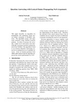
Radiography of the front feet was repeated after
approximately 1 month; perforated soles by the toes of third
phalanges were detected (Fig. 4). The filly retired from race
because of guarded prognosis of laminitis.
Medical history, physical and clinicopathologic findings
suggest that the laminitis and hepatopathy in this horse were
most likely induced by repeated administration of
exogenous corticosteroid. Because of the manifold
F
ig. 1.
Endoscopic findings of urinary bladder, there was flare
in
b
ladder wall.
F
ig. 2.
Endoscopic findings of urinary bladder, there w
as
h
ematuria in bladder.
F
ig. 3.
Lateromedial projection of left 3
rd
phalanx, 17 degrees
of
v
entral deviation of the third phalanx is shown.
Glucocorticoid-induced laminitis with hepatopathy in a Thoroughbred filly 273
physiologic processes affected by glucocorticoids, a wide
variety of clinical and laboratory findings have been
reported including polyuria, polydipsia, laminitis,
hyperglycemia, glucosuria, neutrophilia, lymphopenia [3,8,
15,18]. High activities of AST and GGT and high
concentration of total bilirubin indicated hepatopathy.
Hepatic disease following exogenous administration or
endogenous production of excess glucocorticoids had
developed in people and dogs [7,21]. The condition has been
termed steroid hepatopathy [21]. The condition most often
develops subsequent to administration of exogenous
glucocorticoids. High concentration of glucocorticoids in
blood also was considered to have caused neutrophilia and
lymphopenia in this horse. Corticosteroid-induced
neutrophilia is attributed to decreased ability of neutrophils
to adhere to vascular endothelium [5,16] resulting in
decreased margination of neutrophils in vascular channels
and reduced diapedesis of neutrophils from blood into
tissues [4,5,16]. The mechanism of corticosteroid induced
lymphopenia is believed to be diminished recirculation or
redistribution into lymphoid tissue of recirculating
lymphocytes.
Hematuria was considered to originate from flare of the
mucosa by cystitis rather than nephritis.
Glucocorticoids are known to potentiate vasoconstriction
caused by catecholamines [9] Digital vasoconstriction and
subsequent diminished laminar perfusion is believed to be
an important factor in the pathogenesis of laminitis [14].
Signs of laminitis in this horse developed around 30 days
after initial administration of glucocorticoid, either because
of delayed onset or slow progression of disease. Laminitis is
reportedly more common after triamcinolone acetonide,
compared with other corticosteroids [11]. Adams theorized
laminar necrosis was caused by laminar edema after
demonstrating increased blood flow through the digital
arteries [2]. The edema led to necrosis when the laminae
were compressed between bone and the noncompliant hoof
wall. Using digital angiography, Garner and Coffman
showed decreased arterial flow in the circumflex and
laminar vessels during acute and chronic laminitis [1,6].
Hood and Galey, using scintigraphy, demonstrated
decreased laminar capillary blood flow during acute
laminitis [10,12]. Many of these studies converged upon the
theory that during the development of acute laminitis blood
flow to the digit increased, but was shunted through
arteriovenous shunts resulting in decreased laminar capillary
blood flow. The presence of arteriovenous shunts is
supported by studies of Molyneux and Pollitt, who
demonstrated anatomic arteriovenous shunts in the equine
digit [17,19].
However, guarded prognosis of treating laminitis
undermined the benefit of improvement of hematuria
following EA stimulation. The combined stimulation of
kidney related acupoints (Shen Peng, Shen Shu), lumber
related acupoints (Yao Qian, Yao Zhong) and associate
acupoints (Guan Yuan Shu, Bai Hui) at 5 Hz, 1-2 V, for 40
minutes was of value in the treatment of hematuria. It is
probable that electroacupuncture enhanced the healing rate
of flare. Acupuncture or electroacupuncture by needles
around the edge of trophic ulcers, including postphlebitis
ulcers, cured most cases. Acupuncture cured thromboangitis
obliterans. Acupuncture with anticoagulant therapy
(heparin) resolved thrombophlebitis. Pain and bleeding
improved or stopped after the first session in 82% of cases
[20].
This case shows that horses under steroids may exhibit
laminitis and steroid hepatopathy, and warrants judicious
usage of such agents. Early recognition and good
management of laminitis are important prerequisites in the
limitation of complications.
References
1. Ackerman N, Garner HE, Coffman JR, Clement JW.
Angiographic appearance of the normal equine foot and
alterations in chronic laminitis. J Am Vet Med Assoc 1975,
166, 58-62.
2. Adams OR. Vascular changes in experimental laminitis.
Proc Am Assoc Equine Pract 1972
,
18, 359-373.
3. Beech J. Tumors of the pituitary gland (pars intermedia). In:
Robinson NE (ed), Current Therapy in Equine Medicine 2,
pp. 182-185, Saunders, Philadelphia, 1987.
4. Chiang JL, Patterson R, McGillen JJ, Phair JP, Roberts
M, Harris K, Riesing KS. Long-term corticosteroid effect
on lymphocyte and polymorphonuclear cell function in
asthmatics. J Allergy Clin Immunol 1980, 65, 263-268.
5. Clark RAF, Gallin JI, Fauci AS. Effects of
in vivo
prednisone on in vitro eosinophil and neutrophil adherence
and chemotaxis. Blood 53, 633-41.
6. Coffman JR, Johnson JH, Guffy MM, Finocchio EJ. Hoof
F
ig. 4. Lateromedial projection of left 3
rd
phalanx, perforat
ed
s
oles by the toe of third phalanx is detected (arrow).
274 Seung-ho Ryu
et al.
circulation in equine laminitis. J Am Vet Med Assoc 1970,
156
,
76-83.
7.
DeNovo RC, Prasse KW.
Comparison of serum biochemical
and hepatic functional alterations in dogs treated with
corticosteroids and hepatic duct ligation. Am J Vet Res 1983,
44
, 1703-1709.
8.
Dybdal NO.
Endocrine disorders. In: Smith BP (ed), Large
Animal Internal Medicine, pp. 1296-1302, Mosby, St. Louis,
1990.
9.
Eyre P, Elmes PJ, Strickland S.
Corticosteroid-potentiated
vascular responses of the equine of the equine digit: a
possible pharmacologic basis for laminitis. Am J Vet Res
1979,
40
,
135-138.
10.
Galey FD, Twardock AR, Goetz TE, Schaeffer DJ, Hall
JO, Beasley VR.
Gamma scintigraphic analysis of the
distribution of perfusion of blood in the equine foot during
black walnut (
Juglans nigra
) -induced laminitis. Am J Vet
Res 1990,
51
,
688-695.
11.
Harkins JD, Carney JM, Tobin T.
Clinical use and
characteristics of the corticosteroids. Vet Clin North Am
Equine Pract 1993,
9
,
543-562.
12.
Hood DM, Amoss MS, Hightower D.
Equine laminitis:
Radioisotopic analysis of the hemodynamics of the foot
during acute disease. J Equine Med Surg 1978,
2
,
439-444.
13.
Hunt RJ.
The pathophysiology of acute laminitis. Compend
Contin Educ Pract Vet 1991,
13
,
1003-1011.
14.
Linford RL.
Laminitis. In: Smith BP (ed), Large Animal
Internal Medicine, pp. 1158-1168, Mosby, St. Louis, 1990.
15.
Loeb WF, Capen CC, Johnson LE.
Adenoma of the pars
intermedia associated with hyperglycemia and glycosuria,
two horses. Cornell Vet 1966,
56
,
623-639.
16.
McGillen J, Patterson R. Phair JP.
Adherence of
polymorphonuclear leukocytes to nylon: modulation by
prostacyclin (PGI
2
), corticosteroids ad complement
activation. J Infect Dis 1980,
141
,
382-388.
17.
Molyneux GS, Haller CJ, Mogg K, Pollitt CC.
The
structure, innervation and location of arteriovenous
anastomoses in the equine foot. Equine Vet J 1994,
26
, 305-
312.
18.
Moore J, Steiss J, Nicholson WE, Orth DN.
A case of
pituitary adrenocorticotropin-dependent Cushing’s syndrome
in the horse. Endocrinology 1979,
104
,
576-582.
19.
Pollitt CC, Molyneux GS.
A scanning electron
microscopical study of the dermal microcirculation of the
equine foot. Equine Vet J 1990,
22
, 79-87.
20.
Rogers PAM.
Immunologic effects of Acupuncture In:
Shoen AM. (ed), Veterinary Acupuncture, pp. 245-246,
Mosby, St. Louis, 1994.
21.
Rogers WA, Ruebner BH.
A retrospective study of probable
glucocorticoid induced hepatopathy in dogs. J Am Vet Med
Assoc 1977,
170
,
603-606.