Báo cáo khoa học: "Isolation and identification of Escherichia coli O157:H7 using different detection methods and molecular determination by multiplex PCR and RAPD" docx
Bạn đang xem bản rút gọn của tài liệu. Xem và tải ngay bản đầy đủ của tài liệu tại đây (1.69 MB, 13 trang )
-2851$/ 2)
9H W H U L Q D U \
6FLHQFH
J. Vet. Sci.
(2005),
/
6
(1), 7–19
Isolation and identification of
Escherichia coli
O157:H7 using different
detection methods and molecular determination by multiplex PCR and
RAPD
Ji-Yeon Kim
1,2
, So-Hyun Kim
2
, Nam-Hoon Kwon
2
, Won-Ki Bae
2
, Ji-Youn Lim
2
, Hye-Cheong Koo
2
,
Jun-Man Kim
2
, Kyoung-Min Noh
2
, Woo-Kyung Jung
2
, Kun-Taek Park
2
, Yong-Ho Park
2,
*
1
Department of Animal Disease Diagnosis, National Veterinary Research and Quarantine Service, Anyang 430-824, Korea
2
Department of Microbiology, College of Veterinary Medicine and School of Agricultural Biotechnology, Seoul National University,
Seoul 151-742, Korea
Escherichia coli
O157:H7 is recognized as a significant
food-borne pathogen, so rapid identification is important
for food hygiene management and prompt epidemiological
investigations. The limited prevalence data on Shiga toxin-
producing
E. coli
(STEC) and
E. coli
O157:H7 in foods
and animals in Korea made an assessment of the risks
difficult, and the options for management and control
unclear. The prevalence of the organisms was examined
by newly developed kit-
E. coli
O157:H7 Rapid kit. For the
isolation of
E. coli
O157:H7, conventional culture,
immunomagnetic separation, and
E. coli
O157:H7 Rapid
kit were applied, and multiplex PCR and randomly
amplified polymorphic DNA (RAPD) were performed for
the molecular determination. There was high molecular
relatedness among 11 Korean isolates and 17 U. S. strains
at 63% level. Additionally, distinct differentiation between
pig and cattle isolates was determined. It implied that
RAPD had a capacity to distinguish strains with different
sources, however it could not discriminate among isolates
according to their differences in the degree of virulence.
In antimicrobial susceptibility tests, 45.5% of isolates
showed antibiotic resistance to two or more antibiotics.
Unlike the isolates from other countries, domestic isolates
of
E. coli
O157:H7 was mainly resistant to ampicillin and
tetracylines. In summary, the application of
E. coli
O157:H7 Rapid kit may be useful to detect
E. coli
O157:H7 due to its sensitivity and convenience. Moreover,
combinational analysis of multiplex PCR together with
RAPD can aid to survey the characteristics of isolates.
Key words:
Escherichia coli
O157:H7, multiplex PCR,
RAPD
Introduction
Shiga toxin-producing
Escherichia coli
(STEC) has been
recognized as an important cause of human diseases such as
hemolytic uremic syndrome (HUS) [29,36]. STEC constitute
one of the most important causes of food-borne disease
worldwide. Since the first report by Riley
et al.
[38], STEC
has been associated with outbreaks and sporadic cases of
human diseases, ranging from uncomplicated diarrhea to
hemorrhagic colitis and HUS. Disease in humans following
infection with STEC generally results in either exclusively
intestinal symptom, such as abdominal pain, and bloody or
nonbloody diarrhea, or less frequently, serious systemic
complications. The complications associated with STEC
infection are largely related to the development of thrombotic
microangiopathy in a number of sites. This is especially
prevalent in the kidney, and the end result is the development
of HUS, which is characterized by the triad of acute renal
failure, thrombopenia, and anemia. A number of organs other
than the kidney are often involved in STEC-related
complications. Central nervous system and pancreas are
frequent targets [1]. Besides humans, STEC can cause
damage to animals. For example, STEC develops renal
tubular necrosis in mice and damages certain endothelial cells
in pigs and rabbits. Greyhounds inoculated with STEC
develop vascular lesions in the glomeruli that mimic those
seen in patients with HUS [3].
STEC has been found to produce a family of related
cytotoxins known as Shiga toxins (Stxs). They have been
classified into two major classes, Stx1 and Stx2. Whereas
the Stx1 family is very homogenous, several Stx2 variants
have been identified. These variants are: Stx2c and Stx2d
produced by human STEC isolates, Stx2e typically found in
STEC pathogenic for pigs, and Stx2f, described recently in
STEC isolates from feral pigeons [40]. An STEC can
produce Stx1, Stx2 (or its variants) or both. The Stx2 is
*Corresponding author
Tel: 82-2-880-1257; Fax: 82-2-871-7524
E-mail:
8Ji-Yeon Kim
et al.
responsible for the severe necrotic renal tubular lesions and
death of treated mice fed an EHEC which possesses both
Stx1 and Stx2. This difference in toxicity is also evident
when human renal microvascular endothelial cells are
treated with purified Stx1 or Stx2. They are capable of
crossing an intact polarized epithelium via an energy-
requiring process and, most importantly, the toxin that
moves across this barrier retains its biological activity;
damage to epithelial cells. Except Stxs, there are several
virulence factors can contribute to the pathogenicity in
STEC. The
eae
gene that codes intimin is a 94-to 97-kDa
outer membrane protein produced by all attaching-and-
effacing (A/E) enteric pathogens including STEC O157:H7.
It is the only bacterial adherence factor identified thus far as
important intestinal colonization in animal models. Another
putative virulence factor is RTX toxin designated as EHEC-
hemolysin, coded by the EHEC
hly
operon. There are two
different plasmid-encoded hemolysins, both members of the
RTX toxin family, have been described for STEC. Alpha-
hemolysin is formed by porcine edema disease-causing
STEC serovars which produce Stx variant 2e. Moreover,
STEC serotypes may also possess additional virulence
factors such as secreted proteins for signal transduction
encoded by
esp
A,
esp
B and
esp
D and the translocated
intimin receptor encoded by
tir
[7].
STEC infection has been often associated with the
consumption of contaminated ground beef, raw milk, and
other bovine products, thus cattle are suspected to be a
primary reservoir [15]. But bacteria also have been isolated
from domestic [6] and wild animals [48]. Moreover, recent
outbreaks of foodborne illness associated with eating fresh
products have heightened concerns that these foods
contaminated with STEC may be an increasing source of
illness [43]. In the past decades, outbreaks of diseases
caused by STEC have been associated with the consumption
of leaf lettuce [2], potatoes [9], radish sprouts [50], and raw
vegetables [34]. Fruit-related outbreaks have also been
caused by the consumption of fresh-pressed apple juice [13].
Detection of
E. coli
O157:H7 in the clinical laboratory is
dependent on distinguishing the pathogenic serotypes from
normal fecal flora containing commensal strains of
E. coli.
Fortunately,
E. coli
O157:H7 has two unusual biochemical
markers; delayed fermentation of
D
-sorbitol and lack of
β
-
D-glucuronidase activity, which help to phenotypically
separate O157:H7 isolates from nonpathogenic
E. coli
strains [49]. One of these markers (delayed sorbitol fermentation)
enables to develop several selective media (e.g., Sorbitol-
MacConkey; SMAC) which aid in the initial recognition of
suspicious colonies isolated from bloody stools. The
selectivity of SMAC agar has been improved with the
addition of cefixime-rhamnose (CR-SMAC), cefixime-tellurite
(CT-SMAC), and 4-methylumbelliferyl-
β
-D-glucuronide
(MSA-MUG). In addition to modifying of SMAC agar, new
selective media have been developed to increase the
effectiveness of
E. coli
O157:H7 isolation, including Fluorocult
E. coli
O157:H7 (Merck, Germany), Chromocult agar
(Merck, Germany), Rainbow agar O157 (RB; Biolog,
USA), and Biosynth Culture Media O157:H7 (BCM
O157:H7; Biosynth, Switzerland). Once suspicious colonies
are identified, confirmation of the isolates as
E. coli
O157:H7 is dependent upon biochemical identification and
demonstration of the presence of somatic and flagellar
antigens (O157, H7). These steps are necessary since other
enteric bacteria can be sorbitol-negative and can possess
antigens those are identical to or cross-reactive with O157
antigens. However, Feng [16] reported that sorbitol-
fermenting
E. coli
O157:H7 had been detected from foods
and increased number of such strains has been identified in
Europe. Furthermore, an increasing phenotypic variation in
O157 isolates has been noted in European studies which
could potentially lead to misidentification of O157:H7 as
some other species [49].
Detection of
E.
coli
O157:H7 from food samples requires
enrichment and isolation with selective and/or indicator
media, but lacks specificity to identify STEC [36,39,53].
Thus, more sensitive methods are required to improve the
detectability of STEC O157:H7 from food and environmental
samples. Apart from the traditional culture methods relying
on biochemical characteristics, various genotypic methods
have been proven useful for species identification,
epidemiological typing, and determining genetic relatedness
among pathogenic and nonpathogenic bacteria [44].
Besides, the low infectious dose of
E. coli
O157:H7 (from
50 to 100 organisms) necessities the development of
sensitive detection techniques. For examples, immunomagnetic
separation (IMS) techniques have been employed widely
within routine microbiology testing laboratories for the
isolation of specific microorganisms [9,20]. IMS allows the
rapid capture and concentration of bacteria from a range
matrics. The magnetic beads used for IMS are commercially
available, either pre-coated with antibodies or ready for
antibody conjugation. The beads are typically 2-3
µ
m
spheres containing Fe
2
O
4
and Fe
3
O
4
to make them super-
paramagnetic. They are only magnetic in the presence of a
magnetic field and readily separate from each other when
the magnetic field is removed. By applying a strong
magnetic field to the outside of the reaction vessel, the beads
and captured bacteria can be immobilized against the vessel
wall. This allows selective removal of the remainder of the
samples including non-target bacteria and other organic
particles. The beads are then released by withdrawing the
magnet. This simple step of IMS procedure can help us to
isolate STEC from samples easily. Recently, immunomagnetic
particles for the separation of
E. coli
O26 and O111 have
become commercially available. With the use of IMS, the
isolation rate of
E. coli
O157 has been markedly improved.
Wright
et al.
[51] showed a 100-fold increase in sensitivity
of detection by IMS compared with direct subculture from
Molecular determination of
E. coli
O157:H7 9
enrichment broth. However, manual IMS (MIMS) is very
labor intensive when large numbers of samples have to be
analyzed. So, an automated IMS in combination with an
integrated ELISA (EiaFoss; Foss, Denmark) would increase
efficiency and lighten the workload. This method can test
about 81-108 samples per day, after overnight enrichment
[37]. The latex agglutination method (Verotox F-Assay) for
the Stxs detection has been developed and available [24]. It
is based on the use of latex particles sensitized with
antibodies to these two toxins which are detected by
reversed passive latex agglutination (RPLA). Additionally,
methods to detect Stx-gene or Stx-production have been
proven to be useful for identification of STEC. Among a lot
of commercially available detection techniques, we selected
one of visual immunochromatographic assays,
E. coli
O157:H7 Rapid kit (Dong-A Pharm, Korea). The
effectiveness of the kit has not yet been determined. We
examined its capacity to detect STEC O157:H7 comparing
with IMS which is proven to be one of the most sensitive
detection techniques.
The isolation of
E. coli
belonging to serogroup O157 has
rarely been reported in Asian countries except Japan; though
isolation of
E. coli
O157 from clinical sources in India,
China, Korea, and Hong Kong has been briefly reported
[47]. The limited prevalence data on STEC and
E. coli
O157:H7 in foods and animals in the country made an
assessment of the risks difficult, and the options for
management and control unclear.
The objectives of this study are (i) to examine the
prevalence of
E. coli
O157:H7 in slaughterhouses and retail
markets, (ii) to characterize the isolates by determination of
stx
1,
stx
2,
eae
A
,
and
hly
A
in multiplex PCR assay, (iii) to
compare the genetic patterns of Korean isolates and U.S.
isolates, and (iv) to compare the efficiency among
conventional culture method, IMS, and
E. coli
O157:H7
commercial diagnostic kit, the
E. coli
0157:H7 Rapid kit.
The study will provide information on newly developed
diagnostic kit for its detectability, rapidity and convenience
to perform. The diagnostic procedures examined in this
study can be correctly applied to the areas which require to
supervise the presence of the organism, especially enforced
the Hazard Analysis Critical Control Point (HACCP)
program. And, the result of genotypes of the isolates can
envision the determination of Korean epidemiological
characteristics. All together, we may propose the effective
control strategy against STEC infection in humans and
animals, and food contamination in livestock products.
Materials and Methods
Bacterial strains
E. coli
O157:H7 strains used in this study are listed in
Table 1. Four strains, one produces both Stx1 and Stx2, and
one produces Stx1 only, one produces Stx2 only, and one
non-Stx producing strain, were obtained from American
Type Culture Collection (ATCC). Seven
E. coli
O157:H7
strains were obtained from
E.
coli
reference center
(Pennsylvania State University, USA) and six strains were
obtained from Cornell University. Additionally, eleven
Korean isolates detected in this study were also listed.
Sample collections
From April 2000 to June 2002, a total of 1,682 samples
were collected. Among them, 1,042 fecal samples were
collected from pigs and cattle at 3 slaughterhouses, and from
chicken at meat processing plants. The sponge sampling
method was used to collect 286 pork and beef samples and
homogenization was conducted to process the samples from
retail markets. A total of 355 chicken samples were obtained
from chicken meat processing plants and markets by rinsing
the samples with buffered peptone water (BPW; Becton
Dickinson, USA).
In case of fecal samples, a cup of feces was taken into
each 100 ml of specimen cup, and pork and beef carcasses
from three slaughterhouses were conducted by sponge
sampling method within 24 h after slaughtering [19]. For
each carcass, three sites were investigated; belly, leg, and
hip. For swabbing with sterilized sponge, an area of 5 by
10 cm was delimitated by sterile plastic template. The
delimited area was then swabbed with a sterilized sponge
that had been moistened by being placed in a sterilized vial
with 10 ml of BPW in Meat/Turkey Carcass Sampling Kit
Table 1. Bacterial strains used in this study
Sample No.
a
Bacterial
strains
Stxs genes
b
Sources
A1 43888 - , - ATCC
A2 43889 - , + ATCC
A3 43892 + , - ATCC
A4 43894 + , + ATCC
C1 29 (4-FS) + , - Cornell U.
C2 40 (1398) - , - Cornell U.
C3 41 (973) - , - Cornell U.
C4 42 (75) + ,+ Cornell U.
C5 43 (796) + ,+ Cornell U.
C6 44 (1489) - , + Cornell U.
P1 3009-88 (3D) + , + Penn. Univ.
P2 3077-88 (3E) - , + Penn. Univ.
P3 3104-88 (3C) + , + Penn. Univ.
P4 3299-85 (3A) + , + Penn. Univ.
P5 C7-88 (4E) + , - Penn. Univ.
P6 C681-87 (4D) - , + Penn. Univ.
P7 C999-87 (4B) - , - Penn. Univ.
a
Strains: A1-4 (ATCC strains), C1-6 (strains of Cornell Univ.) and P1-7
(strains from
E. coli
reference center of Pennsylvania State Univ.)
b
The presence of Stx1 and Stx2. “-” and “+” indicate negative and
positive, respectively.
10 Ji-Yeon Kim
et al.
(Nasco, USA), and placed into the icebox. Upon arrival at
the laboratory, samples were either analyzed immediately or
held at 4
o
C for no longer than 24 h before analysis. Each
sample was placed aseptically in a stomacher bag with
90 ml BPW and mixed using a stomacher and incubated at
37
o
C for 6 h and 24 h. In case of meat samples from retail
markets weighed 25 g, then aseptically transferred into
sterile plastic bags (Whirl-Pak, Nasco, USA) and were held
at 4
o
C. After arrival, samples were homogenized with
225 ml of BPW, and incubated at 37
o
C for 6 h and 24 h.
Chicken samples were obtained from two chicken meat
processing plants. Chicken carcasses were collected from
the line at a processing plant after rinsing inside and outside
and immediately before entering the chill tank. All carcasses
had been eviscerated, inspected, and subjected to repeat
wash steps. Each carcass was placed into an individual
sterile plastic bag with 400 ml of BPW. To obtain carcass
rinse, each carcass was massaged thoroughly for 3-5 min.
Then, only 50 ml of the broth was taken in the conical tube
(Becton Dickinson, USA), and placed into the ice for
transport to the laboratory within 4 h. Ten ml of each sample
was transferred into 90 ml of BPW for preliminary
enrichment.
Enrichment Procedures
As described above, 6 h-incubation broth with BPW was
used directly for analysis of IMS. On the other hand, 24 h-
incubation broth with BPW was used for conventional
culture method and analysis of the
E. coli
O157:H7 Rapid
kit. After 24 h-incubation, 10 ml of each broth was
transferred into 90 ml of modified
E. coli
broth (mEC;
Becton Dickinson, USA) supplemented with novobiocin
(20 mg/l) (Difco, USA) for secondary selective enrichment.
Analysis of
E. coli
O157:H7 using IMS
One milliliter portions of the enriched homogenate were
mixed with 20
µ
l magnetic polystyrene beads coated with
E.
coli
O157 antibody (Dynabeads, Norway). Separation and
washing procedures were followed by the manufacturers
instructions. Washed beads were resuspended in 100
µ
l
wash buffer and 50
µ
l were streaked on SMAC agar
supplemented with cefixime (0.05 mg/l) and tellurite
(2.5 mg/l, CT-SMAC, Dynabeads, Norway). CT-SMAC
plates were incubated at 37
o
C for 18-24 h and sorbitol-
negative colonies were streaked for confirmation on
Chromocult agar (Merck, Germany), which were held at
37
o
C overnight. These presumptive
E. coli
O157 isolates
were tested for motility test and agglutination test with O157
and flagellar H7 antiserum (Difco, USA). For motility test,
overnight cultured colonies were inoculated into motility
test medium (Difco, USA) and incubated at 37
o
C for 24 h.
This experiment was repeated 3 times for increase motility
of isolates. And, their biochemical properties were determined
using API 20E (BioMérieux, France). Agglutinating strains
which were serotyped (O157 and H7 antigen) were
performed multiplex PCR for identifying the presence of
several virulence factors.
Conventional Culture Method
After secondary selective enrichment procedures with 90
ml of mEC broth, one loopful of the broth was inoculated
onto CT-SMAC agar. After 24 h- incubation at 37
o
C, up to
five colorless colonies were transferred onto Chromocult
agar and incubated at 37
o
C overnight. The purple colonies
were examined by the standard biochemical tests for
confirmation of
E. coli
[22]. Those identified as
E. coli
were
subjected to motility test and the slide agglutination test
using anti-O157 and flagellar H7 serum as described in
IMS. Presence of virulence genes was examined by the
multiplex PCR method.
Analysis with the
E. coli
O157:H7 Rapid kit
For the
E. coli
O157:H7 Rapid kit assay, 100
µ
l of
secondary enrichment broth culture (as mentioned above)
was added to the sample well and incubated at room
temperature for 5-10 min before recording results. Results
of the assays were interpreted according to the manufacturer’s
instructions. The
E. coli
O157:H7 positive strains were
applied for further determination by multiplex PCR and
PCR for flagellar H7 antigen detection.
DNA preparation for Multiplex PCR, flagellar H7 PCR
and RAPD analysis
E. coli
O157:H7 strains which isolated from three
experiments used in this study were cultured on 5% sheep
blood agar (Korea Media, Korea). The USA standard strains
and ATCC strains were also cultured on 5% sheep blood
agar. After overnight culture, suspected colonies from each
plate were inoculated into Tryptic Soy Broth (TSB; Difco,
USA), and the broth was incubated at 37
o
C for 24 h. Boiling
method was used to obtain DNA template as previously
described [36]. One-milliliter aliquot of broth culture was
centrifuged at 12,000 rpm for 5 min, and the supernatant
was discarded. The cell pellet was resuspended in 1.0 ml of
sterile distilled water. Cells were boiled for 15-20 min, and
the insoluble material was removed by centrifugation for
5 min. The supernatant was collected and used as a
template.
Multiplex PCR for
stx
1,
stx
2,
eae
A, and
hly
A, and the
flagellar H7 gene amplification
Multiplex PCR for the detection of
stx
1,
stx
2,
eae
A, and
EHEC
hly
A gene was performed by a GeneAmp PCR
thermocycler (Model 2400, Perkin-Elmer, USA).
Oligonucleotide primers for
Stx
1,
Stx
2,
eae
A, and
hly
A were
synthesized as previously described [14]. Oligonucleotide
sequence of primers and the predicted sizes of PCR
amplified products are listed in Table 2. Each primer pair
Molecular determination of
E. coli
O157:H7 11
had been determined to be specific for
E. coli
and had been
shown not to amplify products detectable by agarose gel
(Sigma, USA) electrophoresis using DNA templates derived
from a range of Gram-positive and Gram-negative bacterial
species from various food and animal sources.
PCR assays were carried out in a 50
µ
l volume containing
4
µ
l of nucleic acid templates prepared from cultures and
reference strains. And 10 mM Tris-HCl (pH 8.4), 10 mM
KCl, 3 mM MgCl
2
; 20 pmol concentrations of each primer,
0.2 mM dNTPs, and 1 U of
Taq
DNA polymerase
(Promega, USA) were added to the reaction mixtures. PCR
conditions consisted of an initial 95
o
C denaturation step for
3 min followed by 35 cycles of 95
o
C for 20 s, 58
o
C for 40 s,
and 72
o
C for 90 s. The final extension cycle was followed by
at 72
o
C for 5 min. Amplified DNA fragments were resolved
by gel electrophoresis using 1.5% agarose gels in Tris-
acetate-EDTA (TAE) buffer. Gels were stained with 0.5
µ
l
of ethidium bromide (EtBr) per ml, visualized and
photographed under UV illumination.
Another PCR amplification analysis was executed for
confirmation of the presence of the flagellar H7 gene. The
PCR primers for H7 were previously described by Gannon
et al.
[18]. Oligonucleotide sequence of the primer and
expected sizes were listed in Table 2. The flagellar H7 PCR
assay was performed in 100
µ
l reaction volume containing
2.5 U
Taq
DNA polymerase (Promega, USA), 0.2 mM of
dNTPs, 2.5 mM MgCl
2
, 50 mM KCl, and 20 pmol
of
flagellar H7 primer. The reactions were carried out with a
GeneAmp PCR thermocycler. The PCR condition was at
94
o
C for 1 min, 65
o
C for 2 min, and 72
o
C for 2 min. The
final extension cycle was followed by at 72
o
C for 5 min. The
amplified PCR products were separated on 1.5% agarose
gels in TAE buffer, followed by EtBr staining and
photographed under UV illumination.
RAPD fingerprinting
To increase the reproducibility of RAPD analysis, two
kinds of 10-mer random primers (referred as CRA22 and
CRA23) were used for investigation of
E. coli
O157:H7
isolates and reference strains. Based on the results obtained,
primer CRA22 and CRA23 were commercially synthesized
for analysis of
E. coli
O157:H7 strains. Twenty ng of each
primer with 70% G+C content resulted in complicated and
unrepeatable PCR band patterns [31]. Two primers, CRA22
and CRA23, were combined in equimolar ratios and used at
20 pmol per primer per 100
µ
l reaction mixture. Amplification
reactions were performed in a total volume of 100
µ
l
containing 3 mM MgCl
2
, 0.2 mM each dNTPs, 20 pmol of
each PCR primer, 2 U of
Taq
DNA polymerase (Takara,
Japan), and 10
µ
l of templates. Temperature conditions
consisted of an initial 94
o
C denaturation step for 4 min
followed by 30 cycles of 94
o
C for 20 s, 45
o
C for 30 s, and
72
o
C for 1 min. The final extension cycle was followed by at
72
o
C for 10 min. The reaction was conducted with
GeneAmp PCR thermocycler. PCR products were resolved
1% agarose gel in TAE buffer. Agarose gel was stained in
EtBr solution (0.5 mg/ml) to visualize amplified DNA bands.
The banding patterns generated by RAPD-PCR and genetic
distances between strains were analyzed with a Quantity-
One program with Gel-Doc (Bio-Rad, USA). In addition, the
discriminatory power was determined according to the
numerical index method described by Hunter and Gaston
[23]. The
D
-value indicates that two isolates randomly
selected from the test population will be assigned to
different typing groups. The formula of
D
-value is as
follows.
S
= total number of different types, n
j
= number of isolates
representing each type and N = number of isolates within
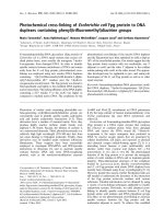
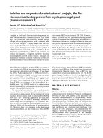
the test population. Overall flow-chart from sampling to
RAPD was shown in Fig 1.
D
11
N
⁄
–
N
1–
()
n
j
n
j
1–
()
j
1=
S
∑
=
Table 2.
Primers used in multiplex PCR, flagellar H7 PCR, and RAPD fingerprinting assay
Primer Oligonucleotide sequences (5
'
-3
'
) Expected size Reference
stx
1-F ACACTGGATGATCTCAGTGG 614 bp Fagan
et al
[14]
stx
1-R CTGAATCCCCCTCCATTATG
stx
2-F CCATGACAACGGACAGCAGTT 779 bp Fagan
et al
[14]
stx
2-R CCTGTCAACTGAGCAGCACTTTG
eae
A-F GTGGCGAATACTGGCGAGACT 890 bp Fagan
et al
[14]
eae
A-R CCCCATTCTTTTTCACCGTCG
hly
A-F ACGATGTGGTTTATTCTGGA 165 bp Fagan
et al
[14]
hly
A-R CTTCACGTGACCATACATAT
H7-F GCGCTGTCGAGTTCTATCGAGC 625 bp Gannon
et al
[18]
H7-R CAACGGTGACTTTATCGCCATTCC
CRA22 CCGCAGCCAA Neilan
et al
[31]
CRA23 GCGATCCCCA Neilan
et al
[31]
12 Ji-Yeon Kim
et al.
Vero cell cytotoxic assay
After confirmation of
E. coli
O157:H7 from isolates in
this study by multiplex PCR and flagellar H7 PCR, the
isolates were carried out by Vero cell cytotoxic assay to
characterize them. The assay was conducted as previously
described by Yoh
et al.
[52] and Kim
et al.
[26]. Briefly,
culture filtrates obtained from the TSB after incubation at
37
o
C for 24 h were used for the assay. Culture supernatants
and extracts were filtered through 0.2
µ
m pore-size sterile
filter (Minisart; Sartorius, Germany). Vero cells were
cultured in Eagles minimum essential medium (EMEM;
Gibco, USA) supplemented with 10% fetal bovine serum
(FBS) and gentamicin (100
µ
g/ml). Two-hundred
µ
l of Vero
cells in EMEM (2.5
×
10
5
cells/ml) were placed in each well
of 96 well tissue culture plate (Costar, USA) and incubated
at 37
o
C for 24 h. Fifty
µ
l of aliquot of the culture filtrates
was added into each well. After incubation at 37
o
C in 10%
CO
2
atmosphere for 3 days, the cytopathic effect (CPE) on
F
ig. 1.
Procedures for the isolation of STEC from fecal and meat samples.
Molecular determination of
E. coli
O157:H7 13
Vero cells was examined under an inverted microscope
(DMIRB/E; Leica, Germany). In this study, we determined
that “weak” was ranging from 0% to 30%, and “strong” was
from 30% to 100% of Vero cells were dead. The result was
shown in Table 5.
Antimicrobial susceptibility test
The antimicrobial susceptibility of 11
E. coli
O157:H7
isolates was determined by Bauer and Kirby method [5]. A
total of 23 concentrated antimicrobial discs tested were
ampicillin (10
µ
g), amikacin (30
µ
g), amoxicillin/clavulanic
acid (20/10
µ
g), carbenicillin (100
µ
g), cefixime (5
µ
g),
cefotaxime (30
µ
g) cephalothin (30
µ
g), chloramphenicol
(30
µ
g), ciprofloxacin (5
µ
g), erythromycin (15
µ
g),
gentamicin (10
µ
g), imipenem (10
µ
g), kanamycin (30
µ
g),
levofloxacin (5
µ
g), nalidixic acid (30
µ
g), norfloxacin
(10
µ
g), ofloxacin (5
µ
g), polymyxin B (300 U), sparfloxacin
(5
µ
g), streptomycin (10
µ
g), tetracycline (30
µ
g),
tobramycin (10
µ
g), and trimethoprim/sulfamethoxazole
(1.25/23.75
µ
g). All antimicrobial discs are purchased from
Becton Dickinson (USA). After 24 h-incubation in TSB,
isolates subcultured in Muller-Hinton broth (MHB, Difco,
USA) for 8 h, diluted to MacFarland scale No. 0.5, and
applied to the surface of Muller-Hinton Agar (MHA, Difco,
USA). The discs were placed using disc dispenser (Becton
Dickinson, USA) and the plates were incubated for 18 h at
37
o
C. Inhibitory zones of the growth were measured. The
results were interpreted by the guideline of National
Committee for Clinical Laboratory Standards (NCCLS).
Results
Isolation of
E. coli
O157:H7
In this study, a total of 1,682 samples were examined.
Nine
E. coli
O157:H7 were isolated from fecal samples, and
two were obtained from meat samples. However, no
E. coli
O157:H7 was detected from chicken rinsing samples.
The detection rates of
E. coli
O157:H7 by the three
different methods were different (Table 3). In conventional
method, seven isolates were obtained through phenotypical
characteristics (non-sorbitol fermenters forming colorless
colonies on CT-SMAC agar and purple colonies on
Chromocult agar). The 11 isolates were detected by the
E.
coli
O157:H7 Rapid kit and 10 suspected isolates in IMS
were further applied to motility and agglutination tests. In
agglutination and motility tests, strains isolated from same
samples showed identical results regardless of different
isolation methods. At motility test, all eleven strains were
positive. In agglutination test against O157 antiserum, all
strains showed positive, but one of them did not agglutinate
against H7 antiserum.
Characterization of
E. coli
O157:H7 isolates by
multiplex PCR for
Stx
1,
Stx
2,
eae
A, and
hly
A genes,
and by flagellar H7 PCR
After identification by motility and agglutination tests
Table 3.
The detection rates of
E. coli
O157:H7 by three different methods
Methods Positive (%) Negative (%)
Conventional culture 0.42 (7/1,682)
a
99.58 (1,675/1,682)
b
Immunomagnetic separation 0.59 (10/1,682) 99.41 (1,672/1,682)
E. coli
O157:H7 Rapid kit 0.65 (11/1,682) 99.35 (1,671/1,682)
a
No. of positive/No. of samples examined
b
No. of negative/No. of samples examined
Table 4.
Antibiotic resistance profiles of isolated
E. coli
O157:H7
Antimicrobial discs Resistant (%) Intermediate (%) Antimicrobial discs Resistant (%) Intermediate (%)
Ampicillin
27.2 54.5
Kanamycin
0 27.3
Amikacin
00
Levofloxacin
00
Amoxicillin/
clavulanic acid
9.1 45.5
Nalidixic acid
09.1
Norfloxacin
00
Carbenicillin
9.1 72.7
Ofloxacin
00
Cefixime
00
Polymyxin B
0 36.4
Cefotaxime
0 18.2
Sparfloxacin
00
Cephalothin
18.2 27.3
Streptomycin
0 36.4
Chloramphenicol
00
Tetracycline
18.2 36.4
Ciprofloxacin
00
Tobramycin
00
Erythromycin
100 0
Trimethoprim/
sulfamethoxazole
00
Gentamicin
00
Imipenem
00
14 Ji-Yeon Kim
et al.
against O157 and H7 antiserum, multiplex PCR and
flagellar H7 PCR were carried out using primers for
stx
1,
stx
2,
eae
A, and
hly
A genes. As shown in Table 5, all eleven
had
stx
1 genes, while six isolates had
stx
2 genes. Eleven
isolates were confirmed as
E. coli
O157:H7 because they all
carried
eae
A and
hly
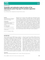
A genes. Specific amplicon sizes of
stx
1,
stx
2,
eae
A, and
hly
A genes were 614 bp, 779 bp, 890
bp, and 165 bp, respectively. The PCR products representing
each of four target STEC virulence factors were amplified
with standard strain, ATCC 43894 as a positive control (lane
12 in Fig. 2).
After confirmation by motility and antiserum tests, the
isolates were further applied to flagellar H7 assay and
multiplex PCR assay to confirm the presence of flagellar
gene. In flagellar H7 PCR assay, all eleven isolates were
found harboring H7 genes. Though one isolate did not react
against H7 antiserum, they all possessed H7 genes (Table 5).
RAPD fingerprinting analysis
Eleven
E. coli
O157:H7 isolates were compared with the
17
E. coli
O157:H7 strains which were obtained from ATCC
(4 strains), Cornell University (6 strains), Pennsylvania State
University (7 strains) using RAPD assay. Representative
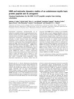
RAPD patterns for all 28 tested strains amplified with two
primers each (CRA22 and CRA23) were shown in Fig. 3.
DNA polymorphism in the isolates was most evident
amongst amplicons in the 2501 bp, 500 bp region. Fig. 3
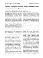
illustrated a dendrogram constructed from amplicon profiles
generated by CRA22 and CRA23. The dendrogram also
contained 5 groups which had coefficient of similarities at
63%. Group A comprised J010703-11-1, E010206 (Korean
pigs) and P6 (USA) which had similarity coefficients
ranging from 65% to 90%. Group B was consisted of only
one strain, P010726-26 (Korean cattle). Group C contained
P1 and P2 (USA), and Group D comprised O157-R1-3-2
(Korean cattle). Group E showed 2 subgroups, E
1
and E
2
.
Subgroup E
1
included two isolates from Korean cattle,
P010726-21 and P010726-24. Subgroup E
2
was broken
down by 5 Korean isolates (P010726-18, P010726-22,
P010726-23, P010726-25, and O157-C-1-2), and 14 USA
isolates; 4 strains of ATCC (A1, A2, A3, and A4), 6 strains
of Cornell University (C1, C2, C3, C4, C5, and C6), and 4
strains of Pennsylvania State University (P3, P4, P5, and
P7). These strains in subgroup E
2
had a similarity coefficient
of about 75%. Conclusively, 2 isolates from pigs in Korea
had distinct genetic patterns from other strains. Three
isolates from Korean cattle (P010726-18, 22, and 23)
showed high similarity with USA isolates at 80% level. The
USA isolates revealed close patterns with each other except
three strains of Pennsylvania State University (P1, P2, and
P6). Among three, P1 and P2 showed 70% similarity, and P6
revealed similar with two pig strains from Korea at 65%
level. Six Korean strains from cattle showed coefficient of
similarities from 63% to 80% level. The discriminatory
power (
D
-value) of this RAPD fingerprinting assay was
0.86.
Vero cell cytotoxic assay
Cytotoxic effects of
E. coli
O157:H7 isolates were
measured by Vero cell cytotoxic assay. CPE of eight isolates
was strong, otherwise three was weak. The results of CPE of
eleven
E. coli
O157:H7 isolates were shown in Table 5.
Antimicrobial susceptibility disc tests
A total of 23 antimicrobial discs were used in this study.
Five of eleven
E. coli
O157:H7 isolates (45.5%) were
resistant to two or more antimicrobial agents (Table 4). All
isolates were resistant to erythromycin (100%) followed by
Table 5.
Results of multiplex PCR, H7 PCR, antiserum tests, motility test, and vero cell assay
Isolates
Presence of
a
Antiserum tests
b
Motility
Test
b
Verocell
Assay
c
stx
1
stx
2
eae
A
hly
A H7 O157 H7
P010726-18 + - + + + + + + ++
P010726-21 + + + + + + - + +
P010726-22 + + + + + + - + +
P010726-23 + - + + + + + + ++
P010726-24 + - + + + + + + ++
P010726-25 + + + + + + + + ++
P010726-26 + - + + + + + + ++
E010206-13-2 + - + + + + + + ++
J010303-11-1 + + + + + + + + ++
O157-R1-3-2 + + + + + + - + ++
O157-C-1-2 + - + + + + + + +
a
+; present, -; absent.
b
+; positive, -; negative.
c
++; strong cytopathic effect (CPE), +; weak CPE.
Molecular determination of
E. coli
O157:H7 15
ampicillin (27.2%), cephalothin (18.2%), and tetracycline
(18.2%), respectively (Table 4).
Discussion
This study was conducted to determine the prevalence of
STEC in cattle, pigs, and chickens using different detection
methods and to define the molecular characteristics of the
isolates using multiplex PCR and RAPD.
The conventional culture method showed the lowest
detection rate. It might be attributable to lack of ability to
detect
E. coli
O157:H7 which showed aberrant biochemical
phenotypes [49]. In the case of IMS method, the detection
rate was relatively high, however IMS was too labor-
intensive when large numbers of samples were subjected to
isolation [37]. The
E. coli
O157:H7 Rapid kit showed
relatively high sensitivity and it only took 10 min to be
proved to positive. Due to its sensitivity and rapidity, this
would be useful to detect
E. coli
O157:H7 from various
sources.
The detection rates of
E. coli
O157:H7 were variable
among countries examined and detection methods they
applied. The prevalence of
E.
coli
O157:H7 from industrial
minced beef was 0.12% in France [46], and other French
researcher reported that there was no
E. coli
O157: H7
isolation in 1,200 samples [7]. In Switzerland, no
E. coli
O157:H7 was detected from 400 samples [15]. Five
E. coli
O157:H7 (3.3%) were isolated from retail beef and bovine
F
ig. 2. Result of multiplex PCR assay for detection of the
Stx
1 (614 bp),
Stx
2 (779 bp),
eae
A (890 bp), and
hly
A (165 bp) genes in
E.
c
oli
O157:H7 isolates. Lane 1, P010726-18; lane 2, P010726-21; lane 3, P010726-22; lane 4, P010726-23; lane 5, P010726-24; lane
6,
P
010726-25; lane 7, P010726-26; lane 8, E010206-13-2; lane 9, J010303-11-1; lane 10, O157-R1-3-2; lane 11, O157-C-1-2; lane 1
2,
A
TCC 43894 (a positive control); M, 100 bp DNA marker.
F
ig. 3. RAPD patterns of 11 Korean isolates and 17 U.S. strains. Lane M, 1 kb DNA marker; lane 1, P010726-18; lane 2, P010726-2
1;
l
ane 3, P010726-22; lane 4, P010726-23; lane 5, P010726-24; lane 6, P010726-25; lane 7, P010726-26; lane 8, E010206-13-2; lane
9,
J
010303-11-1; lane 10, O157-R1-3-2; lane 11, O157-C-1-2; lane 12, A1; lane 13, A2; lane 14, A3; lane 15, A4. lanes 16-21, strains C
1,
C
2, C3, C4, C5, and C6, respectively (Cornell University strains); lanes 22-28, strains P1, P2, P3, P4, P5, P6, and P7, respective
ly
(
Pennsylvania State University strains).
16 Ji-Yeon Kim
et al.
feces in Thailand, and 36 (8.7%) STEC in Spain [33]. The
prevalence of STEC in North American and European cattle
ranged from 0 to 10% [4]. The differences in the detection of
STEC among these studies are probably due to the fact that
the patterns of shedding of STEC are affected by diet, age,
environmental condition, and seasonal variation [27]. The
reasons of low detection rate in this study could be
summarized into three factors. Firstly, limited sampling
sources possibly influenced the detection rate [6,9]. Most
sample sources (80%) in this work were obtained from
bovine fecal and chicken rinsing samples. According to
prevalence surveys about
E. coli
O157:H7 from domestic
animals were less than 0.7% [6,9]. However, the proportions
of STEC in calves and heifers were much higher than those
in adults in other countries [12,21,33,41]. These authors
demonstrated that young animals (calves and heifers) shed
STEC more frequently than adults. In this study, most fecal
samples were obtained from healthy adult cattle. Putting
these studies together, age difference might be attributable to
low detection rate of
E. coli
O157:H7 rather than sample
sources. Secondly, seasonal variation might influence the
detection rate in this study. Though the samples were
collected all the year around, more samples were collected
during January and February (38.3%). The rate of sampling
from July to August was 20.2%. Many reports demonstrated
that the distribution of
E. coli
O157:H7 was peaked between
July and August [21,41]. The warmer and more moist
conditions of the summer months may favor the survival and
growth of STEC [21]. More sampling was conducted during
summer season, more
E. coli
O157:H7 would be detected.
Thirdly, most meat products were obtained from large-
scaled retail markets which have relatively better hygiene
conditions than small-scaled retail markets or meat shops
[10,11].
According to H7 flagellar antiserum test and PCR, one
isolate of Korean strains did not react in antiserum test.
However, it showed positive at PCR assay for H7 gene.
From this result, we could assume that the
E. coli
O157:H7
strain did not express its characteristic though they had H7
gene. Therefore, molecular determination by PCR should be
performed to confirm.
We used RAPD fingerprinting assay to principally
understand the molecular relatedness between the
E. coli
O157:H7 strains isolated from Korea and the USA. Since
PFGE explores the whole length of chromosome whereas
RAPD explores only randomly selected parts of it, RAPD
analysis can be alternative method of PFGE typing method
[36]. In general, high agreement between the results of the
two methods was good for strain differentiation [25].
Moreover, Maurer
et al.
[28] claimed that fingerprinting by
RAPD revealed more genetic differences among avian
E.
coli
strains than restriction fragment length polymorphism
(RFLP) analysis. Therefore, RAPD fingerprinting analysis
was used for this study because its advantages of time and
cost-saving, sensitivity, and no special skills required to
perform.
The results of RAPD patterns in this study compared with
the study of Radu
et al.
[36]. They reported 2 clusters and 22
isolates among 28 strains [36]. Of the 22 isolates, 3
predominant groups were observed and had 3 to 5 different
bands. However, our study has revealed that the RAPD-PCR
patterns were too diverse to differentiate the patterns of each
E. coli
O157:H7 isolates when the patterns were analyzed
based on their band numbers. Using two primers CRA22
and CRA23 at least 7 bands were generated except 4 strains.
Moreover, the discriminatory power (
D
-value) revealed
0.86. These diverse band patterns generated high
D
-value
and differentiation among strains, so these two primers were
recommended to dissect further molecular characteristics
using RAPD analysis. At 63% similarity level, 5 clusters
were generated by RAPD. Except B and D group,
particularly E group showed that high genetic relatedness
between strains at 75% level. Most USA strains had similar
patterns except 3 Pennsylvania State University strains.
More than 50% Korean cattle isolates were genetically
similar to the USA cattle isolates. However, the reason that
distinct genetic pattern between pig and cattle isolates from
Korea may depend on their species difference of sources.
F
ig. 4
. The dendrogram constructed from RAPD data
by
U
PGAMA method.
Molecular determination of
E. coli
O157:H7 17
Several studies demonstrated that source differentiation
could be determined by RAPD [32,35]. Therefore, this
technique could be of use when studying the epidemiology
of
E.
coli
O157:H7. Although RAPD had a capacity to
distinguish strains with different virulence factors from
different sources, we could not define the difference in the
genetic patterns between strains possessing only
stx
1 or
stx
2
and strains possessing both
stx
1 and
stx
2. RAPD has
revealed that it could not discriminate among isolates
according to their differences either in the degree of
virulence in several studies [8,30].
E. coli
O157:H7 was reportedly susceptible to many
antibiotics [42]. Approximately 45.5% of the present strains
showed antibiotic resistance to two or more of the
antimicrobial agents used in this study. Their antibiotic
resistance was against erythromycin (100%), followed by
ampicillin (27.2%), cephalothin (18.2%), and tetracycline
(18.2%). Antimicrobial resistance patterns were observed
most commonly to ampicillin (25.4%), tetracycline (23.8%),
and streptomycin (14.3%) and less frequently to cephalothin
(11,1%) and nalidixic acid (6.4%) in India [25]. The USA
study about antibiotic resistance showed that all isolates
were resistant to tilmicosin, and most isolates were susceptible
to trimethoprim/sulfamethoxazole and ciprofoloxacin [17].
In Malaysia, resistance was observed mostly towards
bacitracin (100%), sulphafurazole (77%), ampicillin (57%),
cephalothin (53%), and carbenicillin (30%) [36]. The
antibiotic resistant patterns to ampicillin, fosfomycin,
kanamycin, and vancomycin were observed in Japan [45].
From these data,
E. coli
O157:H7 was mainly resistant to
ampicillin and tetracycline. Resistance patterns of Korean
isolates were similar to those of Malaysian. The possibility
of the change of resistance patterns could not exclude the
percentage of intermediately resistant group which revealed
relatively high to carbenicillin (72.7%), ampicillin (54.5%),
amoxicillin/clavulanic acid (45.5%), kanamycin (27.3%),
polymyxin B (36.4%), streptomycin (36.4%), tetracycline
(36.4%), and cephalothin (27.3%).
This study has found that the prevalence of
E. coli
O157:H7 was not as high as that of other countries.
However, the
E. coli
O157:H7 has been isolated from
various livestock processing stages from slaughtering to
processing. Therefore, more careful investigation programs
such as HACCP should be applied to establish all dairy
herds, slaughterhouses, and meat processing plants. The
E.
coli
O157:H7 Rapid kit which examined in this study was
apparently useful to detect the contamination of
E. coli
O157:H7 with high accuracy and rapidity. In addition,
RAPD results indicated that Korean cattle isolates were
genetically related with those of the USA strains at 70%
similarity level, which could assume similar mechanism of
contamination in animals and related sources. Continuous
monitoring and surveillance program for examining
microbial contamination of imported feeds should be
performed to minimize the risk of spread of major food-
borne pathogens.
Acknowledgments
This study was supported by the Rural Development
Administration, the Ministry of Agriculture and Forest, and
also supported by the Brain Korea 21 project.
References
1. Acheson DWK, Lincome LL, Jacewicz MS, Keusch GT.
Shiga toxin interaction with intestinal epithelial cells.
Escherichia coli
O157:H7 and Shiga toxin-producing
E. coli
strains. American Society for Microbiology, Washington DC,
1998.
2. Ackers ML, Mahon BE, Leahy E, Goode B, Damrow T,
Hayes PS, Bibb WF, Rice DH, Barrett TJ, Hutwagner L,
Griffin PM, Slutsker L. An outbreak of
Escherichia coli
O157:H7 infectious associated with leaf lettuce consumption.
J Infect Dis 1998, 177, 1588-1593.
3. Angela R, Melton C, O’Brien AD. Structure, biology, and
relative toxicity of Shiga toxin family members for cells and
animals.
Escherichia coli
O157:H7 and Shiga toxin-
producing
E. coli
strains. American Society for Microbiology,
Washington DC, 1998.
4. Armstrong GL, Hollingsworth J, Morris JG Jr. Emerging
foodborne pathogens:
Escherichia coli
O157:H7 as a model
of entry of a new pathogen into the food supply of the
developed world. Epidemiol Rev 1996, 18, 29-51.
5. Bauer AW, Kirby WM, Sherris JC, Turck M. Antibiotic
susceptibility testing by a standardized single disk method.
Am J Clin Pathol 1966, 45, 493-496.
6. Beutin L, Geier D, Steinruck H, Zimmermann S, Scheutz
F. Prevalence and some properties of verotoxin (Shiga-like
toxin)-producing
Escherichia coli
in seven different species
of healthy domestic animals. J Clin Microbiol 1993, 31,
2483-2488.
7. Bouvet J, Bavai C, Rossel R, Le Roux A, Montet MP,
Ray-Gueniot S, Mazuy C, Arquilliere C, Vermozy-
Rozand C. Prevalence of verotoxin-producing
Escherichia
coli
and
E. coli
O157:H7 in pig carcasses from three French
slaughterhouses. Int J Food Microbiol 2001, 71, 249-255.
8. Chansiripornchai N, Ramasoota P, Sasipreeyajan J,
Svenson SB. Differentiation of avian pathogenic
Escherichia
coli
(APEC) strains by random amplified polymorphic DNA
(RAPD) analysis. Vet Microbiol 2001, 80, 75-83.
9. Chapman PA, Siddons CA, Cerdan Malo AT, Harkin
MA. A 1-year study of
Escherichia coli
O157 in cattle,
sheep, pigs and poultry. Epidemiol Infect 1997, 119, 245-
250.
10. Chapman PA, Siddons CA, Cerdan Malo AT, Harkin
MA. A one year study of
Escherichia coli
O157 in raw beef
and lamb products. Epidemiol Infect 2000, 124, 207-213.
11. Chapman PA, Cerdan Malo AT, Ellin M, Ashton R,
Harkin MA.
Escherichia coli
O157 in cattle and sheep at
slaughter, on beef and lamb carcasses and in raw beef and
18 Ji-Yeon Kim
et al.
lamb products in South Yorkshire, UK. Int J Food Microbiol
2001,
64
, 139-150.
12.
Cobbold R, Desmarchelier P.
A longitudinal study of
Shiga-toxigenic
Escherichia coli
(STEC) prevalence in three
Australian dairy herds. Vet Microbiol 2000,
71
, 125-137.
13.
Cody SH, Glynn MK, Farrar JA, Cairns KL, Griffin PM,
Kobayashi J, Fyfe M, Hoffman R, King AS, Lewis JH,
Swaminathan B, Bryant RG, Vugia DJ.
An Outbreak of
Escherichia coli
O157:H7 infection from unpasteurized
commercial apple juice. Ann Intern Med 1999,
130
, 202-209.
14.
Fagan PK, Hornitzky MA, Bettelheim KA, Djordjevic SP.
Detection of shiga-like toxin (
stx
1
and
stx
2), intimin (
eae
A),
and enterohemorrhagic
Escherichia coli
(EHEC) hemolysin
(EHEC
hly
A) genes in animal feces by multiplex PCR. Appl
Environ Microbiol 1999,
65
, 868-872.
15.
Fantelli K, Stephan R.
Prevalence and characteristics of
Shigatoxin-producing
Escherichia coli
and
Listeria
monocytogenes
strains isolated from minces meat in
Switzerland. Int J Food Microbiol 2001,
70
, 63-69.
16.
Feng P.
Escherichia coli
serotype O157:H7: novel vehicles
of infection and emergence of phenotypic variants. Emerg
Infect Dis 1995,
1
, 47-52.
17.
Galland JC, Hyatt DR, Crupper SS, Acheson DW.
Prevalence, antibiotic susceptibility, and diversity of
Escherichia coli
O157:H7 isolates from longitudinal study of
beef cattle feedlots. Appl Environ Microbiol 2001,
67
, 1619-
1627.
18.
Gannon VP, D’Souza S, Graham T, King RK, Rahn K,
Read S.
Use of the flagellar H7 gene as a target in multiplex
PCR assays and improved specificity in identification of
enterohemorrhagic
Escherichia coli
strains. J Clin Microbiol
1997,
35
, 656-662.
19.
Gill CO, Badoni M, McGinnis JC.
Microbiological
sampling of meat cuts and manufacturing beef by excision or
swabbing. J Food Prot 2001,
64
, 325-334.
20.
Hepburn NF, MacRae M, Ogden ID.
Survival of
Escherichia coli
O157 in abattoir waste products. Lett Appl
Microbiol 2002,
35
, 233-236.
21.
Heuvelink AE, van den Biggelaar FL, Zwartkruis-Nahuis
J, Herbes RG, Huyben R, Nagelkerke N, Melchers WJ,
Monnens LA, de Boer E.
Occurrence of Verocytotoxin-
producing
Escherichia coli
O157 on Dutch dairy farms. J
Clin Microbiol 1998,
36
, 3480-3487.
22.
Hitchins AD, Feng P, Watkins WD, Rippey SR, Chandler
LA.
Escherichia coli
and the Coliform bacteria. In: U.S.
Food and Drug Administration (ed.). Bacteriological Analytical
Manual. 8th ed. pp. 4.01-4.29, AOAC International,
Gaithersburg, 1998.
23.
Hunter PR, Gaston MA.
Numerical index of discriminatory
ability of typing systems: An application of Simpson’s index
of diversity. J Clin Microbiol 1988,
26
, 2465-2466.
24.
Karmali MA, Petric M, Bielaszewska M.
Evaluation of a
microplated latex agglutination method (Verotox-F Assay)
for detecting and characterizing Verotoxins (Shiga toxins) in
Escherichia coli
. J Clin Microbiol 1999,
37
, 396-399.
25.
Khan A, Das SC, Ramamurthy T, Sikdar A, Khanam J,
Yamasaki S, Takeda Y, Nair GB.
Antibiotic resistance,
virulence gene, and molecular profiles of Shiga toxin-
producing
Escherichia coli
isolates from diverse sources in
Calcutta, India. J Clin Microbiol 2002,
40
, 2009-2015.
26.
Kim SH, Yang SJ, Koo HC, Bae WK, Kim JY, Park JH,
Baek YJ, Park YH.
Inhibitory activity of
Bifidobacterium
longum
HY 8001 against Vero cytotoxin of
Escherichia coli
O157:H7. J Food Prot 2001,
64
, 1667-1673.
27.
Kudva IT, Hatfield PG, Hovde CJ.
Characterization of
Escherichia coli
O157:H7 and other Shiga toxin-producing
E. coli
serotypes isolated from sheep. J Clin Microbiol 1997,
35
, 892-899.
28.
Maurer JJ, Lee MD, Lobsinger C, Brown T, Maier M,
Thayer SG.
Molecular typing of avian
Escherichia coli
isolates by random amplification of polymorphic DNA.
Avian Dis 1998,
42
, 431-451.
29.
Morgan D, Newman CP, Hutchinson DN, Walker AM,
Rowe B, Majid F.
Verotoxin producing
Escherichia coli
O157 infections associated with the consumption of yoghurt.
Epidemiol Infect 1993,
111
, 181-187.
30.
Motta TR, Moreira-Filho CA, Mendes RP, Souza LR,
Sugizak MF, Baueb S, Calich VL, Vaz CA.
Evaluation of
DNA polymorphisms amplified by arbitrary primers (RAPD)
as genetically associated elements to differentiate virulent
and non-virulent
Paracoccidioides brasiliensis
isolates.
FEMS Immunol Med Microbiol 2002,
33
, 151-157.
31.
Neilan BA, jacobs D, Goodman AE.
Genetic diversty and
phylogeny of toxic cyanobacteria determined by DNA
polymorphisms within the phycocyanin locus. Appl Environ
Microbiol 1995,
61
, 3875-3883.
32.
Nucci C, da Silveira WD, da Silva Correa S, Nakazato G,
Bando SY, Ribeiro MA, Pestana de Castro AF.
Microbiological comparative study of isolates of
Edwardsiella tarda
in different countries from fish and
humans. Vet Microbiol 2002,
89
, 29-39.
33.
Orden JA, Cid D, Ruiz-Santa-Quiteria JA, Garcia S,
Martinez S, de la Fuente R.
Verotoxin-producing
Escherichia coli
(VTEC), enteropathogenic
E. coli
(EPEC)
and necrotoxigenic
E. coli
(NTEC) isolated from healthy
cattle in Spain. J Appl Microbiol 2002,
93
, 29-35.
34.
Pebody RG, Furtado C, Rojas A, McCarthy N, Nylen G,
Ruutu P, Leino T, Chalmers R, de Jong B, Donnelly M,
Fisher I, Gilham C, Graverson L, Cheasty T, Willshaw G,
Navarro M, Salmon R, Leinikki P, Wall P, Bartlett C.
An
international outbreak of Vero cytotoxin-producing
Escherichia coli
O157 infection amongst tourists; a challenge
for the European infectious disease surveillance network.
Epidemiol Infect 1999,
123
, 217-223.
35.
Pereira MS, Leal NC, Leal TCA, Sobreira M, de Almeida
AMP, Siqueira-Junior JP, Campos-Takaki GM.
Typing of
human and bovine
Staphylococcus aureus
by RAPD-PCR
and ribotyping-PCR. Lett Appl Microbiol 2002,
35
, 32-36.
36.
Radu S, Ling OW, Rusul G, Karim MIA, Nishibuchi M.
Detection of
Escherichia coli
O157:H7 by multiplex PCR
and their characterization by plasmid profiling, antimicrobial
resistance, RAPD and PFGE analyses. J Microbiol Methods
2001,
46
, 131-139.
37.
Reinders RD, Barna A, Lipman LJA, Bijker PGH.
Comparison of the sensitivity of manual and automated
immunomagnetic separation methods for detection of Shiga
Molecular determination of
E. coli
O157:H7 19
toxin-producing
Escherichia coli
O157:H7 in milk. J Appl
Microbiol 2002,
92
, 1015-1020.
38.
Riley LW, Remis RS, Helgerson SD, McGee HB, Wells
JG, Davis BR, Herbert RJ, Olcott ES, Johnson LM,
Hargrett NT, Blake PA, Cohen ML.
Hemorrhagic colitis
associated with a rare
Escherichia coli
serotype. N Engl J
Med 1983,
308
, 681-685.
39.
Sanderson MW, Gay JM, Hancock DD, Gay CC, Fox LK,
Besser TE.
Sensitivity of bacteriologic culture for detection
of
Escherichia coli
O157:H7 in bovine feces. J Clin
Microbiol 1995,
33
, 2616-2619.
40.
Schmidt H, Scheef J, Morabito S, Caprioli A. Wieler LH,
Karch H.
A new Shiga toxin 2 variant (Stx2f) from
Escherichia coli
isolated from pigeons. Appl Environ
Microbiol 2000,
66
, 1205-1208.
41.
Shinagawa K, Kanehira M, Omoe K, Matsuda I, Hu D,
Widiasih DA, Sugii S.
Frequency of Shiga toxin-producing
Escherichia coli
in cattle at a breeding farm and at a
slaughterhouse in Japan. Vet Microbiol 2000,
76
, 305-309.
42.
Swerdlow DL, Woodruff BA, Brady RC, Griffin PM,
Tippen S, Donnell HD Jr, Geldreich E, Payne BJ, Meyer
A Jr, Wells JG.
A water borne outbreak in Missouri of
Escherichia coli
O157:H7, associated with bloody diarrhea
and death. Ann Intern Med 1992,
117
, 812-819.
43.
Tauxe RV.
Emerging foodborne diseases: an evolving public
health challenge. Emerg Infect Dis 1997,
3
, 425-434.
44.
Tenover FC, Arbeit RD, Goering RV, Mickelsen PA,
Murray BE, Persing DH, Swaminathan B.
Interpreting
chromosomal DNA restriction patterns produced by pulse-
field gel electrophoresis: criteria for bacterial strain typing. J
Clin Microbiol 1995,
33
, 2233-2239.
45.
Tsuboi I, Ida H, Yoshikawa E, Hiyoshi S, Yamaji E,
Nakayama I, Nonomiya T, Shigenobu F, Shimizu M,
O’Hara K, Sawai T, Mizuoka K.
Antibiotic susceptibility
of enterohemorrhagic
Escherichia coli
O157:H7 isolated
from an outbreak in Japan in 1996. Antimicrob Agents
Chemother 1998,
42
, 431-432.
46.
Vernozy-Rozand C, Ray-Gueniot S, Ragot C, Bavai C,
Mazuy C, Montet MP, Bouvet J, Richard Y.
Prevalence of
Eshcherichia coli
O157:H7 in industrial minced beef. Lett
Appl Microbiol 2002,
35
, 7-11.
47.
Vuddhakul V, Patararungrong N, Pungrasamee P,
Jitsurong S, Morigaki T, Asai N, Nishibuchi M.
Isolation
and characterization of
Escherichia coli
O157 from retail
beef and bovine feces in Thailand. FEMS Microbiol Lett
2000,
182
, 343-347.
48.
Wallace JS, Cheasty T, Jones K.
Isolation of verocytotoxin-
producing
Escherichia coli
O157 from wild birds. J Appl
Microbiol 1997,
82
, 399-404.
49.
Ware JM, Abbott SL, Janda JM.
A new diagnostic
problem: isolation of
Escherichia coli
O157:H7 strains with
aberrant biochemical properties. Diagn Microbiol Infect Dis
2000,
38
, 185-187.
50.
Watanabe Y, Ozasa K, Mermin JH, Griffin PM, Masuda
K, Imashuku S, Sawada T.
Factory outbreak of
Escherichia
coli
O157:H7 infection in Japan. Emerg Infect Dis
1999,
5
,
424-428.
51.
Wright DJ, Chapman PA, Siddons CA.
Immunomagnetic
separation as a sensitive method for isolating
Escherichia coli
O157 from food samples. Epidemiol Infect 1994,
113
, 31-39.
52.
Yoh M, Frimpong EK, Honda T.
Effect of antimicrobial
agents, especially fosfomycin, on the production and release
of Vero toxin by enterohaemorrhagic
Escherichia coli
O157:H7. FEMS Immunol Med Microbiol 1997,
19
, 57-64.
53.
Zhao T, Doyle MP, Shere J, Garber L.
Prevalence of
enterohemorrhagic
Escherichia coli
O157:H7 in a survey of
dairy herds. Appl Environ Microbiol 1995,
61
, 1290-1293.