Báo cáo khoa học: " Comparison of a small volume of hypertonic saline solution and dextran 40 on hemodynamic alternations in conscious calves" ppsx
Bạn đang xem bản rút gọn của tài liệu. Xem và tải ngay bản đầy đủ của tài liệu tại đây (445.67 KB, 6 trang )
JOURNAL OF
Veterinary
Science
J. Vet. Sci. (2005), 6(2), 111–116
Comparison of a small volume of hypertonic saline solution and dextran 40
on hemodynamic alternations in conscious calves
Kazuyuki Suzuki *, Tomoko Suzuki , Mitsuyoshi Miyahara , Shigehiro Iwabuchi , Ryuji Asano
Department of Veterinary Medicine, College of Bioresource Sciences, Nihon University, 1866 Kameino, Fujisawa, Kanagawa 252-
8510, Japan
Central Research Laboratories, Nippon Zenyaku Kogyo, 1-1 Aza Tairanoue, Sasagawa, Asakamachi, Koriyama, Fukushima 963-
0196, Japan
The hemodynamic effects of rapid intravenous (IV)
administration of 10% dextran 40 in saline solution (D40)
and 7.2% hypertonic saline solution (HSS) in calves were
compared. Calves received isotonic saline solution (ISS),
HSS or D40 (3 calves/group) and were monitored of blood
pressure, and cardiac output (CO) for 180 min. HSS and
D40 infusions induced a significant increase in relative
plasma volume reaching 134.9 ± 2.8 and 125.0 ± 1.9%,
respectively at the end of fluid infusion. In the HSS group,
CO, cardiac index (CI) and stroke volume (SV) remained
constant at low levels after 90 minutes despite the
maximal values of CO, CI and SV at the end of infusion,
reaching 21.0 ± 6.3 l/min (p<0.05), 177.8 ± 14.2 ml/min/kg
(p < 0.001) and 0.20 ± 0.03 l/beat (at t = 10 min, p < 0.001),
respectively. In contrast, CI and SV in the D40 group
showed significant increases to 14.7 ± 2.9 l/min and 153.5
± 17.2 ml/min/kg, respectively, at the end of fluid infusion.
And those values remained constant at higher levels than
those of the before infusions values throughout the
experimental periods. Positive effects for hemodynamic
alternations of D40 in calf practice were milder and
longer than those of HSS. Therefore, the D40 infusion
should be explored as a possible treatment for dehydrated
calves, since rapid infusion of D40 may be safe and more
beneficial for rehydrating more than HSS treatment.
Key words: calf, cardiac output, dextran, hemodynamic,
hypertonic saline
Calf diarrhea remains a major cause of economic loss to
cattle industry [1]. The cost-effective use of electrolyte
containing oral rehydration solution for treatment of
diarrheic calves is popular and widely accepted, despite
progress in the understanding of the pathophysiology of the
disease, [1,14]. If treatment with oral fluids is not successful,
intravenous restoration of circulation volume is preferred.
The goal of intravenous fluid therapy in dehydrated calves is
to correct extracellular dehydration and restore circulating
plasma volume.
The colloidal solutions, such as albumin, dextran, and
hetastarch, are extremely effective volume expanders [4]
and used for fluid resuscitations in hypovolemic patients [6],
horses [9] and dogs [17]. Increasing colloidal osmotic
pressure (COP) with colloidal products has remained an
attractive theoretical premise for volume resuscitation.
Especially, dextran has considerable advantage over other
types of colloids for initial hypovolemia treatment due to its
antithrombotic properties whereby cell aggregability is
prevented and the incidence of system complications is
convincingly reduced [6]. Van Den Broke et al. [20]
suggested that dextran 40 (D40) can be recommended as a
plasma substitute due to its higher initial increase in
circulating plasma volume for humans, its moderate
duration of effect and its low incidence of anaphylactic
reaction.
Therefore, administration of a small-volume of D40
should be checked for safety and efficacy of hemodynamic
responses before being recommended for the treatment of
hypovolemic calves. The purposes of this study were to
compare effects of rapid intraveneus infusion (IV) of a small
volume of D40 and those of IV infusion of an equivalent
volume of 7.2% hypertonic (HSS) and 0.9% isotonic saline
solutions (ISS) on plasma volume and hemodynamic status
of calves.
Materials and Methods
All procedures were in accordance with the National
Research Council on Guide for the Care and Use of
Laboratory Animals [13]. Experiments were performed on 9
healthy 3-month-old Holstein calves weighing 106.1 ± 26.0
*Corresponding author
Tel: +81-466-84-3842; Fax: +81-466-84-3842
E-mail:
112 Kazuyuki Suzuki et al.
kg. Healthy calves were selected on the basis of physical
examination, electrocardiography and hematological analysis.
A well-balanced growth diet consisting of pelleted
concentrated ration and mixed grass hay and free access to
fresh water were provided until the day before the
experiment.
The day prior to the experiment, all calves were sedated
with an IV infusion of xylazine hydrochloride (Seduluck-
2%; Nippon Zenyaku, Japan) as a dose of 0.2 mg/kg of body
weight. An 8-F introducer (Fast-Cath; Nihon Koden, Japan)
was placed in the left jugular vein. A 14-gauge, 40-cm
length arterial and 15-cm length venous catheters (Sefelet
Catheter NCKP-14; Nipro, Japan) were inserted in the right
femoral artery and vein, respectively. After 24 h of
catheterization, infusion of resuscitated fluid was initiated.
Approximately 1 hour before fluid infusion, 16-gauge
catheter (Sefelet Catheter NSL-16WOT; Nipro, Japan) was
implanted percutaneously into the right jugular vein for fluid
infusion. A balloon-tipped, flow directed thermo-dilution
catheter (TC-704; Nihon Koden, Japan), which was used for
measurement of cardiac output (CO) and pulmonary arterial
pressure (PAP), was inserted through the introducer into the
left jugular vein. This catheter was positioned with the
proximal port in the right atrium and the distal port and
thermistor in the pulmonary artery. Various pressures
(systemic, central venous pressure (CVP) and PAP) were
measured, using the bedside monitoring system (BP-
308ETI; Nihon Koden, Japan) with a strain-gauge transducer
(DX-360; Nihon Koden, Japan) positioned at the level of the
point of the shoulder. All catheter positions were confirmed
by the evidence of characteristic pressure waveforms. The
transducers were calibrated with a water manometer before
use. A base-apex electrocardiograph was continuously
monitored throughout the experiment to detect any
arrhythmias. Food and water were withheld from calves
during the experiment.
After preparations, calves were monitored for 15 min to
ensure hemodynamic stability. Calves were randomly
divided into 1 of 3 groups (n = 3/group). Calves in each
group were assigned to receive 5 ml/kg of ISS, HSS or D40
infusion at a flow rate of 20 ml/kg/hr via the right jugular
vein catheter using infusion pump (PRS-25; Nikkiso,
Japan). All calves were then monitored for 180 min. Arterial
and venous samples were collected at time 0 (pre), and 5,
10, 15, 30, 45, 60, 90, 120, 150 and 180 minute after
initiation of fluid infusion. Before collection of each blood
sample, systolic (SAP), diastolic (DAP) and mean systemic
pressure (MAP), mean PAP and CVP, heart rate (HR) and
abnormal signs were recorded. Immediately after the
recording was completed, CO was determined after ice-cold
isotonic dextrose (5 ml) was injected into the right atrium at
end-expiration of the ventilatory cycles using a bedside
moritoring system. All CO measurements were performed
in triplicate, and a mean value was determined for the 3
values. Cardiac index (CI: CO/body weight, l/min/kg),
stroke volume (SV: CO/HR, l/beat), systemic vascular
resistance (SVR: [MAP/CO] × 80, mmHg/l/min) and
pulmonary vascular resistance (PVR: [PAP/CO] × 80,
mmHg/l/min) were calculated [14].
Venous samples were collected at each recording point
and used to determine hemoglobin concentration (Hb) and
hematocrit values (Ht) using an automatic cell counter
(MEK-6248; Nihon Koden, Japan). Other blood samples
were centrifuged, and plasma was collected and stored at
–20
C until assay. Changes in relative plasma volume (rPV)
were calculated from Hb and Ht [18,19]. Plasma sodium,
potassium and chloride concentrations were analyzed by
electrode methods, using an automatic analyzer (Model 644;
Bayer Medical, Japan). Plasma osmolarity was determined
by use of the freezing point depression method using an
osmometer (One-Ten Osmometer; Fiske, USA).
To test changes with time, data for each group were
analyzed by repeated-measures ANOVA followed by the
Bonferroni test. To test differences among groups, data for
each time point were analyzed by one-way ANOVA and the
Bonferroni test. Data represent as mean ± SD. For all
analyses, values of p < 0.05 were considered significant
[15].
Results
All calves were verified as clinically normal before the
experiment based upon the assessment of their vital signs,
attrition, food and water intake, and urine and feces
production. Clinical signs, such as moist rales on ausculation,
moist cough, jugular vein congestion, ophthalmoptosis,
salivation or arrhythmia were not observed throughout the
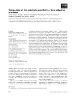
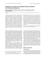
experiment. Sequential change in rPV was monitored in
calves given D40 infusion (Fig. 1). There was a slight
increase in the rPV of the ISS group, reaching 107.3 ± 3.1%.
For the HSS and D40 groups, a progressive and significant
increase in rPV was observed, reaching 134.9 ± 2.8% and
125.0 ± 1.9%, respectively, at the end of fluid infusion.
These increases were greater than that for the ISS group.
The pre-values of HR, SAP, MAP, DAP, PAP and CVP were
86.7 ± 15.9 bpm, 129.3 ± 8.6, 99.2 ± 5.1, 74.7 ± 5.7, 24.1 ±
4.3 and 0.7 ± 2.0 mmHg, respectively (Table 1). There was
slight increase in the CVP of the ISS group, reaching 2.7 ±
0.6 mmHg at the end of fluid infusion. For the HSS and D40
groups, a progressive and significant increases in CVP were
observed, reaching 6.0 ± 0.0 and 5.3 ± 0.6 mmHg, respectively,
at the end of fluid infusion. The CVP increase of the HSS
group was significantly greater than that for the other groups
(p < 0.05). The mean values of HR, SAP, MAP, DAP and
PAP were not affected by ISS, HSS or D40 infusion and
remained constant throughout the experiment in all groups
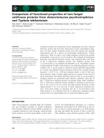
The mean values of CO, CI, SV, SVR and PVR before
infusion were 11.5 ± 1.7 l/min, 113.6 ± 8.6 ml/min/kg, 0.14
Dextran 40 and hypertonic saline in calf 113
± 0.03 l/beat, 700.4 ± 96.6 and 170.2 ± 35.9 mmHg/l/min,
respectively. Those values in ISS group were slightly
increased from the pre-values until the end of fluid infusion.
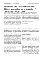
In the HSS group, CO, CI and SV remained constant at low
levels after 90 min despite the increasing maximal values of
CO, CI and SV at the end of infusion, reaching 21.0 ± 6.3 l/
min (p < 0.05), 177.8 ± 14.2 ml/min/kg (p < 0.001) and 0.20
± 0.03 l/beat (at t = 10 min, p < 0.001), respectively (Fig. 2).
In contrast, CI and SV in the D40 group showed significant
increases to 14.7 ± 2.9 l/min and 153.5 ± 17.2 ml/min/kg,
respectively, at the end of fluid infusion. Those values
remained constant at higher levels than those of the pre-
values throughout the experiment.
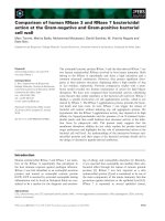
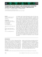
The SVR and PVR in ISS and D40 groups were not
affected by ISS or D40 infusion and remained constant
throughout the experiment (Fig. 3). However, SVR and
PVR in the HSS group were progressively and significantly
decreased from the pre-values, reaching 393.3 ± 95.8 and
100.8 ± 15.5 mmHg/l/min at 10 minutes, respectively. SVR
and PVR were significantly (p < 0.001) lower than the other
groups during HSS infusion.
The mean values of plasma sodium, potassium and
chloride concentrations, and osmolarity were 139.8 ± 2.0,
4.38 ± 0.27 and 102.3 ± 3.0 mEq/l, and 285.7 ± 2.2 mOsm/l,
respectively. Those values remained without changes after
administration of ISS or D40, and remained constant
throughout the experiment. However, plasma sodium and
Fig. 1. Graphs depicting the relative plasma volume (rPV) an
d
osmolarity in calves given 10% dextran 40 in saline or 7.2%
hypertonic saline solution. Levels of significance (p<0.05)
indicated: a: ISS versus HSS, b: ISS versus D40, c: HSS versus
D40 and asterisk: versus pre-values by Bonferroni test.
Fig. 2. Graphs depicting the cardiac index (CI) and stroke
volume (SV) in calves given 10% dextran 40 in saline or 7.2%
hypertonic saline solution.
Fig. 3. Graphs depicting the systemic (SVR) and pulmonary
vascular resistance (PVR) in calves given 10% dextran 40 i
n
saline or 7.2% hypertonic saline solution.
114 Kazuyuki Suzuki et al.
chloride concentrations (Table 1), and osmolarity (Fig. 1) in
HSS group were progressively and significantly increased
from the pre-values until the end of HSS infusion, reaching
155.7 ± 1.5 and 121.3 ± 5.0 mEq/l, and 316.3 ± 0.6 mOsm/l,
respectively (p < 0.001). The sequential changes in plasma
sodium and chloride concentrations, and osmolariy in the
HSS group were significantly greater than those in the other
groups (p < 0.001). In the HSS group, plasma potassium
concentrations were progressively and significantly
decreased from the pre-values until the end of HSS infusion,
reaching 3.61 ± 0.14 l/min (Table 1, p < 0.001).
Discussion
Intravenous infusion of a small volume of 10% dextran 40
in saline or 7.2% hypertonic saline solutions to normal, 3-
months old Holstein calves were found to be effective in
increasing plasma volume. Although the increase in rPV of
D40 group was lower than that of HSS group at the end of
the fluid infusion, the increases in rPV remained up to 10%
Table 1. Hemodynamic and plasma electrolytes alternations of 10% dextran 40 in saline (D40) or 7.2% hypertonic saline solution
(HSS) administered IV in calves (mean±SD)
0 5 10 15 30 45 60 90 120 150 180
Heart Rate (bpm)
ISS 86.7±19.1 093.0±20.9 090.7±24.0 092.7±19.3 091.7±8.9 85.2±23.2 86.7±22.1 83.7±12.5 89.0±17.3 97.0±23.5 94.0±24.3
HSS 95.7±10.7 102.7±10.1 105.7±13.1 111.3±13.9 100.3±11.5 90.3±15.0 88.7±16.7 89.7±15.3 90.7±18.6 89.7±15.7 91.3±11.6
D40 77.7±16.9 084.7±16.7 078.0±16.1 081.7±18.1 078.3±19.9 78.7±16.3 83.0±7.8 72.0±15.4 73.0±23.9 69.7±11.2 74.0±17.3
Cardiac Output (l/min)
ISS 10.6±0.5 11.6±0.9 12.1±0.8* 12.3±0.8* 12.5±0.1* 11.5±1.3 11.5±1.0 11.8±0.5 10.5±0.5 10.5±0.9 11.4±0.3
HSS
12.8±2.4 17.1±3.4 20.9±5.1* 21.0±6.3* 16.4±4.3 13.7±3.2 14.7±3.5 12.7±2.7 11.7±2.7 11.7±2.5 12.5±3.3
D40
10.6±2.4 12.8±1.0 14.2±2.6 14.7±2.9* 13.5±1.8 13.3±2.2 12.8±2.3 13.1±2.5 11.9±2.5 11.9±2.4 12.7±3.0
Systolic Arterial pressure (mmHg)
ISS 128.0±9.5 131.7±12.7 133.3±10.8 131.0±13.2 126.0±16.1 134.0±11.4 128.0±18.2 129.0±11.3 125.0±13.1 126.3±4.2 123.7±5.5
HSS 126.3±6.0 130.0±2.0 122.7±5.9 127.3±4.6 127.3±2.5 127.3±3.1 127.3±7.2 129.0±10.1 131.3±7.5 125.0±10.1 123.7±9.9
D40 134.0±10.8 136.0±9.6 136.3±11.2 138.7±9.3 137.7±3.5 137.3±12.2 132.0±13.1 129.7±13.1 135.0±5.6 133.3±9.6 132.3±16.1
Mean Arterial Pressure (mmHg)
ISS 098.7±5.5 103.7±9.7 105.7±5.9 106.0±8.2 098.7±12.5 103.3±4.6 097.7±10.1 100.0±6.2 099.3±9.3 100.3±2.9 100.7±0.6
HSS 099.0±3.6 105.3±7.6 098.7±8.1 104.0±6.1 102.7±4.2 101.3±3.1 100.0±5.2 101.7±8.3 104.3±11.0 097.0±9.6 096.7±8.3
D40 100.0±7.8 105.0±9.8 108.7±10.0 114.0±6.1 107.7±12.7 107.7±8.6 103.0±10.3 095.7±9.8 104.3±4.5 100.0±6.6 101.3±12.6
Diastolic Arterial Pressure (mmHg)
ISS 75.7±2.1 79.7±8.1 81.3±1.5 81.7±3.5 75.0±11.5 80.7±0.6 73.3±8.1 76.3±6.4 76.0±7.8 82.0±5.6 75.3±0.6
HSS 73.7±4.9 82.3±9.1 72.7±11.7 76.3±9.7 78.7±4.2 77.3±5.5 76.3±4.0 81.7±9.9 84.3±2.2 75.0±10.1 74.0±6.2
D40 74.7±10.0 81.0±14.2 84.3±9.5 88.0±3.5 84.3±16.1 84.3±9.3 79.0±7.2 73.3±8.5 79.3±5.5 73.3±12.2 79.3±14.0
Pulmonary Arterial Pressure (mmHg)
ISS 25.7±1.5 26.7±1.5 26.3±2.9 28.3±2.9 27.3±0.4 25.0±2.0 26.3±3.8 27.0±1.0 27.3±1.5 28.0±3.6 29.3±2.3
HSS 22.3±4.5 23.3±4.2 25.7±3.5 28.7±4.7 26.0±3.0 24.7±5.5 23.7±6.4 22.7±5.5 23.0±6.1 23.7±6.4 25.7±5.0
D40 24.3±6.4 26.3±7.6 24.7±2.5 28.3±4.5 26.3±5.5 24.7±5.5 24.7±6.1 23.3±4.7 24.0±5.0 24.3±5.5 24.0±5.6
Central Venous Pressure (mmHg)
ISS 0.7±2.3 1.0±1.7 1.3±3.1 2.7±0.6 1.3±2.1 -1.0±4.4 -0.7±2.5 0.0±2.0 0.3±1.2 0.3±0.6 -0.3±2.1
HSS
0.3±0.6 4.3±4.0* 4.0±1.0* 6.0±0.0* 4.3±2.1* -4.7±1.5* -5.0±2.6* 2.7±2.1 3.3±3.2 1.7±3.1 -1.0±2.6
D40
0.7±2.0 1.7±1.5 4.0±1.0* 5.3±0.6* 4.3±1.2* -3.3±1.5 -2.3±1.5 1.7±0.6 2.0±1.7 1.7±1.5 -1.7±0.6
Sodium (mEq/l)
ISS 140.3±2.1 140.0±2.0 140.0±2.0 140.0±2.0 140.3±2.1 140.3±2.1 141.7±0.6 141.7±0.6 142.0±1.0 142.0±1.0 142.0±1.0
HSS
139.3±0.6 145.7±1.5* 150.7±2.5* 155.7±1.5* 153.7±1.2* 152.7±1.2* 151.7±1.2* 150.7±1.2* 149.7±1.2* 149.3±1.5* 149.0±2.0*
D40
139.7±3.2 139.7±3.2 139.3±3.1 139.3±2.9 139.7±3.2 139.7±3.2 140.0±3.6 140.7±3.2 141.0±3.6 141.0±3.6 141.0±3.6
potassium (mEq/l)
ISS 4.30±0.14 4.24±0.15 4.21±0.28 4.18±0.17 4.20±0.10 4.25±0.29 4.34±0.24 4.28±0.31 4.41±0.29 4.28±0.20 4.50±0.26
HSS
4.40±0.18 4.05±0.09* 3.80±0.07* 3.61±0.14* 3.83±0.06* 3.94±0.06* 3.94±0.05* 3.98±0.14* 3.93±0.12* 3.98±0.19* 4.02±0.13*
D40
4.45±0.48 4.24±0.42 4.20±0.44 4.23±0.44 4.30±0.43 4.19±0.42 4.33±0.55 4.32±0.50 4.24±0.43 4.29±0.31 4.27±0.34
Chloride (mEq/l)
ISS 103.7±0.6 104.7±1.5 105.7±1.5 105.7±1.5 105.7±1.5 106.7±0.6 106.0±1.0 107.0±1.0 107.7±0.6 109.0±1.0 108.0±1.0
HSS
100.3±4.0* 109.3±5.0* 115.7±5.5* 121.3±5.0* 118.3±3.5* 117.3±3.5* 115.7±3.1* 114.0±2.6* 113.3±2.3* 113.3±2.3* 112.7±2.9*
D40
103.0±3.0 104.0±2.0 105.0±2.0 104.7±2.5 104.7±2.5 105.0±2.0 103.7±2.5 104.0±2.6 105.3±2.1 105.3±2.1 106.0±1.7
Levels of significance indicated (p < 0.05) a: ISS vs HSS, b: ISS vs D40, c: HSS vs D40, *: vs pre-value by Bonferroni test.
Dextran 40 and hypertonic saline in calf 115
higher than pre-values in the D40 group throughout the
experiment. While IV infusion of HSS induced the dramatically
altering hemodynamic status, the positive effects of HSS
were not persistent. In contrast, the positive effects of D40
were mild but persistent, since increases in CI and SV
caused by D40 infusion remained higher than the pre-values
until the end of the experiment. It is suggested that D40
infusion should be explored as a treatment for dehydrated
calves since rapid infusion of D40 may be safer and more
beneficial for rehydrating calves than HSS treatment. In
addition, as HSS travels through the pulmonary artery, a
variety of reflexes are stimulated which result in increased
CO and renal perfusion [1-3,18,19,21]. A number of studies
[1-3,21] have documented clinical benefits of HSS
resuscitation on severely hypovolemic calves with diarrhea.
However, because of an induced natriuresis and rapid
redistribution of sodium molecules, the positive effects of
HSS are short-lived [11]. In this study, while IV infusion of
HSS induced the dramatically altering hemodynamic status,
the positive effects of HSS are short-lived. Although
correcting dehydration with rapid administration of a small
volume of HSS, which successfully restores the circulating
plasma volume of the dehydrated calf, HSS should not be
used in the initial stabilization if dehydration is moderate or
severe. Because it pulls fluid from the interstitial and the
interstitial is already depleted [12] in this dehydrated animals.
Colloids are clearly more efficient than crystalloids in
attaining resuscitation endpoints as judged by the need for
administration of a far smaller fluid volume. Colloid
solutions have been developed and used over the past 70
years as expanders of the intravascular space [16]. Colloid
containing solutions seem superior to crystalloid solutions
due to efficient re-expansion of circulating plasma volume
and enhancement of capillary blood flow [7]. Therefore,
colloids can be considered in hypovolemic calves with
diarrhea resulting from plasma loss, because the fluid
resuscitation of hypovolemia with colloidal solutions
increases COP and requires less volume of resuscitative
fluid. In addition, colloids may be combined with crystalloids
to obviate administration of large crystalloid volumes [5].
Hiippala and Teppo [8] demonstrated that dextran produced
greater plasma volume expansions than hydrocyethyl starch,
and volume effect of Ringer’s solution was clearly exceeded
by both colloids. Van Den Broke et al. [20] suggested that
D40 can be recommended as a plasma substitute due to its
higher initial increase in circulating plasma volume, its
moderate duration of effect and its low incidence of
anaphylactic reaction. Therefore, administration of a small-
volume D40 should be confirmed for safety and efficacy of
hemodynamic responses before being recommended for the
treatment of hypovolemic calves.
More than three times the volume of crystalloids had to be
substituted as compared to Dextran solution for maintenance
of plasma volume and left ventricular filling pressure [10].
In addition, CO remained higher in the treatment with D40
than that with Lactated Ringer’s [10].
In the present, D40
infusion induced significant increases in CO, CI and SV,
reaching 14.7 ± 2.9 l/min, 153.5 ± 17.2 ml/min/kg and 0.18
± 0.04 l/beat, respectively at the end of the fluid infusion.
And those values remained constant at higher levels than
those of pre-values throughout the experimental periods.
Although the increases in plasma volume caused by the 5
ml/kg D40 infusion were lower than that by HSS infusion,
CO and SV remained constant higher during the experimental
periods. In the present, we demonstrated that the positive
effects for hemodynamic alternations of D40 in calf practice
were milder and longer than those of HSS. Therefore, D40
infusion should be explored as a treatment for dehydrated
calves, since rapid infusion of D40 may be safer and more
beneficial for rehydrating calves than HSS treatment.
Acknowledgment
This work was partially supported by the Ministry of
Education, Science, Sports and Culture, Japan through the
Grant-in-Aid (No. 15780204) for scientific research given to
Dr. K. Suzuki.
Refer enc es
1. Berchtold J. Intravenous fluid therapy of calves. Vet Clin
North Am Food Anim Pract 1999, 15, 505-531.1.
2. Constable PD, Gohar HM, Morin DE, Thurom JC. Use of
hypertonic saline-dextran solution to resuscitate
hypovolemic calves with diarrhea. Am J Vet Res 1996, 57,
97-104.
3. Dupe R, Bywater RJ, Goddard M. A hypertonic infusion
in the treatment of experimental shock in calves and clinical
shock in dogs and cats. Vet Rec 1993. 133, 585-590.
4. Griffel MI, Kaufman BS. Pharmacology of colloids and
crystalloids. Crit Care Clin 1992, 8, 235-253.
5. Groeneveld AB. Alubmine and artificial colloids in fluid
management: where does the clinical evidence of their utility
stand? Crit Care 2000, 4 (Suppl 2), S16-S20.
6. Haljamae HH. Rationale for the use of colloids in the
treatment of shock and hypovolemia. Acta Anaesthesiol
Scand 1985, 29, 48-54.
7. Haupt MT, Rackow EC. Colloid osmotic pressure and fluid
resuscitation with hetastarch, albumin, and saline solution.
Crit Care Med 1982, 10, 159-162.
8. Hiippala S, Teppo AM. Perioperative volume effect of HES
120/0.7 compared with dextran 70 and Ringer acetate. Ann
Chir Gynaecol 1996, 85, 333-339.
9. Jones PA, Bain FT, Byars TD, David JB, Boston RC.
Effect of hydroxyethyl starch infusion on colloid oncotic
pressure in hypoproteinemic horses. J Am Vet Med Assoc
2001, 218, 1130-1135.
10. Kreimeier U, Ruiz-Morales M, Messmer K. Comparison
of the effects of volume resuscitation with Dextran 60 vs.
Ringer’s lactate on central hemodynamics, regional blood
116 Kazuyuki Suzuki et al.
flow, pulmonary function, and blood composition during
hyperdynamic endotoxemia. Circ Shock 1993, 39, 89-99.
11. Mandell DC, King LG. Fluid therapy in shock. Vet Clin
North Am Small Anim Pract 1998, 28, 623-644.
12. Meadow WL, Rudinsky BF. Effects of dextran infusion on
cardiac output, oxygen delivery, and oxygen utilization in
piglets during the first month of life. Crit Care Med, 1990,
18, 980-984.
13. National Research Council. Guide for the Care and Use of
Laboratory Animals. pp. 1-70, National Academy Press,
Washington DC, 1996.
14. Naylor JM. A retrospective study of the relationship
between clinical signs and severity of acidosis in diarrheic
calves. Can Vet J 1989, 30, 577-580.
15. Pypendop B, Verstegen J. Cardiorespiratory effects if a
combination of medetomidine, midazolam, and butorphanol
in dog. Am J Vet Res 1999, 60, 1148-1154.
16. Roberts JS, Bratton SL. Colloid volume expanders.
Problems, pitfalls and possibilities. Drugs 1998, 55, 621-630.
17. Smiley LE, Garvey MS. The use of hetastarch as adjunct
therapy in 26 dogs with hypoalbuminemia; a phase two
clinical trial. J Vet Intern Med 1994, 8, 195-202.
18. Suzuki K, Ajito T, Iwabuchi S. Effect of infusion of
hypertonic saline solution on conscious heifers with
hypoxemia caused by endotoxin infusion. Am J Vet Res
1998, 59, 452-457.
19. Suzuki K, Ajito T, Iwabuchi S. Effect of a 7.2% hypertonic
saline solution infusion on arterial blood pressure, serum
sodium concentration and osmotic pressure in normovolemic
heifers. J Vet Med Sci 1998, 60, 799-803.
20. Van den Broke WG, Trouwborst A, Bakker WH. The
effect of iso-oncotic plasma substitutes: gelatine, dextran 40
(50 g/l) and the effect of Ringer’s lactate on the plasma
volume in healthy subjects. Acta Anaesthesiol Belg 1989, 40,
275-280.
21. Walker PG, Constable PD, Morin DE. Comparison of
hypertonic saline-dextran solution and lactated Ringer’s
solution for resuscitating severely dehydrated calves with
diarrhea. J Am Vet Med Assoc 1998, 213, 113-121.