Báo cáo khoa học: "Changes of biomarkers with oral exposure to benzo(a)pyrene, phenanthrene and pyrene in rats" ppt
Bạn đang xem bản rút gọn của tài liệu. Xem và tải ngay bản đầy đủ của tài liệu tại đây (393.89 KB, 8 trang )
JOURNAL OF
Veterinary
Science
J. Vet. Sci. (2007), 8(4), 361
368
*Corresponding author
Tel: +82-31-467-1837; Fax: +82-31-467-1845
E-mail:
Changes of biomarkers with oral exposure to benzo(a)pyrene,
phenanthrene and pyrene in rats
Hwan Goo Kang
1
, Sang Hee Jeong
1,
*
, Myung Haing Cho
2
, Joon Hyoung Cho
1
1
National Veterinary Research and Quarantine Service, Anyang 430-824, Korea
2
College of Veterinary Medicine, Seoul National University, Seoul 151-742, Korea
Polycyclic aromatic hydrocarbons (PAHs) are ubiquitous
environmental contaminants present in air and food.
Among PAHs, benzo(a)pyrene(BaP), phenanthrene (PH)
and pyrene (PY) are considered to be important for their
toxicity or abundance. To investigate the changes of bio-
markers after PAH exposure, rats were treated with BaP
(150
µ
g/kg) alone or with PH (4,300
µ
g/kg) and PY (2,700
µ
g/kg) (BPP group) by oral gavage once per day for 30
days. 7-ethoxyresorufin-O-deethylase activity in liver mi-
crosomal fraction was increased in only BaP groups. The
highest concentration (34.5 ng/g) of BaP, was found in
muscle of rats treated with BaP alone at 20 days of treat-
ment; it was 23.6 ng/g in BPP treated rats at 30 days of
treatment. The highest PH concentration was 47.1 ng/g in
muscle and 118.8 ng/g in fat, and for PY it was 29.7 ng/g in
muscle and 219.9 ng/g in fat, in BPP groups. In urine,
114-161 ng/ml 3-OH-PH was found, while PH was 41-69
ng/ml during treatment. 201-263 ng/ml 1-OH-PY was
found, while PH was 9-17 ng/ml in urine. The level of PY,
PH and their metabolites in urine was rapidly decreased
after withdrawal of treatment. This study suggest that
1-OH-PY in urine is a sensitive biomarker for PAHs; it
was the most highly detected marker among the three
PAHs and their metabolites evaluated during the exposure
period and for 14 days after withdrawal.
Key words: benzo(a)pyrene, biomarker, PAHs, phenanthrene,
pyrene
Introduction
Polycyclic aromatic hydrocarbons (PAHs) are by-prod-
ucts from incomplete combustion or pyrolysis of organic
materials containing carbon and hydrogen and are present
as mixtures in air and food [41]. Some of the PAHs are clas-
sified as possible human carcinogens [18]. The low molec-
ular weight PAHs have been shown to induce immune sup-
pression, phototoxicity, neurological, and reproductive
dysfunction in experimental animals [2,44,39]. Most of the
PAHs are metabolized to hydroxylated compounds by the
liver microsomal NADPH-dependent cytochrome P450
monooxidase system, then they are excreted into the urine
and feces [15,18].
The biotransformation and metabolic activation, of carci-
nogenic PAHs, take place primarily by the action of the
CYP1 family of cytochrome P450 monooxygenase [42].
The cytochrome P450 enzyme CYP1A1 catalyzes the gen-
eration of the reactive metabolites from the PAHs, and the
metabolites subsequently result in the formation of adducts
with DNA and protein [26,33,37]. The induction of the
CYP1A1 enzymes in the liver microsomal fraction has
been used as a biomarker for toxicity screening for many
chemicals in experimental animals [12,38,40] by measure-
ments of 7-ethoxyresorufin-O-deethylase (EROD) activity
[35]. Benzo(a)pyrene (BaP) is the most potent carcinogen;
it is embryo toxic and teratogenic in animals [18]. The level
of BaP may be a good marker for carcinogenic PAH con-
tamination in an environmental sample [7,41]. BaP is me-
tabolized by the liver microsomal mixed function oxidase
system to highly reactive compounds that can bind to spe-
cific target sites of DNA, which is of critical importance in
the initiation of BaP-induced carcinogenesis [3,14].
Phenanthrene (PH) and pyrene (PY) have been found at
high levels in the air and in food [10,11,32,41]. The level of
PY in the air was highly correlated with the total amount of
airborne PAHs; it is composed of a large portion of high
molecular weight PAHs in occupational and environ-
mental samples [5,22]. The 1-OH-pyrene in urine is repre-
sentative of pyrene and the total amount of PAHs in occu-
pational and dermal exposure in humans; it is a good in-
dicator of mutagenic activity in animal and human hepatic
fractions [6,21]. The 3-OH-benzo(a)pyrene and 3-OH-
phenanthrene compounds have been reported to be the
most abundant metabolites after BaP and PH exposure, re-
362 Hwan Goo Kang et al.
Table 1 . Changes of relative liver weight by treatment with a vehicle, benzo(a)pyrene alone (BaP) or benzo(a)pyrene with pyrene an
d
p
henanthrene (BPP)
(Liver weight / body weight, %)
Groups
Treatment (day) Withdrawal (day)
10 20 30 7 14 21
Ve hi cl e
BaP
BPP
3.27 ± 0.14
3.50 ± 0.11
3.31 ± 0.06
2.96 ± 0.13
3.18 ± 0.24
3.13 ± 0.12
3.07 ± 0.10
3.03 ± 0.17
3.02 ± 0.04
3.27 ± 0.14
3.05 ± 0.30
2.96 ± 0.14
3.00 ± 0.29
3.10 ± 0.05
3.00 ± 0.14
2.95 ± 0.13
3.23 ± 0.15
3.14 ± 0.19
Chemicals were treated for 30 days via gavages to 9 week old female SD rats. Values are the mean ± SD (n = 3).
spectively, in human urine [16].
To investigate the changes of biomarkers after exposure
to PAHs, we administered BaP, PH and PY, representative
chemicals of the PAHs, to female rats. We determined the
concentration of BaP, PH and PY in muscle and fat, and
their metabolites in urine; in addition, we studied the
EROD activity in the liver and the DNA adducts of BaP in
blood lymphocytes as well as the serum biochemical pa-
rameters in a time-dependent manner.
Materials and Methods
The chemical BaP, PH, PY, β-glucuronidase, ethoxyr-
esorufin, NADPH, dimethylsulfoxide, and corn oil were
purchased form Sigma-Aldrich (USA), and the 3-OH-ben-
zo(a)pyrene, 1-OH-pyrene, 3-OH-phenanthrene and Ben-
zo(a)pyrene-r-7,t-8,t-9,c-10-tetradyrotetrol(+/-) were pur-
chased from the Midwest Research Institute (USA).
Animals and housing
Eight-week-old female Slc : SD rats (SLC, Japan) were
provided with tap water and commercial diet ad libitum.
The animal room was maintained at a temperature of 22 ±
2
o
C, relative humidity 50 ± 10% and a 12-h light/dark
cycle. All animals were cared for in accordance with the
guidelines established by the National Veterinary Research
and Quarantine Service, Korea.
Experimental design
BaP, PH and PY were dissolved in dimethylsulfoxide and
further diluted with corn oil to the dose of BaP (150 µg/kg),
PH (4,300 µg/kg) and PY (2,700 µg/kg). One group of rats
was treated with BaP alone and another with BaP with PH
and PY simultaneously given by oral gavage once per day
for 30 days at a volume of 2 ml/kg and maintained for an-
other three weeks after withdrawal. Muscle, fat, blood, liv-
er and urine samples were collected every 10 days during
treatment and every 7 days after withdrawal of the treat-
ment. Three rats were housed in a cage specially designed
to collect the overnight urine. The urine samples were kept
at 80
o
C until measurement.
Determination of 7-ethoxyresorufin-O-deethylase
(EROD) activity in liver
Individual liver samples were homogenized in ice-cold
solution (0.25 M sucrose, 0.1 M Tris-HCl, 1 mM EDTA,
pH 7.4). The microsomal fraction was separated by ultra-
centrifugation and the collected pellet was further dis-
solved with 1 ml of 20% glycerol solution (pH 7.4) con-
taining 0.1 M Tris-HCl, 1 mM EDTA and 0.25 M sucrose.
Liver microsomal EROD activity was assayed according
to the method of Pohl and Fouts [35].
Determination of the biochemical parameters in
serum
Alanine aminotransferase (ALT), aspirate aminotrans-
ferase (AST) and alkaline phosphatase (ALP) in serum
were determined using a commercial kit (Bayer, Germany)
and a blood chemical analyzer (Express 550 Ciba Corning,
USA).
Determination of benzo(a)pyrene-r-7,t-8,t-9,c-10-
tetradyrotetrol (+/
) in blood lymphocytes
Blood lymphocyte DNA was isolated with a DNA iso-
lation kit (Roche Molecular Biochemicals, USA). Then
100 µg of DNA was dissolved in 0.1 N HCl solution and in-
cubated at 97
o
C for 30 min to hydrolyze the BaP-DNA
adduct. After cooling to room temperature, methanol was
added to obtain a 10% methanol solution. Benzo(a)pyr-
ene-r-7,t-8,t-9,c-10-tetradyrotetrol (+/) was separated with
a SeP-Pak C18 cartridge (Waters, USA). The purified sam-
ples were analyzed with liquid chromatography according
to the method of Islam et al. [19].
Determination of parent compounds in muscle and
fat, and their metabolites in urine
For the determination of BaP, PH and PY, 3 g of muscle
(0.3 g of fat) was homogenized with liquid nitrogen and ex-
tracted with 50 ml of hexane by sonication for 30 min.
Homogenates were filtered through anhydrous sodium sul-
Changes of biomarkers with oral exposure to benzo(a)pyrene, phenanthrene and pyrene in rats 363
Table 2 . Serum biochemical values by treatment with a vehicle, benzo(a)pyrene alone (BaP) or benzo(a)pyrene with pyrene and phenan-
threne (BPP)
Groups
Treatment (day) Withdrawal (day)
10 20 30 7 14 21
ALT
(unit/l)
Veh ic le
BaP
BPP
28.0 ± 5.9
40.5 ± 15.0
51.5 ± 19.8
23.4 ± 1.9
30.5 ± 3.1
25.6 ± 1.7
29.5 ± 2.4
31.8 ± 1.8
27.7 ± 2.3
25.8 ± 4.0
30.9 ± 1.9
25.8 ± 2.1
21.8 ± 2.1
26.4 ± 1.1
26.1 ± 2.8
25.7 ± 0.5
30.2 ± 3.0
26.0 ± 0.5
AST
(unit/l)
Veh ic le
BaP
BPP
60.7 ± 9.1
75.9 ± 10.5
80.5 ± 13.0
67.6 ± 6.7
83.2 ± 6.6
71.8 ± 3.9
80.5 ± 6.3
80.0 ± 5.5
78.7 ± 0.05
64.0 ± 7.9
72.9 ± 8.5
57.1 ± 0.4
58.2 ± 1.5
68.7 ± 1.7
62.5 ± 6.9
68.1 ± 2.2
69.7 ± 5.5
61.7 ± 0.12
ALP
(unit/l)
Veh ic le
BaP
BPP
158.7 ± 34.7
185.3 ± 29.9
150.0 ± 25.4
154.3 ± 26.2
155.3 ± 9.8
175.0 ± 31.9
135.3 ± 9.0
182.0 ± 25.4
143.5 ± 21.5
117.7 ± 7.8
131.7 ± 3.8
125.0 ± 13.5
122.0 ± 8.0
126.0 ± 8.1
107.0 ± 10.2
109.0 ± 5.0
132.0 ± 2.9
152.0 ± 15.6*
Chemicals were treated for 30 days via gavages to 9 week old female SD rats. Values are the mean ± SD (n = 3). ALT: alanine aminotransferase,
AST: aspartate aminotransferase, ALP: alkaline phosphatase. *Significantly different from vehicle control at p < 0.05.
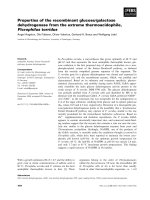
Fig. 1. Change of body weight by treatment with a vehicle (4
ml/kg BW), benzo(a)pyrene 150 µg/kg alone (BaP) and ben-
zo(a)pyrene with pyrene 1,700 µg/kg and phenanthrene 4,300 µ
g
/kg (BPP). The chemicals were used for treatment for 30 days vi
a
gavage in 9-week-old female SD rats. Values are mean ± SD.
fate and concentrated to about 1 ml at 45
o
C with a rotary
evaporator. The concentrated solution was further purified
using an activated Florisil cartridge (Silica cartridge for
fat). The PAHs were eluted with 18 ml of hexane and di-
chloromethane (3 : 1, v/v) solution, and the eluate dried at
45
o
C, then dissolved with 1 ml of acetonitrile by soni-
cation. Next, 20 ul of filtered solution was analyzed using
liquid chromatography with a fluorescence detector ac-
cording to the method of Chen et al. [9]. Analysis of the pa-
rent compound, and their metabolites in urine, was con-
ducted using the method of Jongeneelen et al. with mod-
ifications [21]. A 7 ml sample of 0.2 M sodium acetate buf-
fer (pH 5.0) was added into 5 ml of urine for acidification;
then β-glucuronidase (13,200 unit) and sulfatase (220 unit)
solution was added. The mixture was incubated with a
shaking incubator (37
o
C, 210 rpm) for 16 h. After cen-
trifugation (3,000 × g for 10 min), the supernatant was
loaded onto an activated Sep-Pak C18 cartridge, washed
with 5 ml of 40% methanol, eluted with 8 ml of acetoni-
trile, and 10 ml of hexane and dichloromethane (3 : 1, v/v).
The final eluate was evaporated at 45
o
C and dissolved with
1 ml of acetonitrile.
Statistical analysis
The body weight gain, organ weight, feed consumption,
and blood chemistry parameters were analyzed by break
down and one-way ANOVA, followed by the Duncan test
as a post-hoc comparison using a statistics program (Ver.
5.5; StatSoft, USA).
Results
Body weight, organ weights and blood chemistry
The treatment groups showed no significant difference in
body weight and relative liver weight compared to those in
the vehicle control group (Fig. 1 & Table 2). Rats exposed
to BaP or BPP did not show significant changes in the ac-
tivity of ALT, AST and ALP. However, the ALP activity
was increased by 21 days after the withdrawal of BPP com-
pared to the ALP in the vehicle control group (Table 2).
364 Hwan Goo Kang et al.
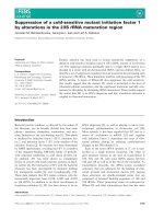
Fig. 2. Liver microsomal EROD activity at different time points
by treatment with a vehicle (4 ml/kg BW), benzo(a)pyrene 150 µ
g
/kg alone (BaP) and benzo(a)pyrene with pyrene 1,700 µg/kg an
d
p
henanthrene 4,300 µg/kg (BPP). Values are mean ± SD.
*Significantly different from vehicle control at p < 0.05.
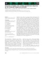
Fig. 3. Changes of PAHs in muscle at different time points by treatment with benzo(a)pyrene 150 µg/kg alone (BaP) and
b
enzo(a)pyren
e
with pyrene 1,700 µg/kg and phenanthrene 4,300 µg/kg (BPP). Values are mean ± SD.
EROD activity in liver and BaP adduct in lympho-
cytes
EROD activity, a major enzyme of CYP1A1 in the liver
microsomal fraction was significantly increased (p <
0.05) only in rats exposed to 30 days of BaP alone. There
were no significant differences in the time course of EROD
activity during BPP treatment and during the withdrawal
periods of BaP and BPP (Fig. 2). Benzo(a)pyrene-r-7,
t-8,t-9,c-10-tetradyrotetrol (+/) was not detected in the
blood lymphocytes of rats exposed to BaP alone or with PH
and PY.
Time-course changes of BaP, PH and PY in muscle
and fat
The highest mean amount of BaP in muscle was 34.4 ng/g
in the BaP treated group; it was 23.6 ng/g in BPP treated
rats, an amount achieved after 20 days of treatment in both
groups. The BaP concentration in muscle showed no sig-
nificant difference in comparisons between both treatment
groups (BaP and BPP). In addition, the amount of BaP in
muscle rapidly decreased after withdrawal of the treat-
ment.
The highest mean amount of BaP in fat was 3.2 ng/g in the
BaP treated group; this amount was obtained by 20 days of
treatment. The highest mean amount of PH was 47.1 ng/g
in muscle and 118.8 ng/g in fat; for PY it was 29.7 ng/g in
muscle and 219.9 ng/g in fat, which was also achieved by
20 days of treatment. Both compounds were rapidly re-
moved from the system after withdrawal of treatment
(Figs. 3&4, Table 3)
Time-course changes of BaP, PH and PY and their
metabolites in urine
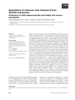
The 3-OH-BaP compound, a major metabolite of BaP,
was not detected in urine during treatment or after with-
drawal of treatment in both the BaP and BPP exposed
groups. The 3-OH-PH compound is a major metabolite of
PH; it was found in the ranges of 114-161 ng/ml and the
highest concentrations were reached by 30 days of treat-
ment. The concentrations of the parent compound (PY)
were 42-69 ng/ml with the highest concentration reached
by 30 days of treatment. The 1-OH-PY compound, a major
metabolite of PY, was found to be in the range of 201-263
ng/ml and the highest concentration was achieved by 10
days of treatment; the parent compound was in the range of
Changes of biomarkers with oral exposure to benzo(a)pyrene, phenanthrene and pyrene in rats 365
Fig. 5. Changes of PAHs and their metabolites in urine at different time points by treatment with benzo(a)pyrene 150 µg/kg alone (BaP)
and benzo(a)pyrene with pyrene 1,700 µg/kg and phenanthrene 4,300 µg/kg (BPP). Values are mean ± SD.
Fig. 4. Changes of PAHs in fat at different time points by treatment with benzo(a)pyrene 150 µg/kg alone (BaP) and
b
enzo(a)pyrene
with pyrene 1,700 µg/kg and phenanthrene 4,300 µg/kg (BPP). Values are mean ± SD.
9-17 ng/ml with the highest concentration reached by 30
days of treatment. The amounts of PY, PH and their metab-
olites were rapidly eliminated from the system after the
withdrawal of treatment (Fig. 5, Table 3).
Discussion
The PAHs are a group of several hundred compounds that
are present as a mixture in air and food. BaP is the most
toxic among the PAHs and wzll known as a carcinogen in
animals. The level of BaP in environmental samples is a
good marker for carcinogenic PAH contamination [7].The
correlation between BaP and the carcinogenic PAHs was
reported to be 0.98 in food [24]. PH and PY are less toxic
than BaP, but they are found in high levels in the air, animal
feed and food compared to the other PAHs [9,30,32].
Therefore, in this study, BaP, PH and PY were selected as
representative PAHs. We determined the treatment dose of
BaP as 150 µg/kg/day based on the reference maximal ex-
posure amount in humans, which was 0.055 µg/kg/day
[41] and multiplying the maximal margin factor by 3,000.
The dosages of PH and PY were determined to be 4,300 µg/
kg/day and 2,700 µg/kg/day, respectively; these doses
were chosen based on the mean contamination ratios of PH
and PY to BaP in air, food and feed.
The metabolic activation of carcinogenic PAHs occurs
primarily through the action of the CYP1A family P450
monooxygenase; CYP1A1 acts mainly by the hydrox-
ylation of BaP [35]. The activity of CYP1A1 was meas-
ured using the EROD activity [8,35,40]. Although the
EROD activity in rats exposed to BaP was increased about
44.7 fold in another reported experiment [28], in this study
366 Hwan Goo Kang et al.
Table 3 . Maximum mean level of benzo(a)pyrene, pyrene and phenanthrene in muscle, fat and urine and their metabolites in the urine
of rats orally exposed to BaP or BPP for 30 days
Treatment group Parent compounds Urine Muscle Fat Metabolites Urine
BaP
BPP
BaP
BaP
PH
PY
ND
ND
69.0 (30)
17.6 (30)
23.6 (20)*
34.5 (20)
47.1 (20)
29.7 (20)
3.2 (30)
1.3 (30)
118.8 (20)
219.9 (20)
3-OH-BaP
3-OH-BaP
3-OH-PH
1-OH-PY
ND
ND
161.6 (30)
263.0 (10)
*Data are maximum mean level (ppb) at date (day) when maximum level was found. BaP: benzo(a)pyrene, BPP: benzo(a)pyrene + phenan-
threne + pyrene.
N
D: not detected.
the increase was three fold compared to the control with the
treatment of BaP alone for 30 days. These differences may
be caused by the treatment dose used. The dose of Moorthy
et al. [28] was more 20 times higher than that used in the
present study. This result is also supported by Nilsen et al.
[29] who showed that mice exposed to 10 mg diesel ex-
hausts for 14 days caused no increase of EROD activity.
The threshold dose for the induction of hepatic EROD ac-
tivity was reported to be about 300 µg/kg for 5 and 6 ring
PAHs [37]. PH and PY did not change the EROD activity
in H4IIE cells, while the cells were sensitive to BaP [4].
Willett et al. [42] reported that fluoranthene inhibited the
BaP induced EROD activity in fish. In this study, EROD
activity in rats exposed to BaP with PH and PY was not
changed. However, it was increased in rats exposed to BaP
alone. PAHs bind to aryl hydrocarbon receptors (AhR) in
the liver and are further metabolized to a hydroxylated
compound. The high carcinogenic potency of certain PAHs
is correlated with the Ah-receptor (AhR) affinity, and the
induction of the CYP1A enzymes leading to increased
rates of formation of reactive metabolites [40]. Therefore,
PH and PY may interrupt BaP binding to AhR; however,
scaled experiments with additional samples as well as in
vitro studies are required for the characterization of the
changes in CYP1A1 activity with BaP with other PAH
chemicals.
DNA adducts of BaP in various organs have been studied
as biomarkers for PAH exposure or toxicity. In addition,
blood lymphocytes have been suggested to be useful bio-
logical targets for DNA adduct formation [19,34,36].
Benzo(a)pyrene-r-7,t-8,t-9,c-10-tetradyrotetrol (+/), one
of the main DNA adducts of benzo(a)pyrene, was not de-
tected in the blood lymphocytes of rats exposed to BaP
alone or to PH in our study. The detection limit for ben-
zo(a)pyrene-r-7,t-8,t-9,c-10-tetradyrotetrol (+/) was about
2.5 pg, which is comparable to that reported by Alexan-
drov et al.[1]. The same metabolite was found as an albu-
min adduct in rats exposed to 100 mg BaP/kg for 3 days or-
ally [19] and 10 mg BaP/kg by intra-peritoneal injection
[13]. Although the dose of BaP in this study was higher
than the maximum exposure level in humans, the dose of
BaP used in this study may not have been enough to induce
EROD activity to form reactive metabolites to bind to
DNA. Our data suggests that DNA adducts in blood lym-
phocytes may have been too low to be detected. Therefore,
a more sensitive method is needed to analyze BaP-specific
DNA adducts.
BaP is known to be readily absorbed from the gastro-
intestinal tract in animals. By contrast, the pretreatment of
BaP, for 7 days, inhibited the accumulation of BaP in the
body fat [41]. PY in an aqueous suspension was poorly ab-
sorbed from the GI tract; the amount observed was highest
in the peritoneal fat compared to the liver, kidney, lung,
heart, testes, spleen, and brain [27,43]. Data from the pres-
ent study showed that the amount of PH and PY in fat also
was higher than in muscle. However, BaP was higher in
muscle than in fat. BaP, PH and PY were rapidly removed
from muscle, consistent with other reports [27,43], show-
ing that the rate of clearance of the PAHs form organs was
very rapid.
Some of the PAHs are quickly metabolized by phase 1 en-
zymes in the liver to a hydroxylated form and they are then
mainly excreted in the urine. Metabolites of PH and PY, or
other PAHs in body fluid, may be useful biomarkers for
PAH exposure in humans and animals [16,17,21,25]. Urine
samples can be obtained quickly and easily in animals and
humans, and may provide a useful biological sample for
the study of exposure and toxicological assessment for en-
vironmental contaminants. I-OH-PY, 3-OH-PH and 3-OH-
BaP are the major hydroxylated metabolites for PY, PH and
BaP, respectively, in humans and animals [16,20,25].
Pyrene is a relatively large portion of the high molecular
weight PAHs, and it is found at high levels in the diet; its
metabolite,1-OH-PY, found in urine has been suggested to
reflect the total PAH contamination and is an indicator of
the mutagenic activity of the PAHs in animals [5,6,22]. In
the human, the ratio of 1-OH-PY to 3-OH-BaP was re-
ported to be 200 times higher [16]. The 1-OH-PY in urine
reflects a recent exposure while the PAH-adduct reflects a
more persistent and long-time exposure [23,31]. In the
Changes of biomarkers with oral exposure to benzo(a)pyrene, phenanthrene and pyrene in rats 367
present study, BaP and its metabolite, 3-OH-BaP, were not
detected and the concentration of PH was five times higher
than PY, while 1-OH-PY was detected at a two fold higher
level than 3-OH-PH in urine. Our data show that the
amount of parent compound in tissue and urine are propor-
tional to the dose used for treatment. However, the level of
the metabolites may not be directly related to the dose; that
is, although the treatment dose of PH was 1.6 times higher
than that of PY, the metabolites of PH were 0.61 times
higher than that of PY. The 1-OH-PY and 3-OH-PH com-
pounds were rapidly excreted into the urine after the with-
drawal of treatment; they were not detected 14 days after
withdrawal.
In conclusion, the results of this study suggest that
1-OH-PY in urine may be a candidate biomarker for ex-
posure to PAHs.
Acknowledgments
This project was supported by Research Funds from
National Veterinary Research and Quarantine Service,
Korea.
References
1. Alexandrov K, Rojas M, Geneste O, Castegnaro M,
Camus AM, Petruzzelli S, Giuntini C, Bartsch H. An im-
proved fluorometric assay for dosimetry of benzo(a)pyrene
diol-epoxide-DNA adducts in smokers' lung: comparisons
with total bulky adducts and aryl hydrocarbon hydroxylase
activity. Cancer Res 1992, 52, 6248-6253.
2. Arfsten DP, Schaeffer DJ, Muneny DC. The effects of near
ultraviolet radiation on the toxic effects of polycyclic aromatic
hydrocarbons in animals and plants: a review. Ecotoxicol
Environ Saf 1996, 33, 1-24.
3. Arif JM, Shappell N, Sikka HC, Kumar S, Gupta RC.
32
P-
Postlabeling analysis of lipophilic DNA adducts resulting
from interaction with (±)-3-hydroxy-trans-7,8-dihydroxy-
9,10-epoxy-7,8,9,10-tetrahydro-benzo[a]pyrene. Chem Biol
Interact 1999, 118, 87-97.
4. Bosveld ATC, de Bie PAF, van den Brink NW, Jongepier
H, Klomp AV. In vitro EROD induction equivalency factor
for the 10 PAHs generally monitored in risk assessment stud-
ies in the Netherlands. Chemosphere 2002, 49, 75-83.
5. Buchet JP, Gennart JP, Mercado-Calderon F, Delavig-
nette JP, Cupers L, Lauwerys R. Evaluation of exposure to
polycyclic aromatic hydrocarbons in a coke production and a
graphite electrode manufacturing plant: assessment of urinary
excretion of 1-hydroxypyrene as a biological indicator of
exposure. Br J Ind Med 1992, 49, 761-768.
6. Buckley TJ, Loiy PJ. An examination of the time course from
human dietary exposure to polycyclic aromatic hydrocarbons
to urinary elimination of 1-hydroxypyrene. Br J Ind Med
1992, 49, 113-124.
7. Bulter JP, Post GB, Lioy PJ, Waldman JM, Greenberg A.
Assessment of carcinogenic risk from personal exposure to
benzo(a)pyrene in the total human environmental exposure
study (THEES). Air Waste 1993, 43, 970-977.
8. Burke MD, Thompson S, Elcombe CR, Halpert J,
Haaparanta T, Mayer RT. Ethoxy-, pentoxy- and benzy-
loxyphenoxazones and homologues: a series of substrates to
distinguish between different induced cytochromes P-450.
Biochem Pharmacol 1985, 34, 3337-3345.
9. Chen BH, Wang CY, Chiu CP. Evaluation of analysis of
polycyclic aromatic hydrocarbons in meat products by liquid
chromatography. J Agric Food Chem 1996, 44, 2244-2251.
10. Dennis MJ, Massey RC, Cripps G, Venn I, Howarth N,
Lee G. Factors affecting the polycyclic aromatic hydro-
carbon content of cereals, fats and other food products. Food
Addit Contam 1991, 8, 517-530.
11. Easton MPL, Luszinak D, Geest EV. Preliminary examina-
tion of contaminant loadings in farmed salmon, wild salmon
and commercial salmon feed. Chemosphere 2002, 46, 1053-
1074.
12. Fouchecourt MO, Berny P, Riviere JL. Bioavailability of
PCBs to male laboratory rats maintained on litters of con-
taminated soils: PCB burden and induction of alkoxyresor-
ufin O-dealkylase activities in liver and lung. Arch Environ
Contam Toxicol 1998, 35, 680-687.
13. Godschalk RWL, Verner ITM, Kriek E, Floot B,
Schilderman PAEL, Moonen EJC, Kleinjans JCS, van
Schooten FJ. Comparison of
32
P-postlabeling and HPLC-
FD analysis of DNA adduct in rat acutely exposed to ben-
zo(a)pyrene. Chem Biol Interact 1997, 104, 41-54.
14. Gr
ä
slund A, Jernstr
ö
m B. DNA-carcinogen interaction:
covalent DNA-adducts of benzo(a)pyrene 7,8-dihydrodiol-
9,10-epoxides studied by biochemical and biophysical
techniques. Q Rev Biophys 1989, 22, 1-37.
15. Gravato C, Santos MA. Juvenile sea bass liver P450,
EROD induction, and erythrocytic genotoxic responses to
PAH and PAH-like compounds. Ecotoxicol Environ Saf
2002, 51, 115-127.
16. G
ü
ndel J, Schaller KH, Angerer J. Occupational exposure
to polycyclic aromatic hydrocarbons in a fireproof stone pro-
ducing plant: biological monitoring of 1-hydroxypyrene, 1-,
2-, 3- and 4-hydroxyphenanthrene, 3-hydroxybenz(a)an-
thracene and 3-hydroxybenzo(a)pyrene. Int Arch Occup
Environ Health 2000, 73, 270-274.
17. Hansen AM, Poulsen OM, Christensen JM. Determina-
tion of 1-hydroxypyrene in human urine by HPLC. J Anal
Toxicol 1993, 17, 38-41.
18. IARC. IARC Monographs on the Evaluation of Carcinogen-
ic Risk to Human. Vol. 32, p. 211, IARC, Lyon, 1983.
19. Islam GA, Greibrok T, Harvey RG,
Ø
vereb
ø
S. HPLC
analysis of benzo[a]pyrene-albumin adducts in benzo[a]pyr-
ene exposed rats. Detection of cis-tetrols arising from hy-
drolysis of adducts of anti- and syn-BPDE III with proteins.
Chem Biol Interact 1999, 123, 133-148.
20. Jacob J, Grimmer G. Metabolism and excretion of poly-
cyclic aromatic hydrocarbons in rat and in human. Cent Eur
J Public Health 1996, 4, 33-39.
21. Jongeneelen FJ, Akker WVD, Bos RP, Anzion RBM,
Theuws JLG, Roelofs HMJ, Henderson PTH. 1-OH-pyr-
ene as an indicator of mutagenicity of coal tar after activation
with human liver preparation. Mutat Res 1988, 204, 195-
201.
368 Hwan Goo Kang et al.
22. Jongeneelen FJ, Anizon RBM, Scheepers PTJ. 1-hydrox-
ypyrene in urine as a biological indicator of exposure to poly-
cyclic aromatic hydrocarbons in several work environments.
Ann Occup Hyg 1988, 32, 35-43.
23. Jongeneelen FJ. Benchmark guideline for urinary 1- as bio-
marker of occupational exposure to polycyclic aromatic
hydrocarbon. Ann Occup Hyg 2001, 45, 3-13.
24. Kazerouni N, Shinha R, Hsu CH, Greenberg A, Rothman
N. Analysis of 200 food items for benzo[a]pyrene and estima-
tion of its intake in an epidemiologic study. Food Chem
Toxicol 2001, 39, 423-436.
25. Keimig SD, Kiby KW, Morgna PP. Identification of 1-hy-
droxypyrene as a major metabolite of pyrene in pig urine.
Xenobiotica 1983, 13, 415-420.
26. Marczynski B, Rihs HP, Rossbach B, Holzer J, Angerer J,
Scherenberg M, Hoffmann G, Bruning T, Wilhelm M.
Analysis of 8-oxo-7,8-dihydro-2'-deoxyguanosine and DNA
strand breaks in white blood cells of occupationally exposed
workers: comparison with ambient monitoring, urinary me-
tabolites and enzyme polymorphisms. Carcinogenesis 2002,
23, 273-281.
27. Mitchell CE, Tu KW. Distribution, retention, and elimi-
nation of pyrene in rats after inhalation. J Toxicol Environ
Health 1979, 5, 1171-1179.
28. Moorthy B, Sriram P, Randerath K. Chemical structures
and time-dependent effects of polycyclic aromatic hydro-
carbon-type inducers on rat liver cytochrome P450, DNA ad-
ducts, and I-compounds. Fundamental Appl Toxicol 1994,
22, 549-560.
29. Nilsen A, Tr
ø
nnes T, Westerholm R, Rannug U, Nilsen
OG, Helleberg H, Kautiainen A, Hedenskog M, T
ö
rn-
qvist M. Short term exposure of rodent to disel exhausts; use-
fulness for studies of genotoxic and immunotoxic effect.
Chem Biol Interact 1999, 118, 19-38.
30. Nisbet ICT, LaGoy PK. Toxic equivalency factors (TEFs)
for polycyclic aromatic hydrocarbons. Regul Toxicol Phar-
macol 1992, 16, 290-300.
31. Ovrebo S, Haugen A, Fjeldstad PE, Hemminkj K,
Szyfter, K. Biological monitoring of exposure to polycyclic
aromatic hydrocarbon in an electrode paste plant. J Occup
Med 1994, 36, 303-310.
32. Park SS, Kim YJ, Kang CH. Atomospheric polycyclic ar-
omatic hydrocarbons in Seoul, Korea. Atmos Environ 2002,
36, 2917-2924.
33. Parvanello S, Zanesi N, Levis AG. BaP metabolism and
DNA -adduct formation in cultured human lymphocytes
treated in vitro with BaP and BaP-7,8-dihydrodiol. ATLA
1992,
20, 126-137.
34. Pavanello S, Favretto D, Brugnone F, Mastrangelo G, Dal
Pra G, Clonfero E. HPLC/fluorescence determination of an-
ti-BPDE-DNA adducts in mononuclear white blood cells
from PAH-exposed humans. Carcinogenesis 1999, 20, 431-
435.
35. Pohl RJ, Fouts JR. A rapid method for assaying the metabo-
lism of 7-ethoxyresorufin by microsomal subcellular frac-
tion. Anal Biochem 1980, 107, 150-155.
36. Rojas M, Alexandrov K, von Schooten FS, Hillebrand M,
Kriek E, Barstch H. Validation of a new fluorimetric assay
for benzo[a]pyrene diolepoxide-DNA adducts in human
white blood cells: comparisons with 32P-postlabeling and
ELISA. Carcinogenesis 1995, 15, 557-560.
37. Roos PH, Tschirbs S, Pfeifer F, Welge P, Hack A, Wilhelm
M, Bolt HM. Risk potentials for humans of original and re-
mediated PAH-contaminated soils: application of biomarkers
of effect. Toxicology 2004, 205, 181-194.
38. Roos PH, van Afferden M, Strotkamp D, Tappe D, Pfeifer
F, Hanstein WG. Liver microsomal levels of cytochrome
P450IA1 as biomarker for exposure and bioavailability of
soil-bound polycyclic aromatic hydrocarbons. Arch Environ
Contam Toxicol 1996, 30, 107-113.
39. Saunders CR, Ramesh A, Shockley DC. Modulation of
neurotoxic behavior in F-344 rats by temporal disposition of
benzo(a)pyrene. Toxicol Lett 2002, 129, 33-45.
40. Sj
ö
gren M, Ehrenbrg L, Rannug U. Relevance of different
biological assays in assessing initiating and promoting prop-
erties of polycyclic aromatic hydrocarbons with respect to
carcinogenic potency. Mutat Res 1996, 358, 97-112.
41. WHO. Evaluation of Certain Food Additives and Contami-
nants; Benzo(a)pyrene. WHO Food Additives Series 28. pp.
27-29, WHO, Geneva, 1998.
42. Willett KL, Wassenberg D, Lienesch L, Reichert W, Di
Giulio RT. In vivo and in vitro inhibition of CYP1A-depend-
ent activity in Fundulus heteroclitus by the polynuclear ar-
omatic hydrocarbon fluoranthene. Toxicol Appl Pharmacol
2001, 177, 264-271.
43. Withey JR, Law FC, Endrenyi L. Pharmacokinetics and bi-
oavailability of pyrene in the rat. J Toxicol Environ Health
1991, 32, 429-447.
44. Yamaguchi K, Near R, Shneider A, Cui H, Ju ST, Sherr
DH. Fluoranthecene-induced apoptosis in murine T cell hy-
bridomas is independent of the aromatic hydrocarbon
receptor. Toxicol Appl Pharmacol 1996, 139, 144-152.