Báo cáo khoa học: " Enhancement of protective immune responses by oral vaccination with Saccharomyces cerevisiae expressing recombinant Actinobacillus pleuropneumoniae ApxIA or ApxIIA in mice" pptx
Bạn đang xem bản rút gọn của tài liệu. Xem và tải ngay bản đầy đủ của tài liệu tại đây (1.22 MB, 10 trang )
JOURNAL OF
Veterinary
Science
J. Vet. Sci. (2007), 8(4), 383
392
†
Present address: Department of Microbiology, Research Institute for
Medical Sciences, College of Medicine, Chungnam National Univer-
sity, Daejeon 301-747, Korea
*Corresponding author
Tel: +82-2-880-1263; Fax: +82-2-874-2738
E-mail:
Enhancement of protective immune responses by oral vaccination with
Saccharomyces cerevisiae expressing recombinant Actinobacillus
pleuropneumoniae ApxIA or ApxIIA in mice
Sung Jae Shin
1,
†
, Seung Won Shin
1
, Mi Lan Kang
1
, Deog Yong Lee
1
, Moon-Sik Yang
2
, Yong-Suk Jang
2
,
Han Sang Yoo
1,
*
1
Department of Infectious Diseases, College of Veterinary Medicine, BK21 for Veterinary Science and KRF Zoonotic Disease
Priority Research Institute, Seoul National University, Seoul 151-742, Korea
2
Division of Biological Science, Institute for Molecular Biology and Genetics, Chonbuk National University, Jeonju 561-756,
Korea
We previously induced protective immune response by
oral immunization with yeast expressing the ApxIIA
antigen. The ApxI antigen is also an important factor in
the protection against Actinobacillus pleuropneumoniae se-
rotype 5 infection; therefore, the protective immunity in
mice following oral immunization with Saccharomyces cer-
evisiae expressing either ApxIA (group C) or ApxIIA
(group D) alone or both (group E) was compared with that
in two control groups (group A and B). The immuno-
genicity of the rApxIA antigen derived from the yeast was
confirmed by a high survival rate and an ApxIA-specific
IgG antibody response (p
<
0.01). The highest systemic
(IgG) and local (IgA) humoral immune responses to
ApxIA and ApxIIA were detected in group E after the
third immunization (p
<
0.05). The levels of IL-1
β
and
IL-6 after challenge with an A. pleuropneumoniae field iso-
late did not change significantly in the vaccinated groups.
The level of TNF-
α
increased in a time-dependent manner
in group E but was not significantly different after the
challenge. After the challenge, the mice in group E had a
significantly lower infectious burden and a higher level of
protection than the mice in the other groups (p
<
0.05).
The survival rate in each group was closely correlated to
the immune response and histopathological observations
in the lung following the challenge. These results suggested
that immunity to the ApxIA antigen is required for opti-
mal protection.
Key words: Actinobacillus pleuropneumoniae, Apx toxins, oral
immunization, protective immunity
Introduction
Most pathogens infect their host across mucosal surfaces,
particularly those of the gastrointestinal tract or respiratory
tract [24]. Immunoglobulin A (IgA) is the most abundant
Ig isotype present in the mucosal tissue during infection
and is crucial as a first line of defense. The main role of se-
cretory IgA in oral immunization [8,22] is to protect the
host by inhibiting pathogen attachment, immune ex-
clusion, and facilitating the clearance of toxic products
[37]. IgA may also function in lung defense by influencing
the trafficking of specific cells through the common mu-
cosal immune system [19]. The important roles that both
specific local IgA and systemic IgG play in the protection
from respiratory diseases have been well documented
[11,12]. Although most bacterial extracts that are com-
monly administered orally produce nonspecific or poor
immune responses, we previously demonstrated that the
protection against Actinobacillus pleuropneumoniae in-
creased with the production of specific IgA in the lung
[34]. In addition, the induction of protective immunity in
A. pleuropneumoniae infection by eliciting specific IgA
and IgG after natural and experimental infection has been
investigated [18].
A. pleuropneumoniae is the etiological agent of porcine
pleuropneumonia, a severe respiratory disease affecting
swine, is characterized by necrotizing fibrinous pneumo-
nia and pleuritis [6]. Although the bacterium produces sev-
eral virulence factors, the virulence of A. pleuropneu-
moniae is strongly correlated with the production of Apx
exotoxins. Four different types of exotoxins, ApxI, ApxII,
ApxIII and ApxIV, have been characterized in this bacte-
rium [15,28]. Both ApxIA and ApxIIA of A. pleuro-
pneumoniae are essential for full virulence in the develop-
384 Sung Jae Shin et al.
ment of clinical signs and typical lung lesions [5,28]. No
preventive strategies have shown complete protection
against the disease to date. Vaccination is thought to be the
most effective way to prevent clinical signs by infection
with the bacterium and many studies have focused on the
development of novel vaccines to prevent A. pleuro-
pneumoniae infection [5,17,18,26,32,39]. However, most
vaccines have taken the form of injections, which are labo-
rious and time-consuming, cause discomfort to the animal,
and may cause adverse effects, such as the induction of an
inflammatory response at the injection site [16,18,26].
Saccharomyces cerevisiae, commonly known as baker's
yeast, has recently been adopted as a delivery vehicle for
oral immunization [3]. This organism can express large
quantities of heterogenous proteins at a relatively low cost
[1,30] and is considered to be safe for human consumption
[31]. In addition, S. cerevisiae has been used as a tracer for
the oral application of vaccines and drugs because it is rela-
tively stable, nonpathogenic, and noninvasive in the gut in
comparison to other biodegradable vehicles [2,30]. The
yeast may also stimulate the host mucosal immune system
by interacting with intestinal epithelial cells in the presence
of butyric acid, a metabolite produced by intestinal bac-
teria [29].
In addition to the induction of a specific antibody re-
sponse, delivery systems and adjuvants are also key factors
in designing an oral vaccine to efficiently induce a mucosal
immune response [19,20,22]. Although several systems
have been developed, they have failed to induce sufficient
immune responses due to antigen dilution or denaturation,
tight immune regulation at mucosal sites, toxicity, or in-
sufficient immunostimulatory effects [27,40]. The recent
success using S. cerevisiae as a delivery vehicle in oral im-
munization [3,4,29,38] led us to choose this yeast system
for the delivery vehicle in our study.
Based on current knowledge, we propose that S. cer-
evisiae expressing Apx toxins is a more effective way to in-
duce protective immunity against A. pleuropneumoniae in-
fection than single administration of the ApxIIA. We first
confirmed the immunogenicity of the yeast-derived
ApxIA antigen. We then investigated the local and sys-
temic immune responses, bacterial clearance, and in-
flammatory responses after oral immunization and
challenge. Finally, we evaluated the protective efficacy of
our vaccine strategy by challenge with a field isolate of A.
pleuropneumoniae serotype 5.
Materials and Methods
Preparation of vaccines
The apxIA and apxIIA genes were cloned from A. pleuro-
pneumoniae serotype 5 isolated from the lungs of Korean
pigs with pleuropneumonia. For the oral vaccine, S. cer-
evisiae expressing ApxIA or ApxIIA antigens were pre-
pared as previously described [34,35].
Experimental animals
Female 5-week-old BALB/c mice (Breeding and Re-
search Center, Seoul National University, Korea) were
used throughout this study in accordance with the policies
and regulations for the care and use of laboratory animals
(Seoul National University, Korea). All animals were pro-
vided with standard mouse chow and water ad libitum.
The immunogenicity of the ApxIA produced in the yeast
was confirmed by subcutaneous immunization with
yeast-derived ApxIA protein, and the survival rate after
challenging with a clinical strain of A. pleuropneumoniae
was determined as previously described [34].
Briefly, 15 mice per group were subcutaneously injected
with 100 µg of protein extract after emulsifying with com-
plete Freund's adjuvant (Sigma, USA). This was then fol-
lowed by a boost immunization with the same amount of
antigens after emulsifying with incomplete Freund's ad-
juvant (Sigma, USA) at 2 weeks after the initial immu-
nization. The final immunization was performed in the
same manner at 2 weeks after the boost immunization.
Blood was drawn to collect serum at 5 days after the final
boost immunization. Finally, a survival test and IgG anti-
body response assays were carried out in order to confirm
the immunogenicity of the yeast-derived ApxIA antigen.
Each experimental group in the oral vaccination study con-
sisted of 40 mice, and each was allocated to one of five im-
munization regimens. Group A (control) received oral ad-
ministration of 500 µl of 10 mM PBS (pH 7.2) and group
B (vector) was orally vaccinated with 20 mg of S. cer-
evisiae powder dissolved into 500 µl of 10 mM PBS (pH
7.2). The vaccinated groups were immunized with 20 mg
of S. cerevisiae expressing either ApxIA (group C),
ApxIIA (group D), or both (10 mg each, group E) dissolved
with the procedures as well.
Delivery of vaccines for immunization and collec-
tion of samples
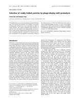
All groups were immunized orally through an oral gavage
with 4 doses at 10-day intervals. Five mice from each im-
munization group were randomly selected after 2 days
(Fig. 1). Samples of lung, intestine, and serum were in-
dividually collected from the mice as described previously
[34]. All serum samples were stored at 20
o
C until use.
Half of the lung and small intestine samples were homo-
genized with 10,000 RPM homogenization (Polytron
PT3000; Kinematica, USA). The homogenized samples
were stored at 4
o
C overnight, then centrifuged at 12,000 ×
g for 10 min at 4
o
C. The supernatants were collected for
subsequent analysis and stored at 20
o
C until use. The total
protein concentration in each sample was measured using
the BCA protein assay kit (Pierce, USA) and normalized to
1 mg immediately before performing the assay.
Immune responses with S. cerevisiae expressing rApxIA or rApxIIA 385
Fig. 1. Schematic of protocols for oral vaccine delivery.
Immune response analysis
Antibody titers (IgA and IgG) against ApxIA or ApxIIA
of A. pleuropneumoniae were measured by ELISA in order
to analyze the immune response in the mice. For this assay,
100 µg of rApxIA and rApxIIA [33] resuspended in 100 µl
of coating buffer (14.2 mM Na
2
CO
3
, 34.9 mM NaHCO
3
,
3.1 mM NaN
3
, pH 9.6) was added to a microplate for
ELISA (Greiner, Australia) and incubated overnight at
4
o
C. The plate was washed three times with PBST (0.05%
Tween 20 in PBS) and blocked with PBST containing 1%
bovine serum albumin by incubation for 1 h at 37
o
C. After
incubation with primary antigens, sera from the immu-
nized mice, lung or intestinal homogenates, were added to
the plate and incubated for 1 h at 37
o
C. After washing three
times with PBST, 100 µl of goat anti-mouse IgG (H + L)-
HRP conjugate (Bio-Rad, USA) or anti-mouse IgA (α
-chain specific)-HRP conjugate (Sigma, USA) was added
to the plate and incubated for 1 h at 37
o
C. Color was devel-
oped by adding 100 µl of ABTS substrate solution (Bio-
Rad, USA) to the plate. After incubation for 20 min at room
temperature, the O.D. was measured at 405 nm using an
ELISA reader (Molecular Device, USA).
Immunohistochemistry
Immunohistochemical staining was followed by our pre-
vious report [34].
Tissue preparation: For tissue preparation, mice from
each group were deeply anesthetized with a mixture of xy-
lazine hydrochloride (Bayer, Korea) and ketamin hydro-
chloride (Yuhan, Korea) and then perfused intracardially
with 0.9% saline, followed by a fixative (4% parafor-
maldehyde in 0.1 M PBS, pH 7.4) at a rate of 70 ml/min
with a perfusion pump (Masterflex, USA). After perfusion,
the lungs and intestines were removed and post-fixed over-
night in the same fixative at 4
o
C. The lungs and intestines
were cryoprotected by transfer to 30% sucrose in 0.1 M
PBS and frozen in OCT embedding medium (Tissue-Tek;
Sakura, USA) for storage at 70
o
C. Tissues were cut into
12 µm thick coronal sections with a cryostat (Reichert-
Jung, Germany), mounted on silane-coated slides (DAKO,
Denmark) and stored at 70
o
C until processing for immu-
nohistochemistry.
Detection of Apx toxin-specific antibody-producing
cells: Tissue sections were rinsed with 0.01 M PBS (pH
7.4) and treated with 0.5% hydrogen peroxide in 0.01 M
PBS for 15 min. The sections were washed three times for
10 min each with 0.01 M PBS, then blocked by incubation
in 10% normal goat serum (DAKO, Denmark) or 10%
skim milk in 0.1 M PBS for 1 h at room temperature. The
sections were incubated with 50 µg/ml of rApxIA or
rApxIIA in 0.1 M PBS overnight at 4
o
C. After incubation
with primary antigens, the sections were washed three
times with 0.01 M PBS for 10 min each and then incubated
with 1 : 200 diluted polyclonal antibodies against a culture
supernatant of A. pleuropneumoniae serotype 2 and 5 in 0.1
M PBS containing 0.3% triton X-100 and 2% normal goat
serum for 2 h at room temperature. After washing with 0.01
M PBS for 10 min, the sections were sequentially reacted
with 1 : 200 diluted goat anti-rabbit IgG (Vector, USA) and
Streptavidin (Vector, USA) in the same solution. Between
386 Sung Jae Shin et al.
sequential reactions, the tissues were washed three times
with PBS for 10 min each. The sections were visualized
with 3'3-diaminobenzidine tetrachloride (Sigma, USA) in
0.1 M Tris buffer (pH 6.8) and mounted with a cover slide
after counterstain with hematoxylin. Immunoreactive pre-
cipitates were observed under an Axioplan microscope
(Carl Zeiss, Germany). Images of IgA immunoreactivity in
ten villi in the small intestine and 10 alveolar spaces in the
lung were randomly chosen from each animal and captured
with an AppleScanner (Apple Computer, USA). The
brightness and contrast of each image file were uniformly
calibrated by Adobe Photoshop version 2.4.1, followed by
analysis using NIH Image 1.59 software. Background
staining values were subtracted from the immunoreaction
intensities. The number of IgA-secreting cells in alveolar
spaces was counted using Optimas 6.5 software (Media-
Cybernetics, USA) by averaging the counts from 10 sec-
tions randomly taken from the same section level of each
group.
Bacterial challenge and survival rate
Mice in each group were challenged by intraperitoneal in-
jection of a field isolate of A. pleuropneumoniae serotype
5 at 1.45 × 10
6
CFU (minimal lethal dose, MLD) in 10 days
after their final immunization, and were then monitored
every 6 h for up to 72 h. During the monitoring, animals
that succumbed to the challenge were dissected and lung
tissues were collected for subsequent analysis of in-
flammatory responses, cytokines, and recovery.
Bacteriological examination
To assess the protective efficacy measured by bacterial
clearance in the lungs, lungs were aseptically removed at
72 h post-challenge. The lungs were homogenized in 5 ml
of PBS using a tissue homogenizer. Each homogenate was
serially diluted in PBS and 50 µl of the homogenate, and
the diluted samples (in triplicate) were then plated on choc-
olate agar plates. The plates were incubated at 37
o
C for 48
h under a 5% (V/V) CO
2
atmosphere. The number of live
bacteria was quantified according to the formula: CFU/ml
= mean no. of colonies × dilution factor × 20. Differences
were considered to be significant if a probability value of p
< 0.05 was obtained when the CFU count of the immu-
nized groups was compared to that of the control groups.
Histological examination
The mice were sacrificed at 72 h after challenge with the
MLD of A. pleuropneumoniae serotype 5, and the lungs
were sliced into pieces and preserved in 10% neutralized
buffer formalin. The tissue samples were embedded in par-
affin, cut into 6 µm sections, assessed by routine staining
with hematoxylin and eosin, and examined by light
microscopy. The inflammatory response was evaluated by
examining the lung tissue for the presence of typical in-
flammatory signs [36]. Inflammatory index was obtained
from the average of the score from each inflammatory re-
sponse in 5 fields of each mouse. The severity of the in-
flammatory response (congestion, neutrophil infiltration,
exudation, consolidation, infiltration of fibrosis and plate-
lets) was ranked using a score of 0 to 3 for each symptom
(0, no sign; 1, mild; 2, notable and local; 3, severe and
spread) based on the size and number of lesions per field.
Cytokine analysis
The levels of TNF-α, IL-1β, and IL-6 in the serum and
lungs were quantified by ELISA (Endogen, USA) accord-
ing to the instructions supplied by the manufacturer. Lung
samples and sera from all experimental groups were pre-
pared as described previously [9]. Briefly, aseptically pre-
pared lungs were homogenized in 3 ml of lysis buffer. Lung
homogenates were incubated on ice for 30 min and then
centrifuged at 2,500 rpm for 10 min. The supernatants were
collected and filtered using 0.45 µm syringe filters (Nalgen,
USA). Before conducting the cytokine assessments, the
protein concentration of each homogenate was normalized
to 1 mg using a BCA protein assay kit (Pierce, USA). The
amount of each cytokine was calculated by comparison
with a standard curve generated by serial dilutions of mur-
ine recombinant cytokines.
Statistical analysis
Changes in IgA-secreting cells according to immuniza-
tion time and treatment group were evaluated with
ANOVA. The antibody titer and cytokine quantification
results were expressed as the mean ± SD. Differences be-
tween control groups and vaccinated groups were analyzed
by a two-tailed independent Student's t-test. Differences
were considered to be significant if probability values of p
< 0.05 were obtained.
Results
Immunogenicity of yeast expressing ApxIA antigen
To initially confirm the immunogenicity of the yeast-de-
rived ApxIA antigen, the production of ApxIA-specific
IgG antibodies and survival rates were investigated as in
our previous study of the yeast-derived ApxIIA antigen
[34]. The levels of ApxIA-specific IgG antibody were sig-
nificantly increased by subcutaneous immunization with
the protein extracted from the yeast expressing ApxIA.
Mice challenged with the MLD of an A. pleuropneumoniae
field isolate had a higher survival rate (70%) than the con-
trol (0%). None of the mice in the control groups showed
significant production of specific antibody or protection
against A. pleuropneumoniae after the challenge (data not
shown).
Immune responses with S. cerevisiae expressing rApxIA or rApxIIA 387
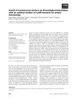
Fig. 2. Specific-IgA antibody responses to Actinobacillus pleu-
ropneumoniea AxpIIA or ApxIA toxin in the lung (A), small in-
testine (B), and sera (C) of mice orally immunized with S. cer-
evisiae (□, group A; ■, group B;
, group C; ▧, group D; ▤,
group E). Bars represent the mean O.D. values at 405 nm. Erro
r
bars represent the standard deviation from the mean. Significant
differences between control groups and vaccinated groups are
expressed as *p < 0.05 and ** p <0.01.
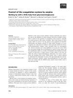
Fig. 3. Systemic specific IgG (A) and specific-IgM antibody re-
sponses (B) against Actinobacillus pleuropneumoniea AxpIIA o
r
ApxIA toxin in the sera of mice orally immunized with S. cer-
evisiae (□, group A; ■, group B;
, group C; ▧, group D; ▤,
group E). Bars represent the mean O.D. values at 405 nm. Erro
r
bars represent the standard deviation from the mean. Significan
t
differences between the control and vaccinated groups are ex-
p
ressed as *p < 0.05 and **p < 0.01.
Induction of specific immune responses
The levels of local and systemic antibodies specific to the
Apx antigens were investigated in mice orally immunized
with Apx antigen-expressing yeast. The antibodies specif-
ic to ApxIA or ApxIIA were produced at similar levels in
the group immunized with both the ApxIA and ApxIIA
antigens. Mucosal immune responses were evaluated in
the lung (Fig. 2A), intestine (Fig. 2B) and sera (Fig. 2C).
Specific IgA responses to ApxIA or ApxIIA in the intes-
tines and lungs from mice immunized with yeast express-
ing Apx antigens were significantly higher than those in
the control groups after the second and third immuniza-
tions, respectively (p < 0.05). In particular, mice immu-
nized with a single antigen (either ApxIA or ApxIIA)
showed significant increases in the level of specific IgA at
the final immunization (day 40) in both the lung and intes-
tine (p < 0.05). However, no significant increases in spe-
cific IgA antibodies were observed in the sera of any ex-
perimental group, even though the levels of specific IgA
were slightly higher in the vaccinated groups (p < 0.05)
(Fig. 2C).
Systemically, the pattern of IgG production to ApxA anti-
gens in the sera was similar to that of IgA. Increases in IgG
antibodies were only observed in the group immunized
with both antigens after the 2nd immunization and were
maintained until the final immunization, while groups vac-
cinated with a single antigen showed no significant differ-
ence during the same period (p > 0.05) (Fig. 3A).
Interestingly, similar levels of IgM antibody responses
were observed in all vaccinated groups during the immuni-
zation period, while those in the two control groups re-
mained unchanged (Fig. 3B).
Changes in IgA-secreting cells in the lung and
intestine
The number of IgA-secreting cells in the lung and intes-
388 Sung Jae Shin et al.
Fig. 4. Representative specimens stained by immunohistochemistry for IgA-secreting cells in the lungs of mice after the final
immunization. A, group B; B, group D; and C, group E. Arrows indicate positive immunoreactive cells. Counterstaining with
hematoxylin. ×400.
Tabl e 1. Number of IgA-secreting cells in the lung following oral immunization in each experimental group
Exp. groups
Days
10 20 30 40
Post-
challenge
A*
B
C
D
E
0.4 ± 0.02
0.2 ± 0.01
1.6 ± 0.042
2.8 ± 0.46
1.3 ± 0.02
0.1 ± 0.01
0.1 ± 0.06
3.2 ± 0.21
5.2 ± 0.64
6.5 ± 0.02
0.3 ± 0.031
0.3 ± 0.013
4.1 ± 1.03
9.8 ± 1.48
14.8 ± 1.06
0.2 ± 0.01
0.2 ± 0.021
4.8 ± 0.16
15.4 ± 1.84
26.8 ± 11.4
5.0 ± 1.02
3.0 ± 0.55
12.5 ± 0.84
22.1 ± 2.23
46.8 ± 5.36
*Group A: PBS control. Group B: S. cerevisiae vector control. Group C: Oral vaccination with S. cerevisiae expressing ApxIA antigen. Grou
p
D: Oral vaccination with S. cerevisiae expressing ApxIIA antigen. Group E: Combined oral vaccination with S. cerevisiae-ApxIA and S. cer-
evisiae-ApxIIA antigen. Values are mean ± SD.
tine was analyzed by counting the number of immunor-
eactive cells and densitometry. Representative specimens
stained by immunohistochemistry for IgA-secreting cells
in the lungs after the final immunization are shown in Fig.
4. The number of IgA-secreting cells significantly in-
creased in the groups immunized with ApxIIA or both anti-
gens after the third immunization, while the number of
IgA-secreting cells in the group immunized with ApxIA
increased only after challenge with A. pleuropneumoniae
(Table 1). However, the relative densities of IgA-secreting
cells in all vaccinated groups gradually increased after ad-
ditional immunizations in comparison to the control
groups. The final relative density of the groups immunized
with ApxIA, ApxIIA, and both antigens were 8.5, 9.5 and
22.5 times higher than in the PBS-treated control group, re-
spectively (Fig. 5).
Bacteriological and histopathological examination
The protective effect of oral immunization with yeast ex-
pressing ApxA antigens was also investigated through his-
topathological scoring and by measuring bacterial clear-
ance at 72 h post challenge. Bacterial clearance was sig-
nificantly enhanced by oral immunization with the anti-
gens in all vaccinated groups (p<0.05) (Table 2).
Moreover, the surviving mice showed significantly better
clearance rates by 36 h post-challenge. The relationship
between ApxA-specific antibody responses and bacterial
counts from mouse lungs was further analyzed in the lung
and sera from the control and vaccinated groups.
Histopathological lesions, as measured by inflammatory
indexes, were significantly reduced after vaccination while
bacterial clearance rates were significantly increased. The
lowest inflammatory index and the highest bacterial clear-
ance rate were observed in the group immunized with both
antigens (Table 2).
Immune responses with S. cerevisiae expressing rApxIA or rApxIIA 389
Tabl e 2 . Bacterial clearance in mice following oral immunizatio
n
with yeast expressing rApxA antigens
Immunization
groups
CFU/mg of
lung
(mean ± SD)
Bacterial
clearance rate
(%)
Inflammatory
index
A
B
C
D
E
1554 ± 284
1526 ± 313
849 ± 300
499 ± 213
230 ± 143
0.0 ± 4.3
1.8 ± 6.8
45.3 ± 10.5
67.9 ± 9.8
85.2 ± 8.4
14.5 ± 0.5
14.0 ± 1.0
9.7 ± 2.4
8.6 ± 2.8
2.2 ± 1.7
*Each group is the same as Table 1.
Fig. 5. Densitometric analysis of IgA immunoreactivity in the
small intestines of mice orally immunized with S. cerevisiae (□,
group A; ■, group B;
, group C; ▧, group D; ▤, group E).
Results are expressed as the mean relative density. Asterisks in-
dicate significant differences from the PBS-treated group, *p <
0.05 and **p < 0.01.
Fig. 6. Comparison of pro-inflammatory cytokines IL-1β (A), IL-6 (B), and TNF-α (C) from the lung and sera of mice following oral
immunization with S. cerevisiae (□, group A; ■, group B;
, group C; ▧, group D; ▤, group E). Bars represent the mean concen-
tration of cytokine proteins. Error bars represent the standard deviation from the mean.
390 Sung Jae Shin et al.
Fig. 7. Survival rates of mice immunized with S. cerevisiae afte
r
being challenged with the minimal lethal dose (MLD) of an A.
p
leuropneumoniae serotype 5 Korean isolate ( , PBS-treated
control;
, vector control; , oral immunization with 20 mg
of S. cerevisiae expressing ApxIA antigen; , oral immuniza-
tion with 20 mg of S. cerevisiae expressing ApxIIA antigen; ,
oral immunization with 10 mg each of S. cerevisiae expressing
ApxIA and S. cerevisiae expressing ApxIIA antigen).
Change in proinflammatory cytokines
The levels of IL-6 and TNF-α significantly increased dur-
ing immunization in the lungs from mice immunized with
both antigens. However, the levels of IL-1β, IL-6 and
TNF-α in the lungs of mice from the immunized groups did
not change significantly after challenge, while the levels of
these cytokines in the mice in the control groups sig-
nificantly increased after challenge (Fig. 6). The cytokine
levels in the sera were similarly raised only after challenge,
with the exception of IL-1β, which did not change sig-
nificantly (Fig. 6A). The production of TNF-α in both the
sera and lung tissue of mice immunized with both antigens
was slightly lower than that of the mice in the other groups
after challenge.
Survival rates
All mice were monitored for up to 72 h after challenge
with the MLD of an A. pleuropneumoniae field isolate.
Overall, the final survival rates of the vaccinated groups
were higher than those of the control groups at each time
point. Notably, all mice in the control groups died at 36 h
after challenge. The highest survival rate was observed in
the group immunized with both antigens (Fig. 7).
The correlation coefficient (r
2
) was calculated by re-
gression analysis in order to determine whether there was
a correlation between survival rate and antibody response
or the levels of bacterial colonization. The results showed
that there was a statistically significant correlation (t test
for correlation, p < 0.001) between the increase in mucosal
IgA (r
2
= 0.84), systemic IgG (r
2
= 0.79), and survival rates.
However, an increase in systemic IgA and IgM did not cor-
relate with the survival rates. Moreover, the number of bac-
teria in the lung correlated negatively with the survival rate
(r
2
= 0.81).
Discussion
Porcine pleuropneumonia caused by A. pleuropneumo-
niae is an important respiratory disease in the swine in-
dustry and has resulted in great economic loss worldwide
[21]. Although the disease is multifactorial, vaccination
has been considered to be the most effective strategy for
protecting swine from A. pleuropneumoniae infection.
Since most current vaccines are injected and may cause
many adverse effects [17,18,26], alternative vaccines, in-
cluding oral vaccines, have been sought after [8,18]. In ad-
dition, the induction of immune responses at remote mu-
cosal effector sites through a common mucosal immune
system has been demonstrated in animal models and has
been partially confirmed in humans [12,13,22]. When de-
veloping an oral vaccine, it is essential to select an effective
immunogen, appropriate adjuvant, and proper vaccine reg-
imen [7,20]. We previously explored oral vaccination us-
ing yeast expressing the ApxIIA antigen as an alternative
and convenient approach against A. pleuropneumoniae in-
fection [34]. However, the protective effect of the oral im-
munization was not sufficient because the bacterium also
produces other exotoxins. In this study, yeast expressing
ApxIA were added as a vaccine component because
ApxIA is also one of the most important factors associated
with pathogenesis and protective immunity [17]. The effi-
cacy of yeast expressing ApxIA or ApxIIA was evaluated
using different vaccination regimens in a mouse model be-
fore being applied to the pigs. Mice immunized with pro-
teins extracted from yeast expressing the ApxIA antigen
produced strong IgG antibody responses and were pro-
tected against challenge, which suggests that the rApxIA
antigen expressed in S. cerevisiae is highly immunogenic.
IgA and IgG immune responses increased following oral
vaccination, and the highest level of response was ob-
served in the group vaccinated with both S. cerevisiae that
expressed ApxIA or ApxIIA. We also observed a large in-
crease in antigen-specific IgA antibodies and the number
of IgA-secreting cells in the intestine and lung. Based on
the findings of other reports [7,8,34], these results strongly
suggest that mucosal immune responses at remote sites in-
duced by oral immunization are directly related to the ef-
fective production of IgA at the target mucosal site.
Only mice immunized with both ApxIA and ApxIIA pro-
duced sufficient humoral immune responses to Apx A tox-
ins and consequently showed the highest survival against
the challenge. These results compliment those of a pre-
vious report showing that exotoxins were required for the
full virulence of A. pleuropneumoniae infection [5].
TNF-α and IL-6 production in the lung increased after
vaccination, and IL-1β, TNF-α, and IL-6 production in the
lung was abrogated only in the vaccinated groups after
challenge with an A. pleuropneumoniae field isolate. This
phenomenon might be due to the involvement of IL-6 in
Immune responses with S. cerevisiae expressing rApxIA or rApxIIA 391
the production of IgA and the induction of TNF-α by IgA
[23]. Moreover, the dual capacities of secreted IgA might
be involved in the mechanism for maintaining balance be-
tween pro-inflammatory and anti-inflammatory activities
[14,23]. In addition, the prevention of IL-1β, TNF-α and
IL-6 production was correlated with a decrease in lung le-
sions in the vaccinated groups after challenge.
The highest bacterial clearance and survival rates were
observed in the group immunized with both antigens.
These results might indicate that oral vaccination using
both antigens could induce more effective protection
against particularly acute infections by decreasing
mortality. It was also possible that IgA contributed to the
protective mechanism by inhibiting the entrance of the
pathogen into the lung and by modulating the pro-in-
flammatory responses [23,25]. The histopathological le-
sions, such as infiltration of inflammatory cells, were pos-
itively correlated with the groups showing high levels of
inflammatory cytokine production. These results are in
good agreement with those of previous studies in which in-
flammatory cell infiltration was mediated by inflammatory
cytokines [9,10]. Although current thinking is that cell-
mediated immunity does not play an important role in pro-
tection against A. pleuropneumoniae infection, the role of
cell-mediated immune responses following oral immuni-
zation needs further investigation.
In conclusion, strains of S. cerevisiae that produce ApxA
antigens could be a promising oral vaccine candidate for
the prevention of A. pleuropneumoniae acute infection in
pigs, alone or in combination with other bacterial compo-
nents, and may provide optimal protection both systemi-
cally and at target mucosal sites.
Acknowledgments
This study was supported by BioGreen 21 (200503013
4414), RDA, Brain Korea 21, and the Research Institute for
Veterinary Sciences, Seoul National University, Korea.
References
1. Bathurst IC. Protein expression in yeast as an approach to
production of recombinant malaria antigens. Am J Trop Med
Hyg 1994, 50, 20-26.
2. Beier R, Gebert A. Kinetics of particle uptake in the domes of
Peyer's patches. Am J Physiol 1998, 275, G130-137.
3. Blanquet S, Antonelli R, Laforet L, Denis S, Marol-Bonnin
S, Alric M. Living recombinant Saccharomyces cerevisiae
secreting proteins or peptides as a new drug delivery system in
the gut. J Biotechnol 2004, 110, 37-49.
4. Blanquet S, Meunier JP, Minekus M, Marol-Bonnin S,
Alric M. Recombinant Saccharomyces cerevisiae expressing
P450 in artificial digestive systems: a model for biodetoxi-
cation in the human digestive environment. Appl Environ
Microbiol 2003, 69, 2884-2892.
5. Boekema BK, Kamp EM, Smits MA, Smith HE,
Stockhofe-Zurwieden N. Both ApxI and ApxII of Actinoba-
cillus pleuropneumoniae serotype 1 are necessary for full
virulence. Vet Microbiol 2004, 100, 17-23.
6. Bosse JT, Janson H, Sheehan BJ, Beddek AJ, Rycroft AN,
Kroll JS, Langford PR. Actinobacillus pleuropneumoniae:
pathobiology and pathogenesis of infection. Microbes Infect
2002, 4, 225-235.
7. Bouvet JP, Decroix N, Pamonsinlapatham P. Stimulation
of local antibody production: parenteral or mucosal vacci-
nation? Trends Immunol 2002, 23, 209-213.
8. Bowersock TL, Shalaby WS, Levy M, Samuels ML,
Lallone R, White MR, Borie DL, Lehmeyer J, Park K.
Evaluation of an orally administered vaccine, using hydrogels
containing bacterial exotoxins of Pasteurella haemolytica, in
cattle. Am J Vet Res 1994, 55, 502-509.
9. Broug-Holub E, Toews GB, van Iwaarden JF, Strieter
RM, Kunkel SL, Paine R, Standiford TJ. Alveolar macro-
phages are required for protective pulmonary defenses in
murine Klebsiella pneumonia: elimination of alveolar macro-
phages increases neutrophil recruitment but decreases bacte-
rial clearance and survival. Infect Immun 1997, 65, 1139-
1146.
10. Choi C, Kwon D, Min K, Chae C. In-situ hybridization for
the detection of inflammatory cytokines (IL-1, TNF-alpha
and IL-6) in pigs naturally infected with Actinobacillus
pleuropneumoniae. J Comp Pathol 1999, 121, 349-356.
11. Cox E, Van der Stedea Y, Verdonck F, Snoeck V, Van den
Broeck W, Goddeeris B. Oral immunisation of pigs with
fimbrial antigens of enterotoxigenic E. coli: an interesting
model to study mucosal immune mechanisms. Vet Immunol
Immunopathol 2002, 87, 287-290.
12. Dietrich G, Griot-Wenk M, Metcalfe IC, Lang AB, Viret
JF. Experience with registered mucosal vaccines. Vaccine
2003, 21, 678-683.
13. Externest D, Meckelein B, Schmidt MA, Frey A. Correlat-
ions between antibody immune responses at different mu-
cosal effector sites are controlled by antigen type and dosage.
Infect Immun 2000, 68, 3830-3839.
14. Fernandez MI, Pedron T, Tournebize R, Olivo-Marin JC,
Sansonetti PJ, Phalipon A. Anti-inflammatory role for in-
tracellular dimeric immunoglobulin a by neutralization of lip-
opolysaccharide in epithelial cells. Immunity 2003, 18,
739-749.
15. Frey J. Virulence in Actinobacillus pleuropneumoniae and
RTX toxins. Trends Microbiol 1995, 3, 257-261.
16. Fuller TE, Martin S, Teel JF, Alaniz GR, Kennedy MJ,
Lowery DE. Identification of Actinobacillus pleuropneu-
moniae virulence genes using signature-tagged mutagenesis
in a swine infection model. Microb Pathog 2000, 29, 39-51.
17. Haga Y, Ogino S, Ohashi S, Ajito T, Hashimoto K,
Sawada T. Protective efficacy of an affinity-purified hemo-
lysin vaccine against experimental swine pleuropneumonia. J
Vet Med Sci 1997, 59, 115-120.
18. Hensel A, van Leengoed LA, Szostak M, Windt H,
Weissenbock H, Stockhofe-Zurwieden N, Katinger A,
Stadler M, Ganter M, Bunka S, Pabst R, Lubitz W.
Induction of protective immunity by aerosol or oral applica-
tion of candidate vaccines in a dose-controlled pig aerosol in-
392 Sung Jae Shin et al.
fection model. J Biotechnol 1996, 44, 171-181.
19. Lauterslager TG, Hilgers LA. Efficacy of oral admin-
istration and oral intake of edible vaccines. Immunol Lett
2002, 84, 185-190.
20. Lauterslager TG, Stok W, Hilgers LA. Improvement of the
systemic prime/oral boost strategy for systemic and local
responses. Vaccine 2003, 21, 1391-1399.
21. Losinger WC. Economic impacts of reduced pork pro-
duction associated with the diagnosis of Actinobacillus pleu-
ropneumoniae on grower/finisher swine operations in the
United States. Prev Vet Med 2005, 68, 181-193.
22. Ogra PL, Faden H, Welliver RC. Vaccination strategies for
mucosal immune responses. Clin Microbiol Rev 2001, 14,
430-445.
23. Olas K, Butterweck H, Teschner W, Schwarz HP, Reipert
B. Immunomodulatory properties of human serum immuno-
globulin A: anti-inflammatory and pro-inflammatory activ-
ities in human monocytes and peripheral blood mononuclear
cells. Clin Exp Immunol 2005, 140, 478-490.
24. Pabst R, Binns RM. The immune system of the respiratory
tract in pigs. Vet Immunol Immunopathol 1994, 43, 151-156.
25. Pascual DW, Trunkle T, Sura J. Fimbriated Salmonella en-
terica serovar typhimurium abates initial inflammatory re-
sponses by macrophages. Infect Immun 2002, 70, 4273-4281.
26. Prideaux CT, Lenghaus C, Krywult J, Hodgson AL.
Vaccination and protection of pigs against pleuropneumonia
with a vaccine strain of Actinobacillus pleuropneumoniae
produced by site-specific mutagenesis of the ApxII operon.
Infect Immun 1999, 67, 1962-1966.
27. Rappuoli R, Pizza M, Douce G, Dougan G. Structure and
mucosal adjuvanticity of cholera and Escherichia coli heat-
labile enterotoxins. Immunol Today 1999, 20, 493-500.
28. Reimer D, Frey J, Jansen R, Veit HP, Inzana TJ.
Molecular investigation of the role of ApxI and ApxII in the
virulence of Actinobacillus pleuropneumoniae serotype 5.
Microb Pathog 1995, 18, 197-209.
29. Saegusa S, Totsuka M, Kaminogawa S, Hosoi T. Candida
albicans and Saccharomyces cerevisiae induce interleukin-8
production from intestinal epithelial-like Caco-2 cells in the
presence of butyric acid. FEMS Immunol Med Microbiol
2004, 41, 227-235.
30. Schreuder MP, Deen C, Boersma WJ, Pouwels PH, Klis
FM. Yeast expressing hepatitis B virus surface antigen deter-
minants on its surface: implications for a possible oral
vaccine. Vaccine 1996, 14, 383-388.
31. Schreuder MP, Mooren AT, Toschka HY, Verrips CT,
Klis FM. Immobilizing proteins on the surface of yeast cells.
Trends Biotechnol 1996, 14, 115-120.
32. Seah JN, Frey J, Kwang J. The N-terminal domain of RTX
toxin ApxI of Actinobacillus pleuropneumoniae elicits pro-
tective immunity in mice. Infect Immun 2002, 70, 6464-
6467.
33. Shin SJ, Cho YW, Yoo HS. Cloning, sequencing and ex-
pression of apxIA, IIA, IIIA of Actinobacillus pleuro-
pneumoniae isolated in Korea. Korean J Vet Res 2003, 43,
247-253.
34. Shin SJ, Bae JL, Cho YW, Lee DY, Kim DH, Yang MS,
Jang YS, Yoo HS. Induction of antigen-specific immune re-
sponses by oral vaccination with Saccharomyces cerevisiae
expressing Actinobacillus pleuropneumoniae ApxIIA. FEMS
Immunol Med Microbiol 2005, 43, 155-164.
35. Shin SJ, Bae JL, Cho YW, Yang MS, Kim DH, Jang YS,
Yoo HS. Expression of apxIA of Actinobacillus pleuro-
pneumoniae in Saccharomyces cerevisiae. J Vet Sci 2003, 4,
225-228.
36. Shin SJ, Wu CW, Steinberg H, Talaat AM. Identification
of novel virulence determinants in Mycobacterium para-
tuberculosis by screening a library of insertional mutants.
Infect Immun 2006, 74, 3825-3833.
37. Silin DS, Lyubomska V. Overcoming immune tolerance
during oral vaccination against Actinobacillus pleuropneu-
moniae. J Vet Med B Infect Dis Vet Public Health 2002, 49,
169-175.
38. Stubbs AC, Martin KS, Coeshott C, Skaates SV,
Kuritzkes DR, Bellgrau D, Franzusoff A, Duke RC,
Wilson CC. Whole recombinant yeast vaccine activates den-
dritic cells and elicits protective cell-mediated immunity. Nat
Med 2001, 7, 625-629.
39. Tonpitak W, Baltes N, Hennig-Pauka I, Gerlach GF.
Construction of an
Actinobacillus pleuropneumoniae sero-
type 2 prototype live negative-marker vaccine. Infect Immun
2002, 70, 7120-7125.
40. Williams AE, Edwards L, Humphreys IR, Snelgrove R,
Rae A, Rappuoli R, Hussell T. Innate imprinting by the
modified heat-labile toxin of Escherichia coli (LTK63) pro-
vides generic protection against lung infectious disease. J
Immunol 2004, 173, 7435-7443.