Báo cáo khoa học: "Molecular characterization and genogrouping of VP1 of aquatic birnavirus GC1 isolated from rockfish Sebastes schlegeli in Korea" potx
Bạn đang xem bản rút gọn của tài liệu. Xem và tải ngay bản đầy đủ của tài liệu tại đây (353.38 KB, 6 trang )
JOURNAL OF
Veterinary
Science
J. Vet. Sci. (2008), 9(1), 85
90
*Corresponding author
Tel: +82-31-467-1805; Fax: +82-31-467-1803
E-mail:
Molecular characterization and genogrouping of VP1 of aquatic
birnavirus GC1 isolated from rockfish Sebastes schlegeli in Korea
Seong Joon Joh
1,
*
, Chae Ik Shon
1
, Sung Won Kang
1
, Byoung Han Kim
1
, Byung Yul Jeong
1
, Kyung Gi Lee
1
,
Jun Hun Kwon
1
, Gang Jun Heo
2
1
National Veterinary Research and Quarantine Service, Anyang 430-824, Korea
2
College of Veterinary Medicine and Research Institute of Veterinary Medicine, Chungbuk National University, Cheongju 361-763,
Korea
The cDNA nucleotide sequence of genome segment B
encoding the VP1 protein was determined for the aquatic
birnavirus GC1 isolated from the rockfish Sebastes schlegeli
in Korea. The VP1 protein of GC1 contains a 2,538 bp open
reading frame, which encodes a protein comprising 846
amino acid residues that has a predicted MW of 94 kDa. The
sequence contains 6 potential Asn-X-Ser/Thr motifs. Eight
potential Ser phosphorylation sites and 1 potential Tyr
phophorylation site were also identified. GC1 contains the
Leu-Lys-Asn (LKN) motif instead of the typical Gly-Asp-
Asp (GDD) motif found in other aquatic birnaviruses. We
also identified the GLPYIGKT motif, the putative GTP-
binding site at amino acid position 248. In total, the VP1
regions of 22 birnavirus strains were compared for
analyzing the genetic relationship among the family
Birnaviridae. Based on the deduced amino acid sequences,
GC1 was observed to be more closely related to the infectious
pancreatic necrosis virus (IPNV) from the USA, Japan, and
Korea than the IPNV from Europe. Further, aquatic
birnaviruses containing GC1 and IPNV have genogroups
that are distinct from those in the genus Avibirnaviruses and
Entomo-birnaviruses. The birnavirusstrains were clustered
into 5 genogroups based on their amino acid sequences. The
marine aquatic birnaviruses (MABVs) containing GC1
were included in the MABV genogroup; the IPNV strains
isolated from Korea, Japan, and the USA were included in
genogroup 1 and the IPNV strains isolated primarily from
Europe were included in genogroup 2. Avibirnaviruses and
entomobirnaviruses were included in genogroup 3 and 4,
respectively.
Keywords: aquatic birnavirus, GC1, genetic characterization,
rockfish Sebastes schlegeli, VP1
Introduction
Members of the family Birnaviridae have 2-segmented
genomes - A and B. This family comprises 3 main genera,
including the genus Aquabirnavirus, Avibirnavirus, and
Entomobirnavirus [4,19]. The type species of the genus
Aquabirnavirus is the infectious pancreatic necrosis virus
(IPNV); the genus comprises marine aquatic birnaviruses
(MABV) of fish and shellfish [3]. Other members of the
family Birnaviridae include infectious bursal disease virus
(IBDV) belongs to the genus Avibirnavirus, and Droso-
phila X virus (DVX) that belongs to the genus Entomobirna-
virus. Aquatic birnaviruses are the largest and most diverse
group of viruses within the family Birnaviridae. The first
reported MABV was isolated from the yellowtail Seriola
quinqueradiata in Japan [22], other MABVs have been
subsequently isolated from various marine fishes in Korea
and Japan, and their characteristics have been investigated
[7,8,14,18,23,24]. The genome segment B of birnaviruses
encodes the VP1 protein, which is the presumptive
virion-associated RNA-dependent RNA polymerase (RdRp)
[13,15]. Some researchers reported the characteristics of
VP1 and compared the VP1 region among birnaviruses [4,
25]. They identified several conserved domains associated
with RdRps and GTP-binding proteinsin the IPNV strains;
these domains were the same as those in other RNA
viruses. However, they also discovered that the typical
Gly-Asp-Asp (GDD) motif that is found in all RNA viruses
was absent in the VP1 region of some IPNV [4] IBDV, and
DXV [2] strains.
The physical, antigenic, and genetic features of the
VP2/NS junction region of the aquatic birnavirus GC1
isolated from the rockfish Sebastes (S.) schlegeli, which is
the second most important in the aquaculture industry in
Korea, has been studied [8,9,20].
In the present study, we investigated the genetic charac-
teristics of the VP1 protein and compared the genetic
relationship between aquatic birnaviruses and other
86 Seong Joon Joh et al.
Table 1. Descriptions of VP1 sequences of aquabirnaviruses cited in this study
Name of virus Genus of virus Geographic origin Host of origin Accession number
GC1 Aquabirna virus Korea Rockfish −
578 Aquabirna virus Spain Turbot AJ489244
1146 Aquabirna virus Spain Trout AJ489238
2290 Aquabirna virus Spain Salmon AJ489240
AM98 Aquabirna virus Japan Amago salmon AY129664
AY98 Aquabirna virus Japan Ayu AY123970
DRT Aquabirna virus Korea Rainbow trout D26527
Jasper Aquabirna virus Canada Turbot M58756
NC1 Aquabirna virus Korea Flounder AY129666
Sp Aquabirna virus Denmark Turbot M58757
WB Aquabirna virus USA Turbot AF078669
Y6 Aquabirna virus Japan Yellowtail AY129662
YT01A Aquabirna virus Japan Yellowtail AY129663
CLV Aquabirna virus Viet Nam Boltched snakehead fish AJ459383
88R Aquabirna virus Spain Oyster AJ489245
24R Aquabirna virus Spain Mussel AJ489243
20Gld Aquabirna virus Spain Deepwater redfish AY780931
19F3b Aquabirna virus Spain Greenland halibut AY780928
6B1a Aquabirna virus Spain Atlantic cod AY780926
H1 Aquabirna virus Japan Flounder AY129665
UPM97/61 Avivirna virus Malaysia Birds AF527040
DXV Entomobirna virus Canada Drosophila AF196645
genuses within family Birnaviridae.
Materials and Methods
Virus and cell
GC1 was isolated from the rockfish S. schlegeli, and it
was grown in the Chinook Salmon Embryo-214 cell line
supplemented with Eagle’s minimum essential medium.
The sources of VP1 sequence cited in this study are listed
in Table 1.
Viral RNA extraction and primers
The viral genomic RNA was extracted using the methods
described by Joh et al. [8]. Briefly, GC1-infected cells were
frozen and thawed 3 times and clarified by centrifugation.
Viral dsRNA was then extracted with phenol and chloroform,
followed by digestion with proteinase K. Seven primer
pairs were used for reverse transcription- polymerase chain
reaction (RT-PCR). The oligonucleotide sequences were
deduced according to the published dsRNA sequences of
the Western Buxton strains (AF078669) (Table 2).
cDNA synthesis by RT-PCR
The RT-PCR procedure used in this study was a
modification of the method previously described by Joh et
al. [10]. The RT-PCR solution was heated to 95
o
C for 3 min
and passed through 35 cycles under the following
conditions: 1 min at 95
o
C for denaturation; 1 min at 54
o
C
to 58
o
C (depending on the primer) to allow annealing; 1
min at 72
o
C for extension and final amplification at 72
o
C
for 3 min. The ethidium bromide-stained PCR products
were electrophoresed on a 1.5% agarose gel and were
visualized by UV fluorescence.
Construction of recombinant plasmids
Each resulting product was gel purified and then cloned into
pCR2.1 TA cloning vectors (Invitrogen, USA) according to
the manufacturer’s instructions. All the clones were amplified
by transformation into competent DH5 α cells. Clones with
correct inserts were confirmed by restriction enzyme
digestion of the recombinant vectors.
Nucleotide sequencing and analysis of the VP1
Nucleotide sequencing was carried out on an ABI 377
sequencer (Applied Biosystems, USA) by the dideoxy-
nucleotide chain termination method by using T7 DNA
and SP6 DNA polymerase. The nucleotide and deduced
amino acid sequences were analyzedby Vector NTI ver
10.0 (Hitachi, Japan) and were compared with the
corresponding sequences of previously reported cite
accession numbers of aquabirnaviruses in Table 1.
Genogrouping of VP1 of aquatic birnavirus
87
Tabl e 2 . RT-PCR primer sets and amplified cDNA fragments used for sequencing
Primers (Sequence) Position* PCR product length
GVP1.1F
GVP1.1R
GVP1.2F
GVP1.2R
GVP1.3F
GVP1.3R
GVP1.4F
GVP1.4R
GVP1.5F
GVP1.5R
GVP1.6F
GVP1.6R
GVP1.7F
GVP1.7R
GGAAACAGTGGGTCAACGTT
AGAAGTGTGATGTCCGGAGC
CCATTCCACAAGCCAGACCA
AGGAGTCAGCCAGTACGAGC
TCCTCAGCCGGCCTACCATA
GAGTACCATGTGTTGTCCTG
AAGAGACAGCCTGGACAATG
GTCTCGACGGCCTCAACGAT
AAGATAGAGCGCGAGCTGAA
ATTCCTTCTAGGTCTCCTCC
CAAGAGGAAGAGACTGGAAG
TGTTGTGCCAGTTCCTCAGT
TACGAGATCAAGCACTAGCG
TCCCTGGCGGAACCGGATGT
1-483
422-908
833-1299
1216-1701
1646-2106
2011-2400
2319-2780
483 bp
486 bp
466 bp
485 bp
460 bp
389 bp
461 bp
*Map position of the primers based on the published sequence of Western buxton (AF078669).
Table 3. Kinds of potential motif exist in VP1 of GC1
Kinds of motif
N
O. of
sites
Position of sites in
amino acid sequences
N
-linked glycosylation site
Serine phosphorylation site
Thyrosine phosphorylation site
GDD motif
GTP-binding site (GLPYIGKT)
6
8
1
1
1
184, 226, 409, 437,
658, 677
13, 21, 236, 245,
375, 635, 701, 802
399
521
248
Results
Nucleotide and amino acid sequences of the VP1
protein
The nucleotide sequence of GC1 was found to be 2,776 bp
long. The VP1 open reading frame (ORF) gene starts at
nucleotide 101 and ends with a single TAA termination
codon at nucleotide 2,638. The predicted molecular weight
of this virus is 94,263 Daltons, and it contains a single large
ORF encoding the 846-amino acid VP1 protein. The VP1
sequence starts with the nucleotide sequence ‘GGAAA’
and contains the inverted terminal repeats ‘GGGTCAA-
GTTGGTGG’ and ‘GTGCCACCAAC-TGACCC’ near
the 5’ and 3’ terminal sequences, respectively.
Characterization of the VP1 protein
The amino acid composition of VP1 was determined. The
VP1 amino acid sequence was scanned for several func-
tional motifs, and the results are summarized in Table 3.
We observed that the VP1 sequence contained 6 potential
Asn-X-Ser/Thr motifs. These motifs were presumed to
contain an N-linked glycosylation site. There were 8
potential Ser phosphorylation sitesand 1 Tyr phophorylation
site. The amino acid sequence of VP1 did not contain the
GDD motif, which exists commonly in the RdRps of RNA
viruses; however, we could identifythe Leu-Lys-Asn
(LKN) motif at position 521 (Table 3). Further, we
confirmed the ‘GLPYIGKT’motif at amino acid position
248; this motif is the putative GTP-binding site that is
commonly found in other aquatic birnaviruses.
Comparative studies of nucleotide and amino acid
sequences of the VP1 protein
On comparing the nucleotide sequences of VP1 in 22
birnavirus strains, it was found that GC1 shares 97-98%
homology with MABVs; 86% homology with the IPNV
strains of aquabirnaviruses isolated mainly from the USA,
Japan, and Korea; 80-82% homology with the IPNV
strains of aquabirnaviruses from Spain; 54-56% homology
with the avibirnaviruses; and 46% homology with
entomobirnaviruses (Table 4). On comparing the amino
acid sequence of VP1, it was found that GC1 shares
97-98% homology with MABVs; 94% homology with the
IPNV strains of aquabirnaviruses found mainly in the
USA, Japan, and Korea; 87-89% homology with the IPNV
strains of aquabirnaviruses from Spain; 46-47% homology
with the avibirnaviruses; and 29% homology with the
entomobirnaviruses (Table 5).
88 Seong Joon Joh et al.
Table 5. Pairwise similarity and distances among the VP1 amino acid sequences of 22 birnavirus strains
Percent identity amino acid sequence of VP1
1 2 3 4 5 6 7 8 9 10111213141516171819202122
1 100 98 95 95 96 96 96 47 47 28 90 90 90 90 90 89 90 90 90 90 90 1
1146
2 0 9895969696464628909090908890889090909090 2
88R
3 2 2 94959595464628898989898987898989898989 3
Sp
4 5 5 6 999898984746278989898990889090909090 4
20Gl
5 5 5 6 1 9999994746278989898990889089909090 5
6Bla
6 4 4 5 2 1 100 100 48 47 28 90 90 90 90 91 89 91 90 91 91 91 6
2290
7 4 4 5 2 1 0 100 48 47 28 90 90 90 90 91 89 91 90 91 91 91 7
24R
8 4 4 5 2 1 0 0 4847289090909091899190919191 8
578
9 53 53 54 53 53 52 52 52 50 28 47 47 47 47 47 47 47 47 47 47 47 9
CLV
10 53 53 54 54 53 53 53 53 50 27 46 46 46 47 46 46 46 46 46 46 46 10
UPM976
11 72 72 72 72 73 72 72 72 72 73 29 29 29 29 29 29 29 29 29 29 29 11
DVX1
12 10 10 11 11 11 10 10 10 53 53 71 10098989794959596969612
19F3b
13 10 10 11 11 11 10 10 10 53 53 71 0 98989794969596969613
WB
14 10 10 11 10 11 10 10 10 53 53 71 2 2 999894969696969614
AM98
15 10 10 11 11 11 10 10 10 53 54 71 2 2 1 9894969696969615
DRT
16 10 10 11 10 10 9 9 9 53 53 71 3 3 2 2 94959596969616
Jasper
17 11 11 12 12 12 11 11 11 53 54 71 6 6 6 6 6 979797979817
GCl
18 9 10 11 10 10 9 9 9 53 53 71 5 4 4 4 5 3 9899999918
NC1
19 10 10 11 10 10 9 9 9 53 54 71 5 4 4 4 5 3 1 100 99 99 19
AY98
20 9 10 11 10 10 9 9 9 53 54 71 4 4 4 4 4 3 1 0 100 100 20
Y6
21 9 10 11 10 10 9 9 9 52 53 71 4 4 4 4 4 3 1 1 0 100 21
H1
22 9 10 11 10 10 9 9 9 52 54 71 4 4 4 4 4 2 1 1 0 022
YT01A
1 2 3 4 5 6 7 8 9 10111213141516171819202122
Table 4. Pairwise similarity and distances among the VP1 nucleotide sequences of 22 birnavirus strains
Percent identity nucleotide sequence of VP1
1 2 3 4 5 6 7 8 9 10111213141516171819202122
1 989889888888885556468081828281808080808080 1
1146
2 2 9889888888885456468080828181808080808080 2
88R
3 2 2 89878787885455468080818180808080808080 3
Sp
4 11 11 11 99 99 98 99 54 55 45 81 81 81 81 81 81 82 82 82 82 82 4
20Gl
5 12 12 13 1 100 100 100 54 56 45 81 81 81 81 81 81 82 82 82 82 82 5
2290
6 12 12 13 1 0 100 100 54 56 45 81 81 81 81 81 81 82 82 82 82 82 6
24R
7 12 12 13 1 0 0 995456458181818181818282828282 7
578
8 12 12 13 1 0 0 0 5355448080818181818282828282 8
6Bla
9 45 45 46 46 45 45 45 46 58 44 54 54 55 55 54 54 55 55 55 54 55 9
UPM976
10 44 44 45 45 44 44 44 45 42 44 56 56 56 56 56 56 56 56 56 56 57 10
CLV
11 54 54 54 55 54 54 54 56 56 56 46 46 47 46 46 46 46 46 46 46 46 11
DVX1
12 19 20 20 19 19 19 19 19 46 44 54 10091919086858586868612
19F3b
13 19 20 20 19 19 19 19 19 46 44 54 0 91919086858586868613
WB
14 18 18 19 18 19 19 19 19 45 44 53 9 9 988986868686868614
AM98
15 18 18 19 18 19 19 19 19 45 44 53 9 9 2 9086868686868615
DRT
16 19 19 19 18 18 18 18 18 46 44 54 10 10 10 10 86 85 85 85 86 86 16
Jasper
17 20 20 20 18 19 19 19 19 46 44 54 14 14 14 14 13 98 97 98 97 98 17
GCl
18 19 20 19 18 18 18 18 18 45 44 54 14 14 14 14 15 2 9899999918
YT01A
19 20 20 20 18 18 18 18 18 45 43 54 14 14 14 14 15 3 2 99999919
NC1
20 20 20 20 18 18 18 18 18 45 43 54 14 14 14 14 15 2 1 1 99 100 20
H1
21 20 20 20 18 18 18 18 18 45 44 54 14 14 14 14 15 2 1 1 0 100 21
AY98
22 20 20 20 18 18 18 18 18 45 43 54 14 14 14 14 15 2 1 1 0 022
Y6
1 2 3 4 5 6 7 8 9 10111213141516171819202122
Genogrouping of VP1 of aquatic birnavirus
89
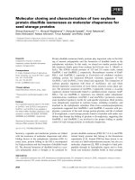
Fig. 1. Cladogram representing phylogenetic relationships between birnaviruses based on deduced amino acid sequences of VP1. The lengt
h
of each pair of branches represents the distance between the sequence pairs, and the numbers in parentheses indicate the bootstrap values.
Phylogenetic relationships
In the phylogenetic cladograms that were based on both
nucleotide and amino acid sequences, the genetic relationships
among the 22 birnaviruses were established and the
viruses, including GC1, were clustered into 5 genogroups
that generally correlated with the geographic origin of the
viruses and the water environment of the host. The MABV
genogroup consisted of strains such as GC1 and NC1 from
Korea and YT01A, H1, AY98, and Y6 from Japan. Geno-
group 1 mostly consisted of strains from the Pacific coastal
nations; DRT is from Korea, WB from the USA, Jasper
from Canada, and AM98 from Japan. The isolates of 1146,
88R, 20G1, 2290, and 6B1A from Spain and Sp from
Denmark comprised genogroup 2. The 2 avibirnaviruses
UPM976/61 from Malaysia and CLV from Vietnamform-
ed genogroup 3, and 1 entomobirnavirus, DVX, formed
genogroup 4 (Fig. 1).
Discussion
The viral B segment encodes VP1, which is approximately
90 kDa in weight [2,11-13]. The estimated molecular weight
of VP1 ranges from 95 kDa for the Jasper isolate [4] to 89
kDa for the Sp and Ab isolates of IPNV [6]. The molecular
weight of GC1 has been estimated as 94 kDa and has been
shown to be similar to that of the Jasper strain.
Some researchers have reported that the sequence
GXXXXGKS/T is a constant motif in GTP-binding
proteins [1,16] and is observed in several viral proteins that
have a tentative role in RNA replication [15]. The same
motif was present in the IPNV strains [4] and in GC1
between residues 248 and 255 (GLPYIGKT). We believe
that this motif represents a potential GTP-binding site in
the VP1 protein, and has been conserved in GC1, including
aquatic birnaviruses.
As reported previously [1,5,17], the GDD sequence is a
highly conserved motif that is present in almost all putative
RdRps. Researchers have found that the Asp-Asp (DD)
sequence lacking Gly, is conserved in IBDV, and also that
IPNV does not contain the typical GDD motif in the
corresponding region of its VP1 [4,21]. Some IPNV strains
containedthe Leu-Lys-Asp (LKD) or LKN motifs instead
of the typical GDD motif [4]. GC1 contains the LKN motif
instead of the typical GDD motif, which is present in other
aquatic birnaviruses.
The study of genetic relationships using a phylogenetic
cladogram revealed that GC1 is more closely related to
genogroup 1 than genogroup 2. This result indicatesthat
genetic relationships may be influenced by the geographical
distributions of the isolates. Aquatic birnaviruses, including
GC1 and IPNV, also belong to genogroups that are distinct
from those of the avibirnaviruses and Entomo-birnaviruses.
This result may thus indicate that the genus Birnavirus has
evolved in different ways resulting in the formation of
distinct genogroups.
90 Seong Joon Joh et al.
References
1. Argos P. A sequence motif in many polymerases. Nucleic
Acids Res 1988, 16, 9909-9916.
2. Azad AA, Barrett SA, Fahey KJ. The characterization and
molecular cloning of the double-stranded RNA genome of
an Australian strain of infectious bursal disease virus.
Virology 1985, 143, 35-44.
3. Dobos P, Roberts TE. The molecular biology of infectious
pancreatic necrosis virus: a review. Can J Microbiol 1983,
29, 377-384.
4. Duncan R, Mason CL, Nagy E, Leong JA, Dobos P.
Sequence analysis of infectious pancreatic necrosis
virusgenome segment B and its encoded VP1 protein: a
putative RNA-dependent RNA polymerase lacking the
Gly-Asp-Asp motif. Virology 1991, 181, 541-552.
5. Gorbalenya AE, Koonin EV, Donchenko AP, Blinov VM.
Coronavirus genome: prediction of putative functional
domains in the non-structural polyprotein by comparative
amino acid sequence analysiss. Nucleic Acids Res 1989, 17,
4847-4861.
6. Hedrick RP, Okamoto N, Sano T, Fryer JL. Biochemical
characterization of eel virus European. J Gen Virol 1983, 64,
1421-1426.
7. Hosono N, Suzuki S, Kusuda R. Genogrouping of
birnaviruses isolated from marine fish: a comparison of
VP2/NS junction regions on genome segment A. J Fish Dis
1996, 19, 295-302.
8. Joh SJ, Heo GJ. Genetic analysis of the VP2/NS junction
region on segment A of marine birnavirus isolated from
rockfish (Sebastes schlegeli) cultured in Korea. Bull Eur Ass
Fish Pathol 1999, 19, 190-195.
9. Joh SJ, Kim DW, Kim JH, Heo GJ. Detection of marine
birnavirus (MBV) from rockfish Sebastes schlegeli using
reverse transcription and nested PCR. J Microbiol 2000, 38,
260-264.
10. Joh SJ, Kim JH, Heo GJ. Genetic analysis of the VP2/NS
junction region on segment A of marine birnavirus isolated
from cultured flounder Paralichthys olivaceous. J Microbiol
2000, 38, 44-47.
11. K
ääriäinen L, Takkinen K, Ker
ä
nen S, S
ö
derlund H.
Replication of the genome of alphaviruses. J Cell Sci Suppl
1987, 7, 231-250.
12. Kamer G, Argos P. Primary structural comparison of
RNA-dependent polymerases from plant, animal and
bacterial viruses. Nucleic Acids Res 1984, 12, 7269-7283.
13. Koonin EV. The phylogeny of RNA-dependent RNA
polymerases of positive-strand RNA viruses. J Gen Virol
1991, 72, 2197-2206.
14. Kusuda R, Kado K, Takeuchi Y, Kawai K. Characteristics
of two virus strains isolated from young Japanese flounder
Paralichthys olivaceus. Suisanzoshoku 1989, 37, 115-120.
15. MacDonald RD, Dobos P. Identification of the proteins
encoded by each genome segment of infectious pancreatic
necrosis virus. Virology 1981, 114, 414-422.
16. M
öller W, Amons R. Phosphate-binding sequences in nu-
cleotide-binding proteins. FEBS Lett 1985, 186, 1-7.
17. Nagy E, Dobos P. Coding assignments of drosophila X virus
genome segments: in vitro translation of native and
denatured virion dsRNA. Virology 1984, 137, 58-66.
18. Oh MJ, Jung SJ, Kim HR. Biological and serological
characteristics of birnavirus isolated from cultured Japanese
flounder in 1999. J Fish Pathol 1999, 12, 56-62.
19. Roberts RJ. Fish Pathology. 3rd ed. p. 211, Saunders,
London, 2001.
20. Seo JJ, Heo GJ, Lee CH. Characterization of aquatic
birnavirus isolated from rockfish Sebastes schlegeli cultured
in Korea. Bull Eur Ass Fish Pathol 1998, 18, 87-92.
21. Shwed PS, Dobos P, Cameron LA, Vakharia VN, Duncan
R. Birnavirus VP1 proteins form a distinct subgroup of
RNA-dependent RNA polymerases lacking a GDD motif.
Virology 2002, 296, 241-250.
22. Sorimachi M, Hara T. Characteristics and pathogenicity of
a virus isolated from yellowtail fingerlings showing ascites.
Fish Pathol 1985, 19, 231-238.
23. Suzuki S, Hosono N, Kusuda R. Detection of aquatic
birnavirus gene from marine fish using a combination of
reverse transcription- and nest PCR. J Mar Biotechnol 1997,
5, 205-209.
24. Suzuki S, Kamakura M, Kusuda R. Isolation of birnavirus
from Japanese pearl oyster Pinctada fucata. Fish Sci 1998,
64, 342-343.
25. Zhang CX, Suzuki S. Comparison of the RNA polymerase
genes of marine birnavirus strains and other birnaviruses.
Arch Virol 2003, 148, 745-758.