Báo cáo khoa học: "Hepatic encephalomyelopathy in a calf with congenital portosystemic shunt (CPSS)" pptx
Bạn đang xem bản rút gọn của tài liệu. Xem và tải ngay bản đầy đủ của tài liệu tại đây (937.35 KB, 3 trang )
JOURNAL OF
Veterinary
Science
J. Vet. Sci. (2008), 9(1), 113
115
Case Report
*Corresponding author
Tel: +41 31 631 24 00; Fax: +41 31 631 26 35
E-mail:
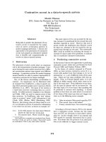
Fig. 1. Simplified drawings illustrating types of portosystemic
shunts. (A) Normal liver. (B) Intrahepatic portosytemic shunt.
(
C
)
Extrahe
p
atic
p
ortos
y
stemic shunt.
Hepatic encephalomyelopathy in a calf with congenital portosystemic
shunt (CPSS)
Valéria Café Mar
ç
al
1,
*
, Anna Oevermann
2
, Tim Bley
3
, Patrizia Pfister
4
, Julien Miclard
1
1
Institute of Animal Pathology, Vetsuisse Faculty, University of Berne, Laenggassstr. 122, CH-3001 Berne, Switzerland
2
Institute of Neuropathology NeuroCenter, Vetsuisse Faculty, University of Berne, Bremgartenstr. 109a, Postfach 8466, CH-3001
Berne, Switzerland
3
Department of Clinical Veterinary Medicine, Division of Clinical Neurology, Vetsuisse Faculty, University of Berne, Laenggassstr.
128, CH-3012 Berne, Switzerland
4
Clinic for Ruminants, Vetsuisse Faculty, University of Berne, Bremgartenstr. 109a, CH-3012 Berne, Switzerland
A 4-month-old female Holstein Friesian calf was referred to
the Veterinary Teaching Hospital, University of Berne,
Switzerland for evaluation of ataxia, weakness, apathy and
stunted growth. Clinical examination revealed generalized
ataxia, propioceptive deficits, decreased menace response
and sensibility. Postmortem examination did not reveal
macroscopic changes of major organs. Histologically, the
brain and the spinal cord lesions were characterized by
polymicrocavitation, preferentially affecting the white matter
fibers at the junction of grey and white matter and by the
presence of Alzheimer type II cells. The liver revealed
lesions consistent with a congenital portosystemic shunt,
characterized by increased numbers of arteriolar profiles
and hypoplasia to absence of portal veins. The pathological
investigations along with the animal history and clinical
signs indicated a hepatic encephalomyelopathy due to a
congenital portosystemic shunt.
Keywords: hepatic encephalopathy, Holstein Friesian cattle,
pathological investigation, portosystemic shunt
Hepatic encephalopathy (HE) refers to a spectrum of
neuropsychiatric abnormalities associated with significant
liver dysfunction and is commonly described in humans
and domestic carnivores [5,6]. In veterinary medicine,
congenital portosystemic shunts (CPSSs) (Fig. 1) are the
most common cause of HE in young dogs and cats [7,11],
while in large animals HE is often described with various
hepatotoxic conditions [14]. In large domestic animals,
CPSSs have only been sporadically reported [2,4,9,10,13].
Generally, the clinical signs associated with CPSS in large
animals are unspecific and mimic those of HE; including
progressive depression, ataxia, lethargy, apparent blindness,
slow proprioception, hindquarters weakness and abnormal
behavior. There are many postulated mechanisms to explain
the pathogenesis of HE, but there is a general consensus
that ammonia plays an important role in the dysfunction of
astroglial cells leading to brain edema [3,5,8]. This report
describes the clinical and neurological investigations, and
pathological findings in a Holstein Friesian calf with
hepatic encephalo-myelopathy due to a CPSS.
A 2-month-old female Holstein Friesian calf was admitted
to the Veterinary Teaching Hospital, University of Berne,
Switzerland for evaluation of ataxia, weakness and stunted
growth. On physical examination the animal was underweight
114 Valéria Café Marçal et al.
Fig. 2. Photomicrographs of calf cerebrum. (A) Capsule interna.
N
ote diffuse severe spongy vacuolation in the adjacent areas
between grey and white matter. H&E stain. Scale bar = 1 mm. (B)
The capsule interna showing severe white matter changes. gm;
g
re
y
matter. wm
;
white matter. H&E stain. Scale
b
ar = 100
µ
m.
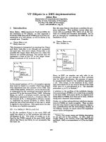
Fig. 3. Photomicrographs of calf spinal cord. (A) Bilateral
symmetric diffuse spongy vacuolation of the grey matter. White
matter funiculi not affected. H&E stain. Scale bar = 500 µm. (B)
The grey matter intermediate horn showing spongy changes
adjacent to neuron. H&E stain. Scale bar = 50 µm.
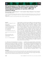
Fig. 4. Photomicrographs of calf liver. (A) Note a hepatic lobule
surrounded by numerous portal spaces with absence of portal
veins. The distance between portal spaces and central vein is
abnormally reduced. H&E stain. Scale bar = 50 µm. (B) The
p
ortal area showing proliferation of arterioles and ductules with
absence of
p
ortal vein. H&E stain. Scale
b
ar = 50
µ
m.
with generalized muscle atrophy. Neurological examina-
tion showed generalized ataxia, propioceptive deficits,
decreased menace response and superficial sensibility.
Temperature, heart and breathing rates were at normal
range. Cerebrospinal fluid analysis, blood and serum bio-
chemical analyses including liver enzymes were also within
normal limits. Electromyographic (EMG) examination
revealed isolated spontaneous activity such as fibrillation
potentials in the proximal musculature of front and hind
limbs, which indicated a nonspecific denervating neuro-
pathic or myopathic disease. The calf was treated during
hospitalization with penicillin (40,000 IU/kg iv), dexa-
methasone (0.03 mg/kg iv; Boehringer Ingelheim, Germany)
and thiamine (4.5 mg/kg; Vétoquinol, France) for 7 days.
The animal recovered and was therefore taken home. Two
months later it was returned to the Veterinary Hospital with
a rapidly progressing debilitation. The animal was in left
lateral recumbency, and dyspneic. Although the rectal
temperature was normal (38.2
o
C), the heart and respiratory
rates were relatively high at 128/beats per min and
80/breaths per min, respectively (normal ranges are 80 to
100 beats per min and 24 to 36 breaths per min) [12]. The
general condition of the calf deteriorated rapidly with
progression to stupor and coma. After poor prognosis was
established, the calf was humanely euthanized.
Subsequently, a complete necropsy was performed, the
analysis of which identified a poor staturoponderal de-
velopment considering the age and the breed of the animal
(measured weight 82 kg, normal weight 136 kg). The
general body condition was reduced with generalized muscle
atrophy. No other macroscopical lesion was observed.
Representative tissue samples were fixed in 10% neutral
buffered formalin, processed by routine methods in
ascending grades of alcohol and embedded in paraffin wax.
Sections were cut (4 µm) and stained with hematoxylin and
eosin. Selected brain and spinal cord tissue samples were
additionally stained with Luxol fast blue. Immunohisto
chemistry was performed on brain and spinal cord sections
with a streptavidin-biotin method using an anti-bovine glial
fibrillary acid protein (GFAP) antibody (1:1,000 DAKO,
Denmark). Paraffin wax-embedded brain and spinal cord
tissues of one normal age-matched calf was used as control.
Histological examination of the CNS revealed an intense
and widespread bilateral-symmetric spongy degeneration,
primarily affecting the white matter with involvement of
transitional areas between grey and white matter (Fig. 2).
The lesion was diffusely distributed throughout the cerebrum,
midbrain, cerebellum, brainstem and spinal cord. Spinal
cord lesions were seen along the whole spinal cord
involving primarily the myelinated fibers in the grey matter
(Fig. 3). In the cerebral cortex and in the adjacent white
matter, Alzheimer type II cells were observed as isolated
clusters. Luxol fast blue staining of spinal cord tissue did not
reveal any changes in myelin compound. Immunohistoche-
mically, GFAP staining did not show any obvious differences
in staining intensity in comparison with a normal control
calf.
Microscopically, the hepatic portal triads presented
numerous prominent proliferating small arterioles,
hypoplastic to absent portal veins, proliferation of bile ducts
and often ectasia of lymphatic vessels (Fig. 4). In
cross-sections, skeletal muscles revealed diffusely atrophic
myofibers (up to 30 µm in diameter) with occasionally
single group of angular and shrunken atrophic myofibers.
Peripheral nerves were histologically unremarkable.
The pathomorphological lesions described herein are
Hepatic encephalomyelopathy in a calf with CPSS 115
compatible with a hepatic encephalomyelopathy due to a
CPSS with secondary generalized muscle atrophy. HE is
described in association with hepatic failure and consists of
bilateral symmetric spongy degeneration of white matter
and the presence of single or small groups of Alzheimer
type II cells [11]. Hepatic myelopathy is rarely described in
veterinary medicine. A similar myelopathy as decribed in
this case has only been described in experimentally induced
hyperammonemic calves [1]. In human medicine, hepatic
myelopathy is defined as a neurological complication of
hepatic cirrhosis [5] with portal hypertension, usually
characterized by symmetrical demyelination of the cortico-
spinal tracts [5]. In this case, the most pronounced spinal
cord changes were restricted to the nerve fibers in the
spinal cord grey matter without obvious demyelination
which differs significantly to that found in human patients.
Hepatic atrophy and intra- or extra-hepatic vascular
anomalies are the main gross findings reported in large
animal species with CPSS [4,13]. Ascites, due to persistent
portal hypertension, is commonly associated with acquired
portosystemic shunts, and is absent in congenital
portosystemic shunts [14]. The histological findings of the
liver together with the absence of ascites, multiple tortuous
shunted vessels or hepatic changes were compatible with a
CPSS, similarly to previous reports [9].
Due to the absence of clinicopathological evidence of
hepatic dysfunction in this calf and to the unspecific
clinical signs, a portosystemic shunt with secondary hepatic
encephalopathy was not included in the differential diagnosis.
However, total serum bile acid concentration, total blood
ammonia values and sulfobromophthalein retention time
tests, normally increased in CPSS, were not measured. The
clinical signs observed in this reported calf (ataxia, depression,
stupor and coma) could be explained by the hepatic
encephalomyelopathy. However, clinical signs were far
unspecific which led to a erroneous diagnosis. The muscle
atrophy in this calf was most likely related to the CPSS, as
severe liver disease are known to induce protein metabolic
perturbations and secondary muscle atrophy [14]. Although
EMG results suggested a peripheral nervous system
involvement, peripheral nerves did not reveal any histopa-
thological changes. However, the presence of single group
of atrophic myofibers in histology suggested a mild
neurogenic atrophy, probably correlated to the severe
spongy changes in adjacent areas of the motor neurons in
the grey matter. The possible explanation for the initial
recovery after antibiotic administration may be that urease-
producing intestinal bacteria were temporarily reduced in
the intestinal tract, hence long-term medical management
using oral antibiotics has been described in small animals
to inhibit both the production and absorption of potential
gut derived CNS toxins [7].
Congenital portosystemic shunt with secondary hepatic
encephalomyelopathy has rarely been described in calves.
However, hepatic encephalomyelopathy due to CPSS might
have heterogeneous manifestations. Because of the non-
specific associated clinical signs, it is likely that CPSS is
often under-diagnosed. Hence, it should be considered in
the differential diagnostic list in young animals exhibiting
stunted growth and history of intermittent neurological
deficits.
Acknowledgments
The authors thank Dr. A. Zakher for reviewing the
manuscript and Mr. Joerg Jenni for technical support.
References
1. Cho DY, Leipold HW. Experimental spongy degeneration
in calves. Acta Neuropathol 1977, 39, 115-127.
2. Fortier LA, Fubini SL, Flanders JA, Divers TJ. The diag-
nosis and surgical correction of congenital portosystemic
vascular anomalies in two calves and two foals. Vet Surg
1996, 25, 154-160.
3. Hooper PT. Spongy degeneration in the central nervous sys-
tem of domestic animals. Part I: Morphology. Acta Neuropathol
1975, 31, 325-334.
4. Keane D, Blackwell T. Hepatic encephalopathy associated
with patent ductus venosus in a calf. J Am Vet Med Assoc
1983, 182, 1393-1394.
5. Lewis M, Howdle PD. The neurology of liver failure. QJM
2003, 96, 623-633.
6. Maddison JE. Canine congenital portosystemic encephalo-
pathy. Aust Vet J 1988, 65, 245-249.
7. Martin RA. Congenital portosystemic shunts in the dog and
cat. Vet Clin North Am Small Anim Pract 1993, 23, 609-623.
8. Norenberg MD. Astroglial dysfunction in hepatic encepha-
lopathy. Metab Brain Dis 1998, 13, 319-335.
9. Olchowy TW, Daniel GB, Tucker RL, Petersen MG.
Hepatic vascular anomaly in a calf. Can Vet J 1992, 33, 131-133.
10. Reimer JM, Donawick WJ, Reef VB, Wagner HR, Divers
TJ. Diagnosis and surgical correction of patent ductus veno-
sus in a calf. J Am Vet Med Assoc 1988, 193, 1539-1541.
11. Summers BA, Cummings JF, de Lahunta A. Degenera-
tive diseases of the central nervous system. In: Summers BA,
Cummings JF, de Lahunta A (eds.). Veterinary Neuropatho-
logy. pp. 189-350, Mosby, St. Louis, 1994.
12. Terra RL. Ruminant history, physical examination and med-
ical records. In: Smith BP (ed.). Large Animal Internal
Medicine: Disease of Horses, Cattle, Sheep and Goats. 2nd
ed. pp. 10-11, Mosby, St. Louis, 1996.
13. Van den Ingh TS, van der Linde-Sipman JS, Berrocal A,
Vos JH. Congenital portosystemic shunts in three pigs and
one calf. Vet Pathol 1990, 27, 56-58.
14. Zachary JF. Nervous system. In: McGavin MD, Zachary JF
(eds.). Pathologic Basis of Veterinary Disease. 4th ed. pp.
883-971, Mosby-Elsevier, St. Louis, 2007.