Báo cáo khoa học: "Pyridoxine induced neuropathy by subcutaneous administration in dogs" ppt
Bạn đang xem bản rút gọn của tài liệu. Xem và tải ngay bản đầy đủ của tài liệu tại đây (3.49 MB, 5 trang )
JOURNAL OF
Veterinary
Science
J. Vet. Sci. (2008), 9(2), 127
131
*Corresponding author
Tel: +82-2-880-1266; Fax: +82-2-880-1266
E-mail:
Pyridoxine induced neuropathy by subcutaneous administration in dogs
Jin-Young Chung
1
, Jung-Hoon Choi
2
, Cheol-Yong Hwang
1
, Hwa-Young Youn
1,
*
1
Department of Veterinary Internal Medicine, and
2
Department of Anatomy and Cell Biology, College of Veterinary Medicine,
Seoul National University, Seoul 151-742, Korea
To construct a sensory neuropathy model, excess pyrido-
xine (150 mg/kg s.i.d.) was injected subcutaneously in dogs
over a period of 7 days. During the administrations peri-
od, the dogs experienced body weight reduction and pro-
prioceptive loss involving the hindquarters. After pyridox-
ine administration was completed, electrophysiological re-
cordings showed that the M wave remained at a normal
state, but the H-reflex of the treated dogs disappeared at 7
days. The dorsal funiculus of L
4
was disrupted irregularly
in the axons and myelin with vacuolation. The dorsal root
ganglia of L
4
, and sciatic and tibial nerves showed degen-
erative changes and vacuolation. However, the lateral and
ventral funiculi of L
4
showed a normal histopathologic
pattern. Although this subcutaneous administration meth-
od did not cause systemic toxicity and effectively induced
sensory neuropathy, this study confirmed the possibility of
producing a pyridoxine-induced sensory neuropathy mod-
el in dogs with short-term administration.
Keywords: dog, H-reflex, pyridoxine, sensory neuropathy
Introduction
Pyridoxine, a form of vitamin B
6
, is an essential dietary
constituent [5]. Vitamin B
6
occurs in three natural forms:
pyridoxol (pyridoxine), pyridoxal, and pyridoxamine.
Pyridoxine is most commonly used as a dietary supplement
and therapeutic agent. High dose pyridoxine is used in con-
ditions such as premenstrual and carpal tunnel syndromes,
and has been prescribed as a treatment for ingestion of the
false morel mushroom, Gyromitra esculenta [4]. The ra-
tionale behind the latter treatment is that the active toxin
monomethylhydrazine competitively inhibits a pyridox-
ine-dependent step in the synthesis of the neurotransmitter
gamma-aminobutyric acid [10]. While pyridoxine defi-
ciency causes distal, predominantly sensory neuropathy,
pyridoxine has also been identified as a neurotoxicant.
During the 1980s, the medical community was alerted to a
neurologic disease occurring in individuals consuming
large quantities of vitamin B
6
for prolonged periods of time
[10]. Investigators described severe sensory neuropathy of
insidious onset and course associated with chronic abuse of
oral pyridoxine supplements. The recommended daily oral
dose is 2-4 mg in human adults. However, with daily oral
doses of up to 6 g for 12-40 months, one may contract pro-
gressive sensory neuropathy manifested by sensory ataxia,
diminished distal limb proprioception, paresthesia, and hy-
peresthesia [3,11].
There are many animal model studies based on these data.
In rat studies, three intraperitoneal dosing regimens are
generally employed: short term/high dose, 1,200 mg/kg/
day for 1-15 days; intermediate dose, 600 mg/kg/day for
1-15 days; and long term/low dose, 100-300 mg/kg/day for
up to 12 weeks [13]. Such experimental studies have con-
firmed the morphologic pattern of peripheral nervous sys-
tem lesions in pyridoxine neurotoxicity, reflecting primary
injury to the cytons of neurons residing in peripheral sen-
sory (dorsal root, trigeminal) ganglia, with secondary de-
generation of axons of affected cells [10].
There have been some experiments wherein sensory neu-
ropathy was induced in dogs with pyridoxine overdose.
However, these previous experiments used extreme doses
of pyridoxine or were performed over a long period of time
[5,8,9].
The incidence of neurodegenerative disorders has been
increasing in the canine population, and this development
requires effective countermeasures. However, canine
models of specific neurodegenerative disorders are pre-
requisite to developing new treatments. The purpose of this
study was to develop a dog model of sensory neuropathy
by administering subcutaneous (SC) injections of pyridox-
ine over a short period of time. This differs from other ex-
periments, in which extreme doses of pyridoxine were de-
livered by oral administration or were delivered over a long
period of time.
128 Jin-Young Chung et al.
Materials and Methods
Animals
Ten dogs (five beagles, three Shih tzus, one Pekingese,
and one mongrel) were used for determining normal elec-
trophysiological values. There were five male and five fe-
male dogs, all of which were around two years of age. Body
weight ranged from 4 to 6 kg.
Eight dogs (four Shih-tzus, two mongrels, one Pekingese,
and one Yorkshire terrier) were used for the pyridoxine-in-
duced neuropathy study. There were four male and four fe-
male dogs, all of which were around two years of age. Body
weight ranged from 4 to 6 kg. Two dogs were in the control
group, and six dogs were in the experimental group.
All dogs were clinically judged to be in good health and to
be neurologically normal. All experimental dogs had their
own admission number issued by the Institute of Laborato-
ry Animal Resources, Seoul National University (Korea).
During the experiment, all dogs were cared for according
to Animal Care and Use Guidelines from the Institute of
Laboratory Animal Resources, Seoul National University.
Body weight and condition were measured every morning
in the test dogs.
Pharmacological treatment
Pyridoxine (Sigma, France) was diluted in a 0.9% sterile
aqueous solution of sodium chloride and administered to
each dog SC, once a day, in the morning, for 7 days. The
pyridoxine solution was prepared immediately before each
injection. Animals in the control group received a vehicle
(iso-osmotic sterile aqueous solution of sodium chloride),
while animals in the experimental group were admini-
stered 150 mg/kg pyridoxine SC, in a concentration of 100
mg/ml. SC injection was chosen as the route of pyridoxine
administration because of convenience. The SC dosage
(150 mg/kg, s.i.d. for 7 days) was decided based on the or-
ally administered dosage of pyridoxine previously shown
to induce peripheral neuropathy [5].
Postural reaction assessments
Postural reaction (wheelbarrowing, hopping, extensor
postural thrust, placing, tonic neck reaction, and proprio-
ceptive positioning) assessments were done on all dogs ev-
ery morning during the test period.
Electrophysiological recordings
All dogs were pre-anesthetized with atropine (0.1mg/kg
of body weight, IM). Anesthesia was induced with dia-
zepam and was maintained with isoflurane through a
semi-closed system. Subcutaneous temperature was main-
tained at 37-38
o
C. Neuropack2 (Nihon Koden, Japan) was
used for all recordings. All measurements were performed
in the left hindlimb of each dog.
Direct-evoked muscle potentials (DEMP, M wave) were
recorded for the tibial nerve using a 1 Hz, 0.5 ms, supra-
maximal stimulus. Stimulating electrodes were positioned
in the distal tibial nerve. A recording electrode was posi-
tioned in the plantar interosseous muscle. The ground elec-
trode was positioned between the stimulating electrode
and recording electrode. The recording electrode was a bi-
polar needle electrode. Reflex-evoked muscle potentials
(REMP, H-reflex) were recorded using a 1 Hz, 0.5 ms, sub-
maximal stimulus. The stimulating electrode was posi-
tioned in the tibial nerve adjacent to the hook, and the re-
cording electrode and ground electrode were positioned in
the same tibial nerve site where the M wave was measured.
All measurements were performed at least eight times.
First, electrophysiological recordings were performed for
determining normal values. Electrophysiological record-
ings were then performed twice more for the experimental
group: once before the experiment and once after the test
period.
Histopathological analysis
After the experimental period (10 days from the start of
the experiment), the dogs were anesthetized with high dose
tiletamine/zolazepam (Virbac, France) and given propofol
(Choongwae, Korea) to induce euthanasia. They were then
perfused transcardially with 0.1 M phosphate-buffered sal-
ine (PBS), followed by 4% paraformaldehyde in 0.1 M
PBS. After perfusion, the lumbar spinal cord (L
4
), left and
right dorsal root ganglia of L
4
, sciatic nerve, and tibial
nerve were quickly removed, post-fixed for 4-6 h in the
same fixative at 4
o
C, and embedded in paraffin. The tissues
were sectioned serially to 5 μm thickness with a micro-
tome (Reichert-Jung, Germany), floated onto gelatin-
coated slides, deparaffinized in xylene, rehydrated in a de-
scending ethanol series, and stained with hematoxylin and
eosin. The sections were examined under an Olympus
BX51 microscope (Olympus, Japan) attached to an
IMT2000 digital camera (iMTechnology, Korea).
Statistical analysis
Paired t-test was done for analysis of body weight and the
M wave and H-reflex amplitudes before and after pyridox-
ine treatment. Statistical significance was set at p < 0.05.
Results
Weight measurement showed that there was weight loss
in the treated group. The weight of control group animals
was maintained at 5.35 ± 0.21 kg for experimental period.
The weight of treated group animals decreased from 4.92 ±
0.40 kg to 4.4 ± 0.43 kg. The difference in body weight be-
fore and after pyridoxine treatment was statistically sig-
nificant (p < 0.05).
All dogs in the treated group developed neurologic abnor-
malities, characterized especially by ataxia involving first,
Pyridoxine-induced neuropathy in dogs 129
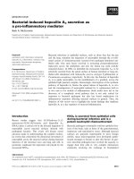
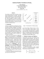
Fig. 1. On electrophysiological analysis, a control dog showed a
n
H-reflex (A), but a pyridoxine model dog showed no H-reflex
(B). Both have normal M waves. The M wave amplitude showe
d
no remarkable change after pyridoxine treatment, a finding
which was confirmed in our statistical analysis (p < 0.05). There
was a remarkable change in the H-reflex after treatment in the
experimental group.
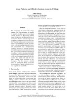
Fig. 2. (A) Normal dorsal funiculus in L
4
. (B) Dorsal funiculus i
n
L
4
showing disruption and irregularity of axons and myelin with
vacuolation. (C) Normal dorsal root ganglia in L
4
. (D) Dorsal
root ganglia in L
4
showing chromatolysis with eccentric locatio
n
of nucleus (arrow). H&E stain, ×200.
Fig. 3. (A) Normal sciatic nerve. (B) Sciatic nerve showing
axonal swelling with vacuolation (arrows). (C) Normal tibial
nerve. (D) Tibial nerve showing axonal swelling with vacuola-
tion (arrows). H&E stain, ×400.
and most prominently, the hindquarters. Five dogs in the
treated group showed proprioceptive abnormalities in-
volving the hindquarters on postural reaction test (wheel-
barrowing, hopping, extensor postural thrust, placing, ton-
ic neck reaction, and proprioceptive positioning), starting
on the third day of pyridoxine injection. On the fourth day
of pyridoxine injection, five affected dogs held their hin-
dlimbs stiffly when standing. These signs remained until
the end of the pyridoxine administration period. One dog in
the treated group showed proprioceptive abnormalities in-
volving the hindquarters on postural reaction test, starting
on the third day of pyridoxine injection. This dog was se-
verely affected. Hindquarter movements were stiff, spas-
tic, and dysmetric, and the dog adopted a broad-based
stance when standing. The lack of coordination in this dog
became so severe that it occasionally fell, especially when
attempting to walk or turn. Except for weight loss and the
neurological problems, there were no changes in body con-
ditions among the test animals.
The results of evaluation of the DEMP (M wave) and
REMP (H-reflex) in clinically normal dogs are as follows.
The mean M wave amplitude (5.4 ± 2.3 mV) was much
higher than the mean H-reflex amplitude (0.5 ± 0.3 mV).
The mean M wave latency (2.8 ± 0.3 ms) showed that the
M wave is an early response, whereas the mean H-reflex la-
tency (16.2 ± 2.5 ms) showed that the H-reflex is a late
response.
Six dogs in the treated group and two dogs in the control
group underwent electrophysiological recordings, through
which the M wave and H-reflex were measured. In the ex-
perimental group, the amplitude of the M wave was 4.89 ±
0.62 mV before treatment. After treatment, the amplitude
of the M wave was 5.04 ± 0.51 mV. M wave amplitude
showed no remarkable change after pyridoxine treatment,
a finding confirmed in our statistical analysis (p < 0.05).
However, there was a remarkable change in the H-reflex
after treatment in the experimental group (Fig. 1). Before
pyridoxine treatment, the amplitude of the H-reflex was
0.54 ± 0.06 mV. After pyridoxine treatment, there was no
130 Jin-Young Chung et al.
consistently detectable H-reflex.
Lesions were observed in the dorsal funiculus of the lum-
bar spinal cord in experimental animals (L
4
). The number
of axons was reduced, and the myelin was irregular and
fragmented, with accompanying vacuolation (Fig. 2B).
However, the lateral funiculi, ventral funiculus, and gray
matter of L4 were histopathologically normal. Vacuolated
myelin was observed in the dorsal root ganglia of L4 (Fig.
2D). Axonal swelling and vacuolation were seen in the sci-
atic and tibial nerves (Figs. 3B and D).
Discussion
Peripheral neuropathies are characterized by motor, sen-
sory, and sympathetic deficits. Sensory neuropathies are
frequently associated with diabetes or anticancer therapies
[2]. Vitamin deficiency, hypothyroidism, uremia, and cha-
racteristically inherited metabolic disorders are also re-
sponsible for sensory neuropathies [1]. Therefore, it is very
important to develop animal models of sensory neuro-
pathies, in order to conduct preclinical studies of putative
neuroprotective and neuroregenerative compounds.
Problematic in the development of experimentally in-
duced sensory neuropathy models are the side effects of the
inducing treatment, such as nephrotoxicity for cisplatin,
cardiotoxicity for taxol, and simultaneous injury of motor
and sensory fibers for acrylamide [7]. Toxicity studies us-
ing those drugs have often successfully induced sensory
neuropathy [1]. However, the animal model of pyridox-
ine-induced peripheral neuropathy established in this
study was found to be selective for sensory fibers and to be
safe for other organs [5,6].
In previous experiments, reduction in body weight was
found to be proportionate to reduction in food consumption
[5]. In this experiment, we did not measure the amount of
food consumption. Therefore, we found no direct correla-
tion in the reduction of food consumption and reduction in
body weight.
Neurologic examination is an earlier indicator of neuro-
toxicity than electrodiagnostic procedures. Therefore, neu-
rologic examinations were performed every day in this
study, and EMG recordings were performed before and af-
ter treatments.
We chose to use a bipolar needle electrode for EMG re-
cordings, in contrast with electrodes used in other experi-
ments. The bipolar needle electrode is best for detecting
correct waves without interfering error waves. However, it
is difficult to use in humans because of the pain it causes.
We were able to use the bipolar needle electrode in animals
because they were anesthetized before the procedure.
The maximal M wave amplitudes were obtained with su-
pramaximal stimulus, and the maximal H-reflex ampli-
tudes were obtained with submaximal stimulus. However,
the H-reflex was not completely cancelled out by stimuli
supramaximal to the direct response. In humans, the H-re-
flex is completely cancelled out when stimulation becomes
supramaximal. This may be due to the fact that the high
spindle density in the interosseous muscles in quadrupeds
induces a stronger reflex [12].
In previous rat studies, the H-reflex was measured to
demonstrate pyridoxine-induced sensory neuropathy. In
dogs, there have been no studies of the H-reflex in pyridox-
ine-induced sensory neuropathy. However, our study did
utilize measurements of the H-reflex.
The EMG recording data obtained during peripheral
nerve stimulation in the experimental group were con-
sistent with selective toxicity to sensory nerves.
An earlier study of pyridoxine toxicity in dogs and rats
demonstrated lesions with a distribution similar to that
seen in the experimental dogs in this study [6,8].
In conclusion, we confirmed that dog models of pyridox-
ine-induced sensory neuropathy can indeed be con-
structed. Our method offers a short-term model of sensory
neuropathy as an alternative to the already existing long-
term model. This was the first trial in which H-reflexes
were measured in dogs with pyridoxine-induced neuro-
pathies. Our method is advantageous in that it did not cause
systemic toxicity. Further study is needed to confirm pyr-
idoxine-induced toxicity using electron microscopic ob-
servation.
Acknowledgments
This work was supported by the Brain Korea 21 program,
Korean Research Foundation Grant (KRF-2006-J02902),
and the Research Institute of Veterinary Science, College
of Veterinary Medicine, Seoul National University.
References
1. Callizot N, Warter JM, Poindron P. Pyridoxine-induced
neuropathy in rats: a sensory neuropathy that responds to
4-methylcatechol. Neurobiol Dis 2001, 8, 626-635.
2. Chaudhry V, Rowinsky EK, Sartorius SE, Donehower
RC, Cornblath DR. Peripheral neuropathy from taxol and
cisplatin combination chemotherapy: clinical and electro-
physiological studies. Ann Neurol 1994, 35, 304-311.
3. Dalton K, Dalton MJ. Characteristics of pyridoxine over-
dose neuropathy syndrome. Acta Neurol Scand 1987, 76,
8-11.
4. Hanrahan JP, Gordon MA. Mushroom poisoning. Case re-
ports and a review of therapy. JAMA 1984, 251, 1057-1061.
5. Hoover DM, Carlton WW. The subacute neurotoxicity of
excess pyridoxine HCl and clioquinol (5-chloro-7-iodo-8-
hydroxyquinoline) in beagle dogs. I. Clinical disease. Vet
Pathol 1981, 18, 745-756.
6. Hoover DM, Carlton WW. The subacute neurotoxicity of
excess pyridoxine HCl and clioquinol (5-chloro-7-iodo-8-
hydroxyquinoline) in beagle dogs. II. Pathology. Vet Pathol
Pyridoxine-induced neuropathy in dogs 131
1981, 18, 757-768.
7. Hopkins AP, Gilliatt RW. Motor and sensory nerve con-
duction velocity in the baboon: normal values and changes
during acrylamide neuropathy. J Neurol Neurosurg Psychia-
try 1971, 34, 415-426.
8. Krinke GJ, Fitzgerald RE. The pattern of pyridoxine-in-
duced lesion: difference between the high and the low toxic
level. Toxicology 1988, 49, 171-178.
9. Krinke G, Naylor DC, Skorpil V. Pyridoxine megavitami-
nosis: an analysis of the early changes induced with massive
doses of vitamin B6 in rat primary sensory neurons. J Neuro-
pathol Exp Neurol 1985, 44, 117-129.
10. Perry TA, Weerasuriya A, Mouton PR, Holloway HW,
Greig NH. Pyridoxine-induced toxicity in rats: a stereo-
logical quantification of the sensory neuropathy. Exp Neurol
2004, 190, 133-144.
11. Schaumburg H, Kaplan J, Windebank A, Vick N,
Rasmus S, Pleasure D, Brown MJ. Sensory neuropathy
from pyridoxine abuse. A new megavitamin syndrome. N
Engl J Med 1983, 309, 445-448.
12. Sims MH, Selcer RR. Occurrence and evaluation of a re-
flex-evoked muscle potential (H- reflex) in the normal dog.
Am J Vet Res 1981, 42, 975-983.
13. Xu Y, Sladky JT, Brown MJ. Dose-dependent expression
of neuronopathy after experimental pyridoxine intoxication.
Neurology 1989, 39, 1077-1083.