Báo cáo khoa học: "The C-terminal variable domain of LigB from Leptospira mediates binding to fibronectin" pptx
Bạn đang xem bản rút gọn của tài liệu. Xem và tải ngay bản đầy đủ của tài liệu tại đây (1.45 MB, 12 trang )
JOURNAL OF
Veterinary
Science
J. Vet. Sci. (2008), 9(2), 133
144
*Corresponding author
Tel: +1-607-253-3675; Fax: +1-607-253-3943
E-mail:
The C-terminal variable domain of LigB from Leptospira mediates
binding to fibronectin
Yi-Pin Lin, Yung-Fu Chang
*
Department of Population Medicine and Diagnostic Sciences, College of Veterinary Medicine, Cornell University, Ithaca, NY
14853, USA
Adhesion through microbial surface components that
recognize adhesive matrix molecules is an essential step in
infection for most pathogenic bacteria. In this study, we
report that LigB interacts with fibronectin (Fn) through
its variable region. A possible role for LigB in bacterial at-
tachment to host cells during the course of infection is sup-
ported by the following observations: (i) binding of the
variable region of LigB to Madin-Darby canine kidney
(MDCK) cells in a dose-dependent manner reduces the ad-
hesion of Leptospira, (ii) inhibition of leptospiral attach-
ment to Fn by the variable region of LigB, and (iii) de-
crease in binding of the variable region of LigB to the
MDCK cells in the presence of Fn. Furthermore, we found
a significant reduction in binding of the variable region of
LigB to Fn using small interfering RNA (siRNA). Finally,
the isothermal titration calorimetric results confirmed the
interaction between the variable region of LigB and Fn.
This is the first report to demonstrate that LigB binds to
MDCK cells. In addition, the reduction of Fn expression in
the MDCK cells, by siRNA, reduced the binding of LigB.
Taken together, the data from the present study showed
that LigB is a Fn-binding protein of pathogenic Leptospira
spp. and may play a pivotal role in Leptospira-host inter-
action during the initial stage of infection.
Keywords: adhesion, Fn, Leptospira, LigB, MDCK cell, siRNA
Introduction
Leptospirosis is a zoonotic disease caused by pathogenic
spirochetes in the genus Leptospira [22]. The disease oc-
curs widely in developing countries and is reemerging in
the United States [29]. The clinical features are variable
and include subclinical infection, a self-limited anicteric
febrile illness and severe, potentially fatal disease [22]. In
the severe form of leptospirosis (Weil's syndrome), the
symptoms include an acute febrile illness associated with
multi-organ damage with liver failure (jaundice), renal
failure (nephritis), pulmonary hemorrhage, and meningitis
[10]. If not treated, the mortality rate may exceed 15% [49].
Furthermore, Leptospira infection can trigger autoimmune
diseases in horses as well as humans [36,41]. Several fac-
tors associated with virulence have been proposed for
Leptospira spp., including the sphingomyelinases, serine
proteases, zinc-dependent proteases, collagenase [3],
LipL32 [59], the novel factor H-binding protein LfhA [54],
and lipopolysaccarides [56].
Pathogenic spirochetes have evolved a variety of strat-
egies to infect host cells such as evasion of the innate as
well as adaptive immunity [54]. Attachment to host cells is
an essential step for colonization by bacterial pathogens.
Leptospira has been shown to bind to mammalian cells,
such as Madin-Darby canine kidney (MDCK) cells [2] via
the extracellular matrix (ECM) [15]. Several adhesion
molecules in the pathogenic spirochetes have been identi-
fied including a Fn binding protein (36 kDa protein) [30],
a laminin binding protein (Lsa24) [1], and Lig proteins
[25,33,34] from Leptospira spp., decorin-binding proteins
(Dbp A and B) [37] and Fn-binding proteins (BBK 32 and
47 kDa) [21,38] from Borrelia spp. and MSP, Tp0155,
Tp0483, Tp0751 from Treponema spp. [4,5,9]. Lig pro-
teins (Lig A, B and C) possess immunoglobulin-like do-
mains with 90 amino acid repeats that have been identified
in other adhesion molecules, such as the intimin of
Escherichia coli and the invasin of Yersinia pseudotu-
berculosis [14,17]. Interestingly, the N-terminal 630 ami-
no acid sequences of LigA and B are identical, but the
C-terminal amino acid sequences are variable with only
34% identitify [33]. ligB also encodes a C-terminal,
non-repeat domain of 771 amino acid residues [33]. On the
other hand, the ligA-ligB intergenic regions from L. kirsch-
neri and L. interrogans are 943 bp and 1347 bp in length re-
spectively, and ligC is not linked to the ligA-ligB locus
[25]. The expression of LigA and LigB is controlled by a
134 Yi-Pin Lin et al
key environmental signal, osmolarity, to enhance the bind-
ing of Leptospira to host cells [26,27].
It has been shown that the lig genes are present ex-
clusively in pathogenic Leptospira spp [25,33]. LigA and
LigB are weakly expressed in low passage, but not in high
passage cultures of this organism [25,33]. Importantly, we
have shown that LigA and LigB expression is upregulated
in vivo in the kidneys of Leptospira-infected hamsters [34].
Recently, LigA and LigB have been reported to bind to ex-
tracellular matrix proteins including collagens type I and
IV, laminin, fibronectin, and fibrinogen [6,24]. These data
indicate that Lig proteins may play an important role in at-
tachment of pathogenic leptospires to host cells.
Although there are three copies of lig genes (ligA, B and
C) in L. interrogans serovar Pomona and L. interrogans se-
rovar Copenhageni [31,33,34], only ligB is present in most
pathogenic Leptospira spp. ligA is absent in L. interrogans
serovar Lai [42], ligC is truncated (a pseudogene) in L.
kirschneri serovar Grippotyphosa [25] and both ligA and
ligC are absent in L. borgpetersenii serovar Harjo [3].
Therefore, we focused on LigB in this study and report that
the variable region of LigB binds with high affinity to Fn,
suggesting that this fragment is crucial for bacterial adhe-
sion to host cells.
Materials and Methods
Bacterial strains and cell culture
L. interrogans serovar Pomona (NVSL1427-35-093002)
was used in this study [35]. All experiments were per-
formed with virulent, low-passage strains obtained by in-
fecting golden syrian hamsters as previously described
[35]. Leptospires were grown in EMJH medium at 30
o
C for
less than 5 passages and growth was monitored by dark-
field microscopy. The MDCK cells (ATCC CCL34) were
cultured in Dulbecco minimum essential medium contain-
ing 10% fetal bovine serum (GIBCO, USA) and were
grown at 37
o
C in a humidified atmosphere with 5% CO
2
.
Reagents and antibodies
Horseradish peroxidase (HRP)-conjugated goat anti-
hamster antibody, HRP-conjugated goat anti-mouse anti-
body and HRP-conjugated goat anti-rabbit antibody were
purchased from Zymed (USA). Rabbit anti-glutathione
S-transferase (GST) antibody, Alexa 594-conjugated goat
anti-hamster antibody, Alexa 488-conjugated goat an-
ti-hamster antibody, and FITC-conjugated goat anti-mouse
antibody were purchased from Molecular Probe (USA).
Anti-Fn (MAB1932) and anti-actin mouse antibodies
(MAB1501) were purchased from Chemicon International
(USA). Human plasma Fn was purchased from GIBCO
(USA). Anti-L. interrogans antibodies were prepared in
hamsters as previously described [35].
Plasmid construction and protein purification
Constructs for the expression of GST, GST fused with the
conserved region of LigB (LigBCon; amino acids 1-630)
and GST fused with the central variable region of LigB
(LigBCen; amino acids 631-1417) were previously gen-
erated using the vector pGEX-4T-2 (Amersham Pharmacia
Biotech, USA) [33]. GST fused with the C-terminal varia-
ble region of LigB (LigBCtv; amino acids 1418-1889) was
generated using the vector pET41A (Novogen, USA).
Relevant fragments of DNA were amplified by PCR using
primers based on the ligB sequence [33]. Primers were de-
signed to introduce a SalI site at the 5' end of each fragment
and a stop codon followed by a NotI site at the 3' end of
each fragment. The PCR products were digested sequen-
tially with SalI and NotI and then ligated into pGEX-4T-2
or pET41A cut with SalI and NotI. We purified the soluble
form of GST-LigBCon, GST-LigBCen and GST-LigBCtv
from E. coli as previously described [34,35].
Binding assays by ELISA
To measure the binding of Leptospira to the ECM compo-
nents, 1 mg of each ECM component (as indicated in Fig.
1A) in 100 μl PBS (pH 7.2) was coated onto microtiter
plate wells. For the dose-dependent binding experiments,
different concentrations of Fn (as indicated in Fig. 1B)
were coated onto the microtiter plate wells. The plates were
incubated at 4
o
C for 16 h and subsequently blocked with
blocking buffer (50 μl/well) containing 3.5% BSA in 50
mM Tris (pH 7.5)-100 mM NaCl-1 mM MgCl
2
, MnCl
2
,
and CaCl
2
at room temperature (RT) for 2 h. Then, the
Leptospira (10
7
) were added to each well and further in-
cubated at 37
o
C for 6 h. To determine the inhibition of
Leptospira binding to the MDCK cells by Fn, the Leptospi-
ra (10
7
) were pre-incubated at 37
o
C for 1 h with various
concentrations of Fn (as indicated in Fig. 1C) prior to the
addition of the MDCK cells (10
5
) and finally incubated for
6 h at 37
o
C. The percentage of adhesions was determined
relative to the attachment of the untreated Leptospira bind-
ing to the MDCK cells. For all experiments, the same con-
centration of BSA was used as a negative control. To de-
termine the binding of LigBCen or LigBCtv to Fn, 10 nM
of GST-LigBCen, GST-LigBCtv or GST (negative con-
trol) was added to 96 well microtiter plates coated with var-
ious concentrations of Fn (as indicated in Fig. 3A) or BSA
(negative control and data not shown) in 100 μl PBS for 1
h at 37
o
C.
To measure the binding inhibition of Leptospira to Fn,
various concentrations of GST-LigBCen, GST-LigBCtv
(as indicated in Fig. 3B) or GST (negative control) in 100
μl PBS was added to Fn or BSA (negative control and data
not shown) (1 mg in 100 μl PBS) coated wells at 37
o
C for
1 h, then the Leptospira (10
7
) were added to each well and
incubated at 37
o
C for 6 h. To measure the binding of
LigBCen or LigBCtv to the MDCK cells, the MDCK cells
LigB-Fn interaction mediates cell adhesion 135
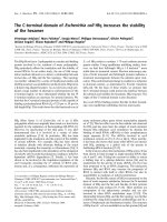
Fig. 1. The binding of L. interrogans serovar Pomona (NVSL 1427-35-093002) to Fn (A). Binding of Leptopsira to various
immobilized ECM components. Leptospira (10
7
) were added to wells coated with each ECM (1 mg in 100 μl PBS) including Fn,
chondroitin-6-sulfate (C6S), chondroitin sulfate A (CSA), chondroitin sulfate B (CSB), gelatin A (GA), gelatin B (GB), heparin (HP),
keratin (KR), or BSA (negative control). (B). Binding of Leptospira (10
7
) to various concentrations of Fn (0, 10, 20, 100 or 1,000 μ
g
in 100 μl PBS). BSA served as a negative control. (C). Fn inhibits the binding of Leptospira to the MDCK cells. Leptospira (10
7
) were
treated with various concentrations of Fn (0, 0.01, 0.1, 0.2, 1, 2, or 10 μg) or BSA (negative control) prior to addition to the MDCK cell
s
(10
5
). The percentage adhesion was determined relative to the attachment of untreated Leptospira onto the MDCK cells. (D). Binding
of Leptospira to immobilize Fn. Leptospira (10
8
) were cultured in Fn or BSA (negative control) coated (1 mg in 100 μl PBS) or
un-coated wells (negative control). (E). Fn inhibited the binding of Leptospira to the MDCK cells. Leptospira (10
8
) were pre-treate
d
with 10 μg of Fn or BSA (negative control) prior to addition to the MDCK cells (10
6
). Un-treated Leptospira was used as a negative
control. The binding of Leptospira to ECMs or Fn or the adhesion of Leptospira to the MDCK cells was measured by ELISA (A, B,
and C) or EPM (D and E). For all experiments, each value represents the mean ± SE of three trials performed in triplicate samples.
Statistically significant (p < 0.05) differences are indicted by an asterisk. The EPM settings were identical for all captured images (D
and E).
(10
5
) were incubated with various concentrations (as in-
dicated in Fig. 4A) of GST-LigBCen, GST-LigBCtv or
GST (negative control) in 100 μl PBS for 1 h at 37
o
C. To
measure the binding inhibition of Leptospira to the MDCK
cells treated with LigBCen or LigBCtv, the MDCK cells
(10
5
) were pretreated with various concentrations (as in-
dicated in Fig. 4B) of GST-LigBCen, GST-LigBCtv or
GST (negative control) in 100 μl PBS for 1 h at 37
o
C.
Then, the Leptospira (10
7
) were added to each well and in-
cubated for 6 h at 37
o
C. Following the incubation, the
plates were washed three times with phosphate-buffered
saline (PBS) containing 0.05% Tween-20 (PBST). To
measure the binding of the Leptospira, hamster anti-
Leptospira (1:200) and HRP-conjugated goat anti-ham-
ster IgG (1:1,000) were used as primary and secondary
antibodies, respectively. To detect the binding of GST-
136 Yi-Pin Lin et al
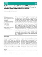
Fig. 2. The interaction between LigB and Fn by the GST-pull
down assay (A) A schematic diagram showing the structure o
f
LigB and the truncated LigB protein used in this study. (B).
Human plasma Fn ( lane 2 to lane 5 ) or cell lysates of the MDC
K
cells (lane 7 to lane 10) was applied to the GST beads pre-
immobilized by GST, GST-LigBCon, GST-LigBCen, or GST-
LigBCtv at 4
o
C for 3 h. The pull down complex was analyzed
by
immunoblot analysis using Fn antibodies. Lane 1 and lane 6
contain 1 μg of human plasma Fn and the cell lysate from 1 × 10
6
MDCK cells, respectively, to serve as a positive reference. Lane
2 and lane 7 are GST-LigBCen, lane 3 and lane 8 are GST-
LigBCtv, lane 4 and lane 9 are GST-LigBCon, and lane 5 and lane
10 are GST. The molecular mass of the human Fn and canine Fn
(MDCK cells) was 261 kDa and 271 kDa, respectively, and the
relative positions of the standards are given in kDa on the left.
LigBCen, GST-LigBCtv, or GST to Fn or the MDCK cells,
rabbit anti-GST (1:200) and HRP-conjugated goat an-
ti-rabbit IgG (1:1,000) were used as primary and secon-
dary antibodies, respectively. After washing the plates
three times with PBST, 100 μl of TMB (KPL, USA) was
added to each well and incubated for 5 min. The reaction
was stopped by adding 100 μl of 0.5% hydrofluoric acid in
each well. Each plate was read at 630 nm by an ELISA
plate reader (Bioteck EL-312; BioTeck, USA). Each value
represents the mean ± standard error of the mean (SEM)
of three trials performed in triplicate samples. Statistically
significant (p < 0.05) differences are indicated by asterisks.
Binding assays by epifluorescence microscopy (EPM)
and confocal laser-scanning microscopy (CLSM)
To measure the binding of Leptospira to Fn by EPM,
Leptospira (10
8
) were added to each well (eight well cul-
ture slides) coated with 1 mg Fn or BSA (negative control)
in 100 μl of PBS and incubated at 37
o
C for 6 h (Fig. 1D).
To measure the binding inhibition of Leptospira to the
MDCK cells by Fn, 10
8
Leptospira were pre-incubated
with 10 μg of Fn or BSA (negative control) in 100 μl of
PBS for 1 h at 37
o
C prior to the addition of 10
6
MDCK cells
and incubated 6 h at 37
o
C (Fig. 1E). To measure the binding
inhibition Leptospira to Fn by LigBCen or LigBCtv by
EPM, 50 nM of GST-LigBCen, GST-LigBCtv or GST
(negative control) in 100 μl PBS was added to each of the
Fn or BSA (negative control and data not shown) (1 mg per
100 μl) coated wells for 1 h at 37
o
C. Then, the Leptospira
(10
8
) were added to each well and incubated for 6 h at 37
o
C
(Fig. 3C). To determine the binding inhibition of Leptospi-
ra to the MDCK cells by LigBCen or LigBCtv by CLSM,
the MDCK cells (10
6
) were preincubated with 50 nM of
GST-LigBCen, GST-LigBCtv or GST (negative control) in
100 μl of PBS for 1 h at 37
o
C respectively. Then, the
Leptospira (10
8
) were added to each well and incubated for
6 h at 37
o
C (Fig. 4C). For the detection of Leptospira bind-
ing in Figs. 1D, E, and Fig. 3C, hamster anti-Leptospira an-
tibodies (1:100) and Alexa 488-conjugated goat an-
ti-hamster IgG (1:250) were used as primary and secon-
dary antibodies, respectively. To determine the attachment
of Leptospira and the binding of GST-LigBCen, GST-
LigBCtv or GST, Fig. 4C, rabbit anti-GST (1:250) and
hamster anti-Leptospira antibodies (1:100) served as pri-
mary antibodies, and FITC conjugated goat anti-rabbit IgG
(1:250) and Alexa 594-conjugated goat anti-hamster IgG
(1:250) were used as secondary antibodies. Fixation and
immunofluorescence staining were performed as pre-
viously described [44] with slight modifications. Briefly,
Leptopsira and the MDCK cells were fixed in 2% paraf-
ormaldehyde for 60 min at RT. For the antibody labeling,
fixed bacteria were incubated in PBS containing 0.3%
BSA for 10 min at RT. The primary and secondary anti-
bodies, in the PBS containing 0.3% BSA, were incubated
sequentially for 60 min at RT. After incubation with the pri-
mary and secondary antibodies, the glass slides were
mounted with coverslips using Prolong Antifade (Molecu-
lar Probe, USA) and viewed with a 60 × objective by EPM
(Nikon, Japan) or CLSM (Olympus, Japan). An Olympus
Fluoview 500 confocal laser-scanning imaging system,
equipped with krypton, argon and He-Ne lasers on an
Olympus IX70 inverted microscope with a PLAPO 60 ×
objective, was used. The settings were identical for all cap-
tured images. Images were processed using Adobe
Photoshop CS2. For counting the attachment of Leptospira
to the MDCK cells or Fn, three fields were selected to
count the number of binding organisms. All studies were
repeated three times and the number of Leptospira attached
to the MDCK cells were counted by an investigator blinded
to the treatment group.
GST pulldown assay
The GST pull-down assay was performed as previously
described [57]. Purified proteins or GST (negative control)
were loaded onto 0.5 ml glutathione-Sepharose beads
LigB-Fn interaction mediates cell adhesion 137
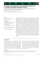
Fig. 3. LigBCen or LigBCtv binds to Fn and inhibits the binding of Leptospira to Fn (A). Binding of LigBCen or LigBCtv to various
concentrations of immobilized Fn. Ten nM of GST-LigBCen, GST-LigBCtv or GST (negative control) was added to wells coated wit
h
various concentrations of Fn (0, 0.27 μM, 0.45 μM, 2.7 μM, 4.5 μM, 27 μM, or 45 μM) in 100 μl PBS. The binding of each of these
p
roteins to Fn was measured by ELISA. (B) LigBCen or LigBCtv inhibited the binding of Leptospira to immobilized Fn. Various
concentrations (0, 2, 4, 6, or 8 nM) of GST-LigBCen, GST-LigBCtv, or GST (negative control) were added to each well coated with F
n
(1 mg in 100 μl PBS) prior to the addition of Leptospira (10
7
). The attachment of Leptopsira to wells was measured by ELISA. The
p
ercentage of attachment was determined relative to the attachment of Leptopsira in the untreated Fn. (C) LigBCen or LigBCtv
inhibited the binding of Leptospira to Fn. Fifty nM of GST-LigBCen, GST-LigBCtv or GST (negative control) was added to wells
coated with Fn (1 mg in 100 μl PBS) prior to the addition of Leptospira (10
8
). The binding of Leptospira to wells was detected by EPM.
In (A) and (B), each value represents the mean ± SE of three trials performed in triplicate samples. Statistically significant differences
(p < 0.05) are indicted by *. In (C), The EPM settings were identical for all captured images. Images were processed using Adobe
Photoshop CS2.
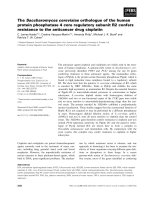
Fig. 4. Isothermal titration calorimetry (ITC) profile of LigBCtv with Fn as a typical ITC profile in this studyA: heat differences obtaine
d
from 25 injections. B: Integrated curve with experimental point () and the best fit (−). The thermodynamic parameters are show
n
in Table 1.
138 Yi-Pin Lin et al
Table 1. Thermodynamic parameters for the interaction of Fn and truncated LigB
Macromolecule LigB Residues [Macromolecule] [Fn] ΔH ΔSK
d
μM μM kcal mol
−1
cal mol
−1
K
−1
μM
LigBCon 1-630 1.25 25 n/f
*
n/f
*
n/f
*
LigBCen 631-1,417 2 40 −2,002.67 ± 14 −6.68 0.011 ± 0.003
LigBCtv 1,418-1,889 2.82 56.4 −12,140 ± 557 −40.71 8.55 ± 0.75
*
n/f: non-fittable.
(Amersham Biosciences Piscataway, USA) at 4
o
C over-
night. The beads were then washed three times with the ly-
sis buffer containing 30 mM Tris acetate, 10 mM sodium
phosphate, pH 7.4, 0.1% Tween 20, 1 mM EDTA, 2 μg/ml
leupeptin, 4 μg/ml aprotinin, 1 μg/ml pepstatin A, and 1
mM phenylmethylsulfonyl fluoride (PMSF). The MDCK
cells (10
6
) were lysed in the lysis buffer and used immedi-
ately after lysis. A 500 μl aliquot of cell lysate or human
plasma Fn (40 μg/ml) was incubated with purified pro-
teins immobilized on glutathione-Sepharose beads at 4
o
C
for 3 h. After incubation, the beads were separated by cen-
trifugation, washed three times with the lysis buffer and
boiled in Laemmli sample loading buffer consisting of 50
mM Tris-HCl (pH 6.8), 100 mM dithiothreitol, 2% sodium
dodecyl sulfate, 0.25 mM PMSF, and 0.1% bromophenol
blue in 20% glycerol. The eluted proteins were subjected to
6% SDS-PAGE and electroblotted onto polyvinylidene di-
fluoride membranes. The membranes were incubated in
5% skim milk in PBS/T overnight and then incubated with
mouse anti-Fn antibody (1:1,000). The immunocom-
plexes were detected with an HRP-conjugated goat an-
ti-mouse IgG antibody (1:5,000).
Small interfering RNA (siRNA) inhibition of LigB
binding
The siRNA duplexes directed against the sequence 5'-
gcagcacaacuuccaauua-3' of Fn and negative siRNA du-
plex, 5'-auucuaucacuagcgugac-3', were selected by the
software, siDESIGN [43] and synthesized by Dharmacon
(USA). The RNA duplexes were introduced into the
MDCK cells by the method of lipofection [18], and 8 × 10
5
cells were transfected with 0.4 μg negative siRNA and
Fn-siRNA. Adhesion assays were performed 72 h after lip-
ofection [51]. The knockdown efficiency of endogeneous
Fn expression was determined as previously described [57]
with slight modification. The total protein contents of the
MDCK cells (10
6
) were analyzed using Western immuno-
blotting as described under 'GST pulldown assays'. The
protein bands of actin derived from the MDCK cells were
measured as a control using a mouse anti-actin antibody
(1:5,000). The band intensity was measured by densi-
tometry using the Image J software (National Institutes of
Health, Bethesda, MD, USA) [53]. A LigB binding assay
was performed 72 h after lipofection. To determine the
binding of LigB fragments to Fn, each fragment (50 nM)
was added to the MDCK cells (10
6
) transfected with Fn or
negative siRNA. To determine the binding of each frag-
ment and the expression of Fn in the MDCK cells, rabbit
anti-GST (1:250) and mouse anti-Fn (1:250) served as
the primary antibodies, and FITC-conjugated goat an-
ti-mouse IgG (1:250) and Texas Red-conjugated goat an-
ti-rabbit IgG (1:250) were used as secondary antibodies.
Fixation, immunofluorescence staining, image detection,
and processing were carried out as described in previous
sections. All experiments were performed in triplicate.
Isothermal titration calorimetry
The experiments were carried out with CSC 5300 micro-
calorimeter (Calorimetry Science, USA) at 25
o
C as pre-
viously described [47]. In a typical experiment, the cell
contained 1 ml of a solution of proteins, and the syringe
contained 250 μl of a solution of Fn at a concentration that
was 20 times higher than the protein concentration in the
cell. Both solutions were in PBS pH 7.5. The titration was
performed as follows: 15 to 25 injections of 10 μl (Table 1)
with a stirring speed of 250 rpm, and the delay time be-
tween the injections was 5 min. Data were analyzed using
Titration BindingWork 3.1 software (Calorimetry Science,
USA) that was fit to an independent binding model. The
concentration of Fn and LigB used in this study was based
on our preliminary titration experiments (data not shown).
Statistical analysis
Statistically significant differences between samples
were determined using the Student's t-test following loga-
rithmic transformation of the data. Two-tailed p-values
were determined for each sample, and a p < 0.05 was con-
sidered significant. Each data point represents the mean ±
SE of a sample tested in triplicate. An asterisk indicates
that the result was statistically significant.
Results
Attachment of Leptospira to the MDCK cells was
mediated by fibronectin
The binding of leptospiral cells to various ECM compo-
LigB-Fn interaction mediates cell adhesion 139
Fig. 5. The binding of LigBCen or LigBCtv to the MDCK cells reduced leptospiral adhesion (A) Binding of LigBCen or LigBCtv to
the MDCK cells. Various concentrations (0, 2, 4, 6, or 8 nM) of GST-LigBCen, GST-LigBCtv or GST (negative control) was added to
the MDCK cells (10
5
). The binding of each of these proteins to the MDCK cells were measured by ELISA. (B) LigBCen or LigBCt
v
inhibits the binding of Leptopsira to MDCK cells. The MDCK cells were incubated with various concentrations (0, 2, 4, 6, or 8 nM)
of GST-LigBCen, GST-LigBCtv or GST (negative control) prior to the addition of Leptopsira (10
7
). The adhesion of Leptospira to the
MDCK cells (105) was detected by ELISA. The reduced percentage of attachment was determined relative to the attachment o
f
L
eptopsira in the untreated MDCK cells. (C). LigBCen or LigBCtv inhibited the binding of Leptopsira to the MDCK cells. The MDC
K
cells (10
6
) were pre-treated with 50nM of GST-LigBCen, GST-LigBCtv and GST (negative control) prior to the addition of the
L
eptopsira (10
8
). The adhesion of Leptospira or the binding of these proteins to the MDCK cells were detected by CLSM. In (A) an
d
(B), each value represents the mean± SEM of three trials in triplicate samples. Statistically significant values (p < 0.05) are indicte
d
by *. In (C), the CLSM settings were identical for all the captured images. Images were processed using Adobe Photoshop CS2.
nents was determined by ELISA. As shown in Fig. 1A,
Leptospira were strongly bound to Fn, but not to other
ECM molecules (Fig. 1A). Furthermore, the binding of
Leptospira to Fn was dose dependent (Fig. 1B). When
Leptospira were pretreated with Fn, binding to the MDCK
cells was decreased (Fig. 1C). There was an approximately
3.5-fold increase in the immobilization of Leptospira in the
Fn-coated wells compared to the controls (Fig. 1D). More-
over, Fn was observed to block the attachment of Leptospi-
ra, by approximately 47%, when the Fn treated Leptospira
were added to the MDCK cells (Fig. 1E). Thus, Fn appears
to mediate the attachment of Leptospira to the MDCK
cells.
Interaction between LigB and Fn
To determine whether LigB interacts with Fn, we trun-
cated the LigB protein into three parts, LigBCon, LigBCen
and LigBCtv, (Fig. 2A) due to the difficulty of expressing
and purifying the full length LigB [33]. First, we analyzed
the interaction of each LigB fragment with Fn using a
GST-pull down assay. Our results showed that both human
plasma Fn and Fn derived from the MDCK cell lysates
could bind both LigBCen and LigBCtv, but not LigBCon
(Figs. 2B and C). Since LigBCen and LigBCtv showed a
positive pull down result, the interaction between LigBCen
and LigBCtv with Fn was further studied by ELISA. We
found that both the binding of LigBCen and LigBCtv to Fn,
140 Yi-Pin Lin et al
Fig. 6. The binding of LigBCen or LigBCtv to Fn siRN
A
transfected MDCK cells was reduced (A). Detection of the
expression of Fn and actin in the MDCK cells 72 h after
transfected by Fn or negative siRNA. Fn and α-actin were
detected by immunoblotting probed by actin antibody or Fn
antibody. (B) Binding of GST-LigBCen or (C) GST-LigBCtv
was reduced by the siRNA transfected cells. (D) GST served as
a
negative control. Fifty nM of GST-LigBCen, GST-LigBCtv o
r
GST was added to Fn or the negative siRNA transfected MDC
K
cells. Expression of Fn and the binding of these proteins to the
MDCK cells were detected by CLSM. The CLSM settings were
identical for all the captured images. Images were processe
d
using Adobe Photoshop CS2.
and the inhibition of the attachment of Leptospira to Fn by
LigBCen and LigBCtv, were dose-dependent (Figs. 3A
and B). Moreover, the EPM images revealed an up to 40%
reduction in the attachment of Leptospira to Fn in the pres-
ence of LigBCen and LigBCtv (Fig. 3C). Finally, in order
to quantitatively evaluate the binding affinity between Fn
and LigB fragments, the dissociation constants (K
d
) were
measured by ITC (Table 1). Fig. 4 shows the data from a
typical ITC experiment. The interaction appears to be exo-
thermic with a favorable enthalpy and unfavorable
entropy. The calculated K
d
values for Fn binding to
LigBCen and LigBCtv were 0.01 μM and 8.55 μM, re-
spectively (Table 1). The binding of LigBCon could not be
detected by ITC (data not shown). These findings are in
agreement with our previous results. Altogether, these data
indicate that Fn specifically interacts with LigBCen and
LigBCtv fragments.
LigBCen and LigBCtv mediate the attachment of
Leptospira to the MDCK cells
To determine if LigB is used by Leptospira to adhere to
the MDCK cells, various concentrations of LigBCen or
LigBCtv were added to the MDCK cells, and binding was
detected by ELISA and immunofluorescence staining. Our
results clearly showed that LigBCen and LigBCtv were
bound to the MDCK cells in a dose dependent manner (Fig.
5A). Pretreatment of the MDCK cells with LigBCen or
LigBCtv reduced the attachment of Leptospira by ∼32%.
The reduction of Leptospira attachment was also dose-de-
pendent (Figs. 5B and C). We further elucidated the re-
ceptor role of Fn in the MDCK cells for its possible ligand,
LigB on the surface of Leptospira, by RNA interference to
decrease the endogeneous Fn expression in the MDCK
cells. As shown in Fig. 6A, transfection of the cells with
siRNA duplex specific for canine Fn resulted in a ∼36%
reduction of the Fn expression, relative to the control cells.
The binding of LigBCen and LigBCtv to Fn siRNA-trans-
fected MDCK cells was significantly reduced (Figs. 6B
and C). These results suggest that Fn serves as a receptor
for LigB that mediates Leptospira adhesion.
Discussion
Adhesion to host cells is pivotal for many pathogenic bac-
teria including Leptospira spp. Since pathogenic Leptospi-
ra spp. can infect a variety of tissues including liver, kidney
and lung, study of the host-pathogen interaction is ex-
tremely important for improved understanding of lepto-
spirosis. Recently, the leptospiral genome has been se-
quenced and a number of tentative virulence factors have
been proposed [3,31,42]. However, their exact roles in lep-
tospiral pathogenesis remain to be established. To date,
several leptospiral adhesion molecules have been identi-
fied. These include a 36 kDa Fn-binding protein [30], a 24
LigB-Fn interaction mediates cell adhesion 141
kDa laminin-binding protein [1] and LigA, LigB and LigC
proteins [25,33,34]. These molecules may play an im-
portant role in the pathogenesis of leptospiral infection
since they are able to bind to ECMs such as collagens I and
IV, laminin and fibronectin [6,24].
Pathogenic Leptospira spp. have been previously reported
to adhere to extracellular matrices [15,16] including Fn.
Fns are dimers of two similar peptides linked at their C-ter-
mini by two disulfide bonds [8] and serve as receptors for
several bacteria, including spirochetes [7,11,12,19,20,
23,28,32,38,40,46,50,55]. Our results showed that Fn im-
mobilized Leptospira. In addition, Fn was observed to
block the attachment of Leptospira to MDCK cells if the
Leptospira were pre-treated with Fn. These results support
the recent report that Fn might be an important molecule
involved in the pathogenic adherence of Leptospira spp. to
host cells [6,24].
We demonstrated the interaction between LigB and Fn. It
was shown that the LigBCen and LigBCtv fragments were
bound to Fn, by GST-pulldown assays, ELISA and ITC
measurements. The low K
d
values for LigBCen indicated
that the LigB-Fn interaction was specific. This evidence
strongly suggests that LigB is a Fn-binding protein. A
study reported by Choy et al. [6] showed that LigB U1 and
LigB U2 (LigBCen equivalent) could strongly bind to Fn,
while the LigB CTD (LigBCtv equivalent) binds weakly to
Fn. However, the Kd values of LigBCen and LigBCtv to Fn
that we obtained were slightly different than those reported
by Choy et al. [6]. The differences in the obtained K
d
val-
ues could be explained by (i) the protein fragments eval-
uated in this study (LigBCen and LigBCtv) were not ex-
actly the same length fragments (LigBU1, LigBU2 and
LigBCTD) and (ii) the method we used (ITC) to measure
the K
d
differed from that of Choy et al. [6].
Since pathogenic Leptospira spp. adheres to renal tubular
epithelial cells and induces a severe tubulointerstitial
nephritis leading to renal failure [58], it is possible that
LigB is responsible for the binding of Leptospira to the re-
nal tubular epithelium. Our results indicated that LigB
binds to the MDCK cells via the LigBCen or the LigBCtv
fragments. However, the LigBCen was observed to bind to
both the MDCK cells and Fn with a greater affinity than the
LigBCtv. The microscopic images also showed that not all
of the Fn was co-localized with the LigB. This result sug-
gests that LigB might bind to two or more receptors. Our
results elucidate the process of Leptospira attachment to
the MDCK cells, as noted in a previous study [52], and
demonstrated how Fn can block leptospiral attachment to
the MDCK cells.
Our results clearly confirm that LigB is one of the micro-
bial surface components that recognize adhesive matrix
molecules (MSCRAMM) members that bind to the ECM
including Fn. The transmembrane domain of LigB is pre-
dicted to reside within the conserved region, with only the
variable region exposed on the surface [33,34]. These re-
sults support our data that Fn-binding domains of LigB are
localized in the variable regions. This is not surprising
since similar findings have been reported for other
MSCRAMMs [13,37,39]. In Borrelia, the binding motifs
in the decorin-binding proteins, DbpA and B, are located in
the central regions, which vary among the different
Borrelia strains (B. burgdorferi, B. garnii, and B. afzeli)
[37]. The Fn-binding domain of the Fn-binding protein,
BBK32 is also variable among the different Borrelia
strains [39]. The repetitive D1, D2 and D3 elements of
Staphylococcus aureus Fn-binding protein, which bind the
N-terminal 29 kDa of Fn, also vary [13].
Since both LigBCen and LigBCtv bind to Fn, but with dif-
ferent affinities, this suggests that there is more than one
potential Fn-binding domain. In Mycobacterium avium,
two Fn-binding domains are located on two non-con-
tiguous segments of 24 amino acids in the Fn attachment
protein-A [45]. The FnBPA of Staphylococcus aureus con-
tains three repetitive elements, D1, D2 and D3 and each
binds the N-terminal 29 kDa fragment of Fn [13]. Seven
additional Fn-binding elements are located in the N-termi-
nal of the D repeats [48]. In Streptococcus dysgalactiae,
there are five Fn-binding segments within the C-terminus
of the Fn binding protein F1/(FnBB) [47,48]. Therefore, it
is likely that several binding sites might be present in the
LigB variable region. However, we were unable to identify
a similar Fn-binding motif in the other known Fn-binding
proteins.
In conclusion, we have shown that LigBCen and LigBCtv
bind to Fn and have confirmed that LigB is a member of the
MSCRAMMs. Since pathogenic Leptospira spp. initially
attaches to mucosal epithelial cells prior to entry into the
bloodstream and subsequent dissemination to multiple or-
gans such as the kidney, liver and lung, Lig proteins may
play a pivotal role in the pathogenesis of leptospirosis. Fn
is one of the most important ECMs on epithelial cells and
serves as a receptor for leptospiral adherence [6,15,24].
Thus, further studies into the interaction of Lig proteins
and ECMs are warranted.
Acknowledgments
This work was supported in part by the Harry M. Zweig
Memorial Fund for Equine Research, the New York State
Science and Technology Foundation (Center for Ad-
vanced Technology) and the Biotechnology Research and
Development Corporation. We would also like to thank Dr.
Marci Scidmore for help with the epifluorescence micro-
scope and confocal laser florescence microscope techni-
ques and Dr. Bhargavi Jayaraman and Charlene Mottler for
their help with the isothermal titration calorimetry
techniques. We also thank our laboratory members, espe-
cially Drs. Syed Faisal and Tavan Janvilisri, for their sug-
142 Yi-Pin Lin et al
gestions during the course of this study and to Drs. Marci
Scidmore, Linda Nicholson, and Sean McDonough for the
critical reading of this manuscript.
References
1. Barbosa AS, Abreu PA, Neves FO, Atzingen MV,
Watanabe MM, Vieira ML, Morais ZM, Vasconcellos
SA, Nascimento AL. A newly identified leptospiral adhesin
mediates attachment to laminin. Infect Immun 2006, 74,
6356-6364.
2. Barocchi MA, Ko AI, Reis MG, McDonald KL, Riley LW.
Rapid translocation of polarized MDCK cell monolayers by
Leptospira interrogans, an invasive but nonintracellular
pathogen. Infect Immun 2002, 70, 6926-6932.
3. Bulach DM, Zuerner RL, Wilson P, Seemann T, McGrath
A, Cullen PA, Davis J, Johnson M, Kuczek E, Alt DP,
Peterson-Burch B, Coppel RL, Rood JI, Davies JK,
Adler B. Genome reduction in Leptospira borgpetersenii re-
flects limited transmission potential. Proc Natl Acad Sci
USA 2006, 103, 14560-14565.
4. Cameron CE, Brouwer NL, Tisch LM, Kuroiwa JM.
Defining the interaction of the Treponema pallidum adhesin
Tp0751 with laminin. Infect Immun 2005, 73, 7485-7494.
5. Cameron CE, Brown EL, Kuroiwa JM, Schnapp LM,
Brouwer NL. Treponema pallidum fibronectin-binding
proteins. J Bacteriol 2004, 186, 7019-7022.
6. Choy HA, Kelley MM, Chen TL, Moller AK, Matsunaga
J, Haake DA. Physiological osmotic induction of Leptospi-
ra interrogans adhesion: LigA and LigB bind extracellular
matrix proteins and fibrinogen. Infect Immun 2007, 75,
2441-2450.
7. Coburn J, Fischer JR, Leong JM. Solving a sticky problem:
new genetic approaches to host cell adhesion by the Lyme
disease spirochete. Mol Microbiol 2005, 57, 1182-1195.
8. Darnell J, Lodish H, Baltimore D. Molecular Cell Biology.
2nd ed. pp. 802-824, Scientific American Books, New York,
1990.
9. Edwards AM, Jenkinson HF, Woodward MJ, Dymock D.
Binding properties and adhesion-mediating regions of the
major sheath protein of Treponema denticola ATCC 35405.
Infect Immun 2005, 73, 2891-2898.
10. Faine SB, Adher B, Bolin C, Perolat P. Leptospira and
Leptospirosis. 2nd ed. pp. 67-71, MedSci, Medbourne, 1999.
11.
Fischer JR, LeBlanc KT, Leong JM. Fibronectin binding
protein BBK32 of the Lyme disease spirochete promotes
bacterial attachment to glycosaminoglycans. Infect Immun
2006, 74, 435-441.
12. Grab DJ, Givens C, Kennedy R. Fibronectin-binding ac-
tivity in Borrelia burgdorferi. Biochim Biophys ACTA
1998, 1407, 135-145.
13. Ingham KC, Brew S, Vaz D, Sauder DN, McGavin MJ.
Interaction of Staphylococcus aureus fibronectin-binding
protein with fibronectin: affinity, stoichiometry, and modu-
lar requirements. J Biol Chem 2004, 279, 42945-42953.
14. Isberg RR, Voorhis DL, Falkow S. Identification of in-
vasin: a protein that allows enteric bacteria to penetrate cul-
tured mammalian cells. Cell 1987, 50, 769-778.
15. Ito T, Yanagawa R. leptospiral attachment to extracellular
matrix of mouse fibroblast (L929) cells. Vet Microbiol 1987,
15, 89-96.
16. Ito T, Yanagawa R. Leptospiral attachment to four struc-
tural components of extracellular matrix. Nippon juigaku
zasshi1987, 49, 875-882.
17. Jerse AE, Yu J, Tall BD, Kaper JB. A genetic locus of en-
teropathogenic Escherichia coli necessary for the production
of attaching and effacing lesions on tissue culture cells. Proc
Natl Acad Sci USA 1990, 87, 7839-7843.
18. Jiang ST, Chiang HC, Cheng MH, Yang TP, Chuang WJ,
Tang, MJ. Role of fibronectin deposition in cystogenesis of
Madin-Darby canine kidney cells. Kidney Intl 1999, 56, 92-
103.
19. Kim JH, Singvall J, Schwarz-Linek U, Johnson BJ, Potts
JR, Hook M. BBK32, a fibronectin binding MSCRAMM
from Borrelia burgdorferi, contains a disordered region that
undergoes a conformational change on ligand binding. J Biol
Chem 2004, 279, 41706-41714.
20. Konkel ME, Christensen JE, Keech AM, Monteville MR,
Klena JD, Garvis SG.
Identification of a fibronectin-bind-
ing domain within the Campylobacter jejuni CadF protein.
Mol Microbiol 2005, 57, 1022-1035.
21. Kopp PA, Schmitt M, Wellensiek HJ, Blobel H. Isolation
and characterization of fibronectin-binding sites of Borrelia
garinii N34. Infect Immun 1995, 63, 3804-3808.
22. Levett PN. Leptospirosis. Clin Microbiol Rev 2001, 14,
296-326.
23. Li X, Liu X, Beck DS, Kantor FS, Fikrig E. Borrelia burg-
dorferi lacking BBK32, a fibronectin-binding protein, re-
tains full pathogenicity. Infect Immun 2006, 74, 3305-3313.
24. Lin YP, Chang YF. A domain of the Leptospira LigB con-
tributes to high affinity binding of fibronectin. Biochem
Biophys Res Commun 2007, 362, 443-448.
25. Matsunaga J, Barocchi MA, Croda J, Young TA,
Sanchez Y, Siqueira I, Bolin CA, Reis MG, Riley LW,
Haake DA, Ko AI. Pathogenic Leptospira species express
surface-exposed proteins belonging to the bacterial im-
munoglobulin superfamily. Mol Microbiol 2003, 49, 929-
945.
26. Matsunaga J, Lo M, Bulach DM, Zuerner RL, Adler B,
Haake DA. Response of Leptospira interrogans to Physiol-
ogic Osmolarity: Relevance in Signaling the Environment-
to-Host Transition. Infect Immun 2007, 75, 2864-2874.
27. Matsunaga J, Sanchez Y, Xu X, Haake DA. Osmolarity, a
key environmental signal controlling expression of lep-
tospiral proteins LigA and LigB and the extracellular release
of LigA. Infect Immun 2005, 73, 70-78.
28. May M, Papazisi L, Gorton TS, Geary SJ. Identification
of fibronectin-binding proteins in Mycoplasma gallisepti-
cum strain R. Infect Immun 2006, 74, 1777-1785.
29. Meites E, Jay MT, Deresinski S, Shieh WJ, Zaki SR,
Tompkins L, Smith DS. Reemerging leptospirosis, Califor-
nia. Emerg Infect Dis 2004, 10, 406-412.
30. Merien F, Truccolo J, Baranton G, Perolat P. Identifica-
tion of a 36-kDa fibronectin-binding protein expressed by a
virulent variant of Leptospira interrogans serovar icterohae-
morrhagiae. FEMS Microbiol Lett 2000, 185, 17-22.
31. Nascimento AL, Ko AI, Martins EA, Monteiro-Vitorello
CB, Ho PL, Haake DA, Verjovski-Almeida S, Hartskeerl
LigB-Fn interaction mediates cell adhesion 143
RA, Marques MV, Oliveira MC, Menck CF, Leite LC,
Carrer H, Coutinho LL, Degrave WM, Dellagostin OA,
El-Dorry H, Ferro ES, Ferro MI, Furlan LR, Gamberini
M, Giglioti EA, Goes-Neto A, Goldman GH, Goldman
MH, Harakava R, Jeronimo SM, Junqueira-de-Azevedo
IL, Kimura ET, Kuramae EE, Lemos EG, Lemos MV,
Marino CL, Nunes LR, de Oliveira RC, Pereira GG, Reis
MS, Schriefer A, Siqueira WJ, Sommer P, Tsai SM,
Simpson AJ, Ferro JA, Camargo LE, Kitajima JP,
Setubal JC, Van Sluys MA. Comparative genomics of two
Leptospira interrogans serovars reveals novel insights into
physiology and pathogenesis. J Bacteriol 2004, 186, 2164-
2172.
32. Nyberg P, Sakai T, Cho KH, Caparon MG, Fassler R,
Bjorck L. Interactions with fibronectin attenuate the viru-
lence of Streptococcus pyogenes. EMBO J 2004, 23, 2166-
2174.
33. Palaniappan RU, Chang YF, Hassan F, McDonough SP,
Pough M, Barr SC, Simpson KW, Mohammed HO, Shin
S, McDonough P, Zuerner RL, Qu J, Roe B. Expression of
leptospiral immunoglobulin-like protein by Leptospira in-
terrogans and evaluation of its diagnostic potential in a ki-
netic ELISA. J Med Microbiol 2004, 53, 975-984.
34. Palaniappan RU, Chang YF, Jusuf SS, Artiushin S,
Timoney JF, McDonough SP, Barr SC, Divers TJ,
Simpson KW, McDonough PL, Mohammed HO. Cloning
and molecular characterization of an immunogenic LigA
protein of Leptospira interrogans. Infect Immun 2002, 70,
5924-5930.
35. Palaniappan RU, McDonough SP, Divers TJ, Chen CS,
Pan MJ, Matsumoto M, Chang YF. Immunoprotection of
recombinant leptospiral immunoglobulin-like protein A
against Leptospira interrogans serovar Pomona infection.
Infect Immun 2006, 74, 1745-1750.
36. Parma AE, Cerone SI, Sansinanea SA. Biochemical anal-
ysis by SDS-PAGE and western blotting of the antigenic re-
lationship between Leptospira and equine ocular tissues. Vet
Immunol Immunopathol 1992, 33, 179-185.
37. Pikas DS, Brown EL, Gurusiddappa S, Lee LY, Xu Y,
Hook M. Decorin-binding sites in the adhesin DbpA from
Borrelia burgdorferi: a synthetic peptide approach. J Biol
Chem 2003, 278, 30920-30926.
38. Probert WS, Johnson BJ. Identification of a 47 kDa fi-
bronectin-binding protein expressed by Borrelia burgdor-
feri isolate B31. Mol Microbiol 1998, 30, 1003-1015.
39. Probert WS, Kim JH, Hook M, Johnson BJ. Mapping the
ligand-binding region of Borrelia burgdorferi fibronectin-
binding protein BBK32. Infect Immun 2001, 69, 4129-4133.
40. Raibaud S, Schwarz-Linek U, Kim JH, Jenkins HT,
Baines ER, Gurusiddappa S, Hook M, Potts JR. Borrelia
burgdorferi binds fibronectin through a tandem beta-zipper,
a common mechanism of fibronectin binding in staph-
ylococci, streptococci, and spirochetes. J Biol Chem 2005,
280, 18803-18809.
41. Rathinam SR, Rathnam S, Selvaraj S, Dean D, Nozik RA,
Namperumalsamy P. Uveitis associated with an epidemic
outbreak of leptospirosis. Am J Ophthalmol 1997, 124, 71-
79.
42. Ren SX, Fu G, Jiang XG, Zeng R, Miao YG, Xu H, Zhang
YX, Xiong H, Lu G, Lu LF, Jiang HQ, Jia J, Tu YF, Jiang
JX, Gu WY, Zhang YQ, Cai Z, Sheng HH, Yin HF,
Zhang Y, Zhu GF, Wan M, Huang HL, Qian Z, Wang
SY, Ma W, Yao ZJ, Shen Y, Qiang BQ, Xia QC, Guo XK,
Danchin A, Saint Girons I, Somerville RL, Wen YM, Shi
MH, Chen Z, Xu JG, Zhao GP. Unique physiological and
pathogenic features of Leptospira interrogans revealed by
whole-genome sequencing. Nature 2003, 422, 888-893.
43. Reynolds A, Leake D, Boese Q, Scaringe S, Marshall WS,
Khvorova A. Rational siRNA design for RNA interference.
Nature biotechnol 2004, 22, 326-330.
44. Rzomp KA, Scholtes LD, Briggs BJ, Whittaker GR,
Scidmore MA. Rab GTPases are recruited to chlamydial in-
clusions in both a species-dependent and species-indepen-
dent manner. Infect Iimmun 2003, 71, 5855-5870.
45. Schorey JS, Holsti MA, Ratliff TL, Allen PM, Brown EJ.
Characterization of the fibronectin-attachment protein of
Mycobacterium avium reveals a fibronectin-binding motif
conserved among mycobacteria. Mol Microbiol 1996, 21,
321-329.
46. Schroder A, Schroder B, Roppenser B, Linder S, Sinha
B, Fassler R, Aepfelbacher M. Staphylococcus aureus fi-
bronectin binding protein-A induces motile attachment sites
and complex actin remodeling in living endothelial cells.
Mol Biol Cell 2006, 17, 5198-5210.
47. Schwarz-Linek U, Pilka ES, Pickford AR, Kim JH, Hook
M, Campbell ID, Potts JR. High affinity streptococcal
binding to human fibronectin requires specific recognition of
sequential F1 modules. J Biol Chem 2004, 279, 39017-
39025.
48. Schwarz-Linek U, Werner JM, Pickford AR, Gurusi-
ddappa S, Kim JH, Pilka ES, Briggs JA, Gough TS, Hook
M, Campbell ID, Potts JR. Pathogenic bacteria attach to
human fibronectin through a tandem beta-zipper. Nature
2003, 423, 177-181.
49. Segura ER, Ganoza CA, Campos K, Ricaldi JN, Torres S,
Silva H, Cespedes MJ, Matthias MA, Swancutt MA,
Lopez Linan R, Gotuzzo E, Guerra H, Gilman RH,
Vinetz JM. Clinical spectrum of pulmonary involvement in
leptospirosis in a region of endemicity, with quantification of
leptospiral burden. Clin Infect Dis 2005, 40, 343-351.
50. Seshu J, Esteve-Gassent MD, Labandeira-Rey M, Kim
JH, Trzeciakowski JP, Hook M, Skare JT. Inactivation of
the fibronectin-binding adhesin gene bbk32 significantly at-
tenuates the infectivity potential of Borrelia burgdorferi.
Mol Microbiol 2006, 59, 1591-1601.
51. Shi J, Scita G, Casanova JE. WAVE2 signaling mediates
invasion of polarized epithelial cells by Salmonella typhimu-
rium. J Biol Chem 2005, 280, 29849-29855.
52. Thomas DD, Higbie LM. In vitro association of leptospires
with host cells. Infect Immun 1990, 58, 581-585.
53. Vendrame F, Segni M, Grassetti D, Tellone V, Augello G,
Trischitta V, Torlontano M, Dotta F. Impaired caspase-3
expression by peripheral T cells in chronic autoimmune thy-
roiditis and in autoimmune polyendocrine syndrome-2. J
Clin Endocrinol Metabol 2006, 91, 5064-5068.
54. Verma A, Hellwage J, Artiushin S, Zipfel PF, Kraiczy P,
Timoney JF, Stevenson B. LfhA, a novel factor H-binding
protein of Leptospira interrogans. Infect Immun 2006, 74,
144 Yi-Pin Lin et al
2659-2666.
55. Wann ER, Gurusiddappa S, Hook M. The fibronectin-
binding MSCRAMM FnbpA of Staphylococcus aureus is a
bifunctional protein that also binds to fibrinogen. J Biol
Chem 2000, 275, 13863-13871.
56. Werts C, Tapping RI, Mathison JC, Chuang TH,
Kravchenko V, Saint Girons I, Haake DA, Godowski PJ,
Hayashi F, Ozinsky A, Underhill DM, Kirschning CJ,
Wagner H, Aderem A, Tobias PS, Ulevitch RJ. Leptospi-
ral lipopolysaccharide activates cells through a TLR2-de-
pendent mechanism. Nature immunol 2001, 2, 346-352.
57. Xu Q, Yan B, Li S, Duan C. Fibronectin binds insulin-like
growth factor-binding protein 5 and abolishes Its ligand-de-
pendent action on cell migration. J Biol Chem 2004, 279,
4269-4277.
58. Yang CW, Wu MS, Pan MJ. Leptospirosis renal disease.
Nephrol Dialysis Transplant 2001, 16 (Suppl 5), 73-77.
59. Yang CW, Wu MS, Pan MJ, Hsieh WJ, Vandewalle A,
Huang CC. The Leptospira outer membrane protein LipL32
induces tubulointerstitial nephritis-mediated gene expres-
sion in mouse proximal tubule cells. J Am Soc Nephrol 2002,
13, 2037-2045.