Báo cáo khoa học: "Macrophages, myofibroblasts and mast cells in a rat liver infected with Capillaria hepatica" ppsx
Bạn đang xem bản rút gọn của tài liệu. Xem và tải ngay bản đầy đủ của tài liệu tại đây (3.07 MB, 3 trang )
JOURNAL OF
Veterinary
Science
J. Vet. Sci. (2008), 9(2), 211
213
Case Report
*Corresponding author
Tel: +82-53-950-5975; Fax: +82-53-950-5955
E-mail:
Macrophages, myofibroblasts and mast cells in a rat liver infected with
Capillaria hepatica
Won-Il Jeong
1
, Sun-Hee Do
2
, Il-Hwa Hong
1
, Ae-Ri Ji
1
, Jin-Kyu Park
1
, Mi-Ran Ki
1
, Seung-Chun Park
1
,
Kyu-Shik Jeong
1,
*
1
Department of Veterinary Pathology, College of Veterinary Medicine, Kyungpook National University, Daegu 702-701,
Korea
2
College of Veterinary Medicine, Konkuk University, Seoul 143-701, Korea
We trapped a rat (Rattus norvegicus) infected with Capi-
llaria hepatica. At necropsy, grossly yellowish-white nod-
ules (2-3 mm in diameter) were noted to be scattered on
the liver's surface. Microscopically, granulomatous and fi-
brotic nodules that contained the eggs and/or adult worms
of Capillaria hepatica were detected in the liver. Septal fib-
rosis was diffusely formed throughout the liver. There
were a number of ED1-positive macrophages located in
the sinusoids of the pseudolobules. On the double staining,
myofibroblasts and mast cells were generally observed
within the fibrous septa with the mast cells in close prox-
imity to the myofibroblasts. We suggest that the inter-
actions between macrophages, myofibroblasts and mast
cells play a role in the septal fibrosis observed in rats in-
fected by Capillaria hepatica.
Keywords: Capillaria hepatica, fibrosis, liver, rat
Capillaria (C.) hepatica is a zoonotic liver nematode of
mammals and this disease has a global distribution [1]. C.
hepatica is found primarily in rodents, although it has been
reported in other animals and humans as well [1,2]. As the
parasite's name implies, the worms live in the host's liver.
The female produces eggs in the liver parenchyma and all
the worms die inside the liver around the 30th to 40th day
after infection [3]. Breakdown of the dead worms and lease
of eggs cause focal necrosis and encapsulation. When all
the worms are dead and being disintegrated, the focal en-
capsulating fibrous response eventually progresses to sep-
tal fibrosis, and cirrhosis has also been reported [3].
A feral rat (Rattus norvegicus) was trapped and necro-
psied. For histopathological observations, pieces of its liv-
er were immediately fixed in 10% neutral buffered formal-
in; they were then routinely processed and embedded in
paraffin. Tissue sections 4 μm in thickness were cut and
stained with hematoxylin and eosin and toluidine blue, and
Azan stain was used for staining the collagen. For im-
munohistochemistry, sections of liver were deparaffinized
in xylene, rehydrated in a series of graded alcohol solutions
and then incubated in a solution of 3% hydrogen peroxide
in methanol for 10 min. The tissue sections were washed
with PBS and then immunostained with the primary anti-
bodies, anti-rat monocyte/macrophage at a dilution of 1:
600 (clone ED-1; Chemicon, USA) and monoclonal anti-
α-smooth muscle actin (α-SMA) at a dilution of 1:800
(clone 1A4; Sigma, USA). The antigen-antibody complex
was visualized by the labelled streptavidin-biotin (SAB)
method and using a Histostatin-plus bulk kit (Zymed
Laboratories, USA) with 3,3-diamino-benzidine (Zymed
Laboratories, USA). Non-immunized goat sera, which
were used instead of the primary antibody, served as the
negative control. The tissue sections were then rinsed in
distilled water and counterstained with Mayer's hematox-
ylin or toluidine blue for simultaneous observation of the
myofibroblasts and mast cells in the liver.
At necropsy, grossly yellowish-white nodules containing
the eggs and/or the adult worms of C. hepatica were readily
identified microscopically as granulomas, and these were
scattered throughout the liver (Fig. 1). Both viable and de-
generate adult nematodes and eggs were present. Some ne-
crotic worms were mineralized. Others appeared focally
accumulated, and these formed discrete inflammatory nod-
ules surrounded by connective tissue, macrophages, eosi-
nophils, lymphocytes and plasma cells. Increased numbers
of mast cells were detected around the central veins, portal
areas and fibrous septa. Clusters of eggs and worms were
surrounded by collagen fibers. Septal fibrosis that formed
pseudolobules was observed as a diffuse change through-
out the liver. Even in areas unassociated with granulomas,
212 Won-Il Jeong et al.
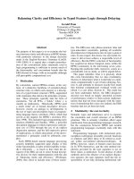
Fig. 1. Collagen fibers are detected in the portal areas and note the
fibrous septa that form pseudolobules. Azan stain. ×13.
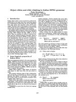
Fig. 2. There are a number of ED1-positive macrophages located
in the sinusoids of the pseudolobules. SAB method. ×13.
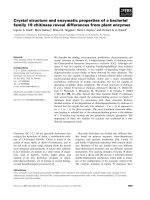
Fig. 3. α-SMA positive myofibroblasts are observed in the
fibrous septa and they form pseudolobules. SAB method. ×33.
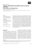
Fig. 4. Mast cells (arrow head) and myofibroblasts (arrow) are
simultaneously detected within the fibrous septa (F) and aroun
d
the central vein (C). Mast cells are largely observed along the
myofibroblasts. SAB method and toluidine blue stain. ×66.
delicate fibrous septa bridged between the central veins to
the portal areas (Fig. 1). There were a number of ED1- pos-
itive cells located in the sinusoids of the pseudolobules
(Fig. 2). Their morphology suggested that most of them
were Kupffer cells. α-SMA positive myofibroblasts were
observed within the fibrous septa (Fig. 3) and within the
walls of the fibrotic granulomas. They were also detected
in the connective tissues surrounding the fibrotic nodules.
On double staining, myofibroblasts and mast cells were
generally observed within the fibrous septa with mast cells
being in close proximity to the myofibroblasts (Fig. 4).
Septal fibrosis is a commonly seen variant of hepatic fib-
rosis, and this is represented by thin, straight fibrosis tissue
septa that dissect the liver parenchyma and sometimes cir-
cumscribe the hepatocellular nodules in cirrhotic livers [4].
Septal fibrosis of the liver can be experimentally produced
in rats by intraperitoneal injection of porcine serum or C.
hepatica infection [5], although its etiology and patho-
genesis are poorly understood [1,6,7]. The pathogenesis of
septal fibrosis associated with C. hepatica might be related
to the slow release of sequestered antigenic materials from
the dead worms and eggs [3,5]. The feature of septal fib-
rosis in C. hepatica infected rats is similar to that seen in
rats repeatedly injected with porcine serum. Its etiology
and pathogenesis are poorly understood. The pathogenesis
of septal fibrosis has been considered to have an immuno-
logical basis in both cases [6,8].
The resident macrophages of the liver, i.e., the Kupffer
cells, normally protect the hepatocytes by phagocytosing
any incoming particles. However, in the liver, xenobiotics
induce localized Kupffer cell accumulation and the release
of mediators, which have the potential to kill the hep-
atocytes [9]. It has been recently reported that myofibro-
blasts (activated hepatic stellate cells) are a major producer
of the extracellular matrix and the myofibroblasts play a
pivotal role in liver fibrosis [10]. Furthermore, many stud-
ies have reported that macrophages, myofibroblasts and
mast cells are closely related to the liver fibrosis induced
by CCl
4
[10,11] and porcine serum [1,6]. Our previous re-
port demonstrated that the number of macrophages, myofi-
Capillaria hepatica infection in a rat liver 213
broblasts and mast cells were increased in animals with
hepatic fibrosis and these cells might play a role in the for-
mation of hepatic fibrosis [5]. However, no previous stud-
ies have reported on the relationship and role of macro-
phages, myofibroblasts and mast cells in the hepatic septal
fibrosis induced by C. hepatica. We hypothesize that a sim-
ilar mechanism involving macrophages, myo-fibroblasts
and mast cells might be associated with the septal fibrosis
in this rat infected with C. hepatica. Moreover, by double
staining via immunohistochemistry and the toluidine blue
stain method, we observed the close proximity of myofi-
broblasts and mast cells in the liver in the hepatic fibrous
septa of an animal infected with C. hepatica. This finding
is consistent with that previously seen in a rat model of
CCl
4
-induced hepatic fibrosis; Akiyoshi et al. [11] re-
ported that in this rat model, the myo-fibroblasts were
closely associated with mast cells, and nerves and mast
cells were largely observed along the myofibroblasts. As a
possible mechanism for cirrhosis [12], the relationship be-
tween the development of myofibroblasts and mast cell-re-
leased fibrogenic cytokines (platelet-derived growth factor
and transforming growth factor-beta) might be involved in
the progress of hepatic inflammation, fibrosis and cirrhosis
in animals with C. hepatica infection. Our observations
closely parallel their findings. Thus, we believe that mac-
rophages, myo-fibroblasts and mast cells might play a role
in the septal fibrosis induced by C. hepatica infection in rat
liver.
Acknowledgments
The authors wish to thank the veterinarians and pet own-
ers for help us collect material for this study. The research
was supported by a grant (CBM32-B3003-01-01-00) from
the Center for Biological Modulators of the 21st Century
Frontier R&D Program, the Ministry of Science and
Technology, Korea.
References
1. Akiyoshi H, Terada T. Mast cell, myofibroblast and nerve
terminal complexes in carbon tetrachloride-induced cirrhotic
rat livers. J Hepatol 1998, 29, 112-119.
2. Bhunchet E, Eishi Y, Wake K. Contribution of immune re-
sponse to the hepatic fibrosis induced by porcine serum.
Hepatology 1996, 23, 811-817.
3. de Souza MM, Silva LM, Barbosa AA Jr, de Oliveira IR,
Parana R, Andrade ZA. Hepatic capillariasis in rats: a new
model for testing antifibrotic drugs. Braz J Med Biol Res
2000, 33, 1329-1334.
4. Ferreira LA, Andrade ZA. Capillaria hepatica: a cause of
septal fibrosis of the liver. Mem Inst Oswaldo Cruz 1993, 88,
441-447.
5. Jeong WI, Lee CS, Park SJ, Chung JY, Jeong KS. Kinetics
of macrophages, myofibroblasts and mast cells in carbon tet-
rachloride-induced rat liver cirrhosis. Anticancer Res 2002,
22, 869-877.
6. Laskin DL. Non parenchymal cells and hepatotoxicity.
Semin Liver Dis 1990, 10, 293-304.
7. Li CY, Baek JY. Mastocytosis and fibrosis: role of
cytokines. Int Arch Allergy Immunol 2002, 127, 123-126.
8. Neafie RC, Connor DH, Cross JH. Capillariasis (intestinal
and hepatic). In: Binford CH, Connor DH (eds.). Pathology
of Tropical and Extraordinary Diseases. Vol. 2. pp. 481-492,
Armed Forces Institute of Pathology, Washington DC, 1976.
9. Oliveira RF, Andrade ZA. Worm load and septal fibrosis of
the liver in Capillaria hepatica-infected rats. Mem Inst
Oswaldo Cruz 2001, 96, 1001-1003.
10. Ramos SG, Montenegro AP, Goissis G, Rossi MA.
Captopril reduces collagen and mast cell and eosinophil ac-
cumulation in pig serum-induced rat liver fibrosis. Pathol Int
1994, 44, 655-661.
11. Santos AB, Tolentino M Jr, Andrade ZA. Pathogenesis of
hepatic septal fibrosis associated with Capillaria hepatica in-
fection of rats. Rev Soc Bras Med Trop 2001, 34, 503-506.
12. Shiga A, Shirota K, Ikeda T, Nomura Y. Morphological
and immunohistochemical studies on porcine serum-in-
duced rat liver fibrosis. J Vet Med Sci 1997, 59, 159-167.