Báo cáo khoa học: "Malignant mixed tumor in the salivary gland of a cat" potx
Bạn đang xem bản rút gọn của tài liệu. Xem và tải ngay bản đầy đủ của tài liệu tại đây (1.55 MB, 3 trang )
JOURNAL OF
Veterinary
Science
J. Vet. Sci. (2008), 9(3), 331
333
Case Report
*Corresponding author
Tel: +81-83-933-5898; Fax: +81-83-933-5930
E-mail:
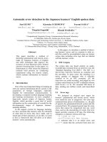
Fig. 1. Radiographic images of the head showing a large
tissue-dense mass (A) with faint circular calcific opacity (B).
Malignant mixed tumor in the salivary gland of a cat
Heejaung Kim
1,2
, Munekazu Nakaichi
3,
*
, Kazuhito Itamoto
2
, Yasuho Taura
2
1
Department of Neurology, College of Medicine, Hanyang University, Seoul 133-792, Korea
Departments of
2
Veterinary Surgery and
3
Veterinary Radiology, Faculty of Agriculture, Yamaguchi University, Yamaguchi
753-8515, Japan
The presence of a malignant mixed tumor, also known as a
carcinosarcoma, in the salivary gland is very rare. Such
tumors, which are typically aggressive, are characterized by
the presence of carcinomatous and sarcomatous components.
A 9-year-old neutered female domestic short-haired cat
presented with swelling in the right mandibular lesion that
had rapidly enlarged over the previous 3 weeks. Physical
examination revealed a large, fluctuated and painless
subcutaneous swelling that was associated with a firm mass.
Radiographs of the head revealed a soft-tissue density that
involved faint circular calcific opacity. Contrast-enhanced
computed tomography revealed that the peripheral capsulated
cystic area had a contrast enhanced region without bone
lysis. The cat received a total excision of the mass and
postoperative radiotherapy. Histopathological analysis of
the mass revealed that it was a malignant mixed tumor.
Metastasis to the lung was discovered 7 weeks later, at which
time treatment was stopped.
Keywords:
carcinosarcoma, cat, computed tomography, malignant
mixed tumor, salivary gland
Salivary gland tumors are rare in dogs and cats, with a
reported overall incidence of 0.17% [1]. Most cases are
reported in older patients and generally affect the
mandibular and parotid glands [6,17]. Clinical signs
include the presence of an asymptomatic mass in the gland
that is often locally invasive [1,6,17]. An adenocarcinoma
is the most common type of tumor in the salivary glands,
whereas malignant mixed tumors, especially carcinosarcomas,
are uncommon both in veterinary [2,16] and human
medicine [1,9]. Here, we describe a case involving a cat
with a carcinosarcoma in the mandibular gland.
A 9-year-old neutered female domestic short-haired cat
with no past trauma or medical history besides cough and
eye trauma presented to the Veterinary Medical Center of
Yamaguchi University with swelling near the base of the
right ear. The swelling had been discovered approximately
3 weeks earlier, at which time the area was drained of
purulent matter and treated with antibiotics at the referring
veterinary hospital. The purulent matter was found to
primarily consist of inflammatory cells and the cat showed
signs of hypersalivation, halitosis and gingivitis, which
were especially severe in the right side region of the head.
With the exception of the aforementioned symptoms, the
cat appeared well.
Upon admission, a large fluctuated and painless
subcutaneous swelling was palpated and found to contain a
firm mass in the right mandibular region. In addition, the
left mandibular lymph node was enlarged and the right
mandibular lymph node could not be found because of the
swelling. There were no neurological problems or facial
nerve disorders. Hematology and biochemical analysis of
the serum revealed neutrophilia (differential counting
84/100), low MCH (11.8 pg, reference range 13 to 17 pg)
and MCHC (24.4 g/dl, reference range 30 to 36 g/dl) levels.
Head radiographs revealed the presence of a soft-tissue
density that involved faint circular calcific opacity (Fig. 1).
There was also a bony reaction in the mandible and maxilla,
which was likely caused by chronic gingivitis. In addition,
thoracic radiographs revealed no abnormalities.
332 Heejaung Kim et al.
Fig. 4. Thoracic radiographs showed a diffuse increase in the
distribution of pulmonary interstitial densities and a poorly
circumscribed-nodular mass in the caudodorsal lung lobe
(arrow), suggesting lung metastasis of the tumor (A:
ventrodorsal view, B: lateral view).
Fig. 2. Post-contrast CT images showing enlargement of the righ
t
submandibular gland with a peripheral capsulated cystic area (A:
tympanic bulla level), and contrast enhanced region (B: occipita
l
bone level, C: first cervical vertebra level) that was believed to
b
e
a tumor.
Fig. 3. Adenocarcinomatous (A) and chondrosarcomatous (B)
elements were observed in the specimen. H&E stain. ×400.
However, the contrast-enhanced computed tomography (CT)
revealed the presence of a peripheral capsulated cystic area
(Fig. 2A) in the right mandibular gland (Figs. 2B and C).
Fine-needle aspiration revealed reddish brown exudates in
the fluctuated region and the presence of some
undifferentiated epithelial cells, suggesting that the mass
was of a neoplastic nature. Based on these findings, this
region was suspected to be a tumor that originated from the
mandibular gland.
Two weeks after admission, the cystic mass was surgically
removed. The mass was excised as widely as possible, and
total removal of the mass was grossly achieved. In
addition, the right mandibular lymph node was also
removed because it showed signs of metastasis. The cat
showed no major side effects following surgery. The mass
contained both cystic and firm portions. Histopathological
examination of the surgically removed tumor tissue
revealed simultaneous proliferation of both the malignant
epithelial and mesenchymal components, suggesting that
the mass was a malignant mixed tumor, also known as a
carcinosarcoma (Fig. 3). Histopathological analysis of the
enlarged lymph node revealed that it contained metastatic
tumor cells.
The cat received post-operative radiotherapy that was
administered using an orthovoltage X-ray radiation unit
(Hitachi Medico, Japan). The treatment was divided into
doses of 4Gy. Four weeks after surgery, after only 2 of the
radiation fractions had been administered, a firm mass (9 ×
7 × 5 mm) was found in the caudal base of the right ear. The
mass was surgically removed and histopathologically
confirmed as a malignant mixed tumor, suggesting local
recurrence of the primary tumor. The radiotherapy was
continued and two additional fractions were delivered to
this patient. However, thoracic radiography conducted
three weeks after the second surgery strongly suggested
diffuse metastasis of the tumor to the lung (Fig. 4), even
though the cat remained in stable physical condition. The
owner did not allow any additional treatment, and the cat
died one month later. Necropsy was not performed.
In human medicine, there are three types of malignant
mixed tumors of the salivary glands, carcinoma ex mixed
tumors, carcinosarcomas, and metastatic mixed tumors
with a benign appearance [3]. Nearly 99% of malignant
mixed tumors are carcinoma ex mixed tumors, which are
also known as carcinoma ex pleomorphic adenoma [11].
Based on the 60 reported cases of true malignant mixed
tumors of the salivary gland in humans, the most common
epithelial-origin tumor was squamous cell carcinoma or
adenocarcinoma, whereas the most common non-
epithelial tumor was chondrosarcoma [10]. Conversely,
carcinosarcoma comprises less than 0.2% of all salivary
tumors and 0.4% of malignant salivary neoplasms reported
in human medicine [5]. Histologically, such tumors are
characterized by the contemporary presence of epithelial
(carcinomatous) and mesenchymal (sarcomatous) com-
ponents [4].
In veterinary practice, tumors that originate from the
salivary gland are also classified into several categories
[7]. Malignant mixed tumors, carcinomas and sarcomas
have been observed in veterinary cases of pleomorphic
Malignant mixed tumor in the salivary gland of a cat
333
adenoma, and several case reports describing such tumors
are available; however, the incidence of this type of tumor
in cats is believed to be lower than in other animals. In the
case reported here, the mass had adenocarcinomatous and
chondrosarcoma elements (Fig. 3), which was compatible
with typical histopathological findings of malignant mixed
tumors in salivary glands.
The cat was referred due to swelling in the right
mandibular lesion that had been rapidly enlarging without
any associated facial palsy. Hammer et al. [6] reported that
the most common presenting complaint for animals with
salivary gland neoplasia was the discovery of a mass by the
owner, followed by other signs of local invasion, including
dysphagia, halitosis, and exophthalmia. However, in this
case, the owner did not realize there was a problem prior to
the sudden increase in the size of mandibular gland.
Salivary gland carcinosarcomas are extremely rare;
therefore, there is no well-established therapeutic
approach to their treatment [8]. In this case, the cat received
postoperative radiotherapy until pulmonary metastasis
was observed. Because it was inconvenient for the owner
to visit the clinic, the dosage of the cats radiotherapy was
limited to 16 Gy; therefore, it is difficult to evaluate the
effectiveness of the radiotherapy in this case. However,
surgical removal followed by radiotherapy seems to be the
most rational approach despite the fact that the available
data are not prospective or statically significant and vary
widely among studies [12,13].
Salivary gland carcinosarcomas are typically aggressive.
In humans, the 5-year survival rate for patients with
carcinosarcoma is reportedly 0% [15]. Conversely, the
survival of patients with malignant tumors is reportedly
54% [12], with an average survival that ranges from 29.3
months to 3.6 years [5,14]. Metastasis is most often to the
lung and liver and the incidence of bone, brain and lymph
node metastasis is low [13]. In the case described here, the
mass did not invade the skull, despite the extensive tumor
size, and distant metastases were found only in the lungs.
Although tumors of the salivary glands occur less
frequently in animals than in humans, benign tumors in
animals are rare. Therefore, it is important that the tumor
be resected as early as possible to prevent it from evolving
into a highly aggressive tumor [17].
References
1. Carberry CA, Flanders JA, Harvey HJ, Ryan AM.
Salivary gland tumors in dogs and cats: a literature and case
review. J Am Anim Hosp Assoc 1988, 24, 561-567.
2. Carpenter JL, Bernstein M. Malignant mixed (pleomorphic)
mandibular salivary gland tumors in a cat. J Am Anim Hosp
Assoc 1991, 27, 581-583.
3. Ellis GL, Gnepp DR. Unusual salivary gland tumors. In:
Gnepp DR (ed.). Pathology of the Head and Neck. pp.
623-627, Churchill Livingstone, New York, 1988.
4. Galli J, Parrilla C, Corina L, Ricci R, Paludetti G.
Carcinosarcoma of the submandibular salivary gland:
clinical case and review of the literature. J Otolaryngol 2005,
34, 66-69.
5. Gnepp DR. Malignant mixed tumors of the salivary glands:
a review. Pathol Annu 1993, 28, 279-328.
6. Hammer A, Getzy D, Ogilvie G, Upton M, Klausner J,
Kisseberth WC. Salivary gland neoplasia in the dog and cat:
survival times and prognostic factors. J Am Anim Hosp
Assoc 2001, 37, 478-482.
7. Head KW, Cullen JM, Dubielzig RR, Else RW, Misdorp
W, Patnaik AK, Tateyama S, van der Gaag I. Histologic
Classification of Tumors of the Alimentary System of
Domestic Animals. 2nd series. pp. 58-72, Armed Forces
Institute of Pathology, Washington DC, 2003.
8. Horky JK, Chaloupka JC, Putman CM, Roth TC,
Weaver EM, Sasaki CT. True malignant mixed tumor
(carcinosarcoma) of tonsillar minor salivary gland origin:
diagnostic imaging and endovascular therapeutic embolization.
AJNR Am J Neuroradiol 1997, 18, 1944-1948.
9. King AD, Ahuja AT, To EW, Chan EC, Allen PW.
Carcinosarcoma of the parotid gland: ultrasound and
computed tomography findings. Australas Radiol 1999, 43,
520-522.
10. Kwon MY, Gu M. True malignant mixed tumor
(carcinosarcoma) of parotid gland with unusual mesenchymal
component. Arch Pathol Lab Med 2001, 125, 812-815.
11. Lai PH, Chang JM, Hou YY, Chu ST, Lin SL, Yang CF.
Carcinosarcoma of the salivary gland on CT. AJNR Am J
Neuroradiol 1995, 16, 1733-1735.
12. Magnano M, Gervasio CF, Cravero L, Machetta G,
Lerda W, Beltramo G, Orecchia R, Ragona R, Bussi M.
Treatment of malignant neoplasms of the parotid gland.
Otolaryngol Head Neck Surg 1999, 121, 627-632.
13. Owaki S, Kitano H, Hanada M, Asada Y, Sugihara H,
Moritani S, Banba M. Carcinosarcoma of the submandibular
gland: an autopsy case. Auris Nasus Larynx 2003, 30,
439-442.
14. Stephen J, Batsakis JG, Luna MA, von der Heyden U,
Byers RM. True malignant mixed tumors (carcinosarcoma)
of salivary glands. Oral Surg Oral Med Oral Pathol 1986, 61,
597-602.
15. Tortoledo ME, Luna MA, Batsakis JG. Carcinomas ex
pleomorphic adenoma and malignant mixed tumors.
Histomorphologic indexes. Arch Otolaryngol 1984, 110,
172-176.
16. Wells GAH, Robinson M. Mixed tumor of salivary gland
showing histological evidence of malignancy in a cat. J
Comp Pathol 1975, 85, 77-85.
17. Withrow SJ. Cancer of the salivary glands. In: Withrow SJ,
MacEwen EG (eds.). Small Animal Clinical Oncology. 2nd
ed. pp. 240-241, Saunders, Philadelphia, 1996.