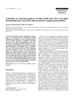
Báo cáo khoa học: " Evaluation of partial cranial cruciate ligament rupture with positive contrast computed tomographic arthrography in dogs" ppt
Bạn đang xem bản rút gọn của tài liệu. Xem và tải ngay bản đầy đủ của tài liệu tại đây (1.52 MB, 6 trang )
JOURNAL OF
Veterinary
Science
J. Vet. Sci. (2008), 9(4), 395
400
*Corresponding author
Tel: +82-43-261-3372; Fax: +82-43-261-3224
E-mail:
Evaluation of partial cranial cruciate ligament rupture with positive
contrast computed tomographic arthrography in dogs
Sungyoung Han
1
, Haengbok Cheon
1
, Hangmyo Cho
1
, Juhyung Kim
1
, Ji-Houn Kang
1
, Mhan-Pyo Yang
1
,
Youngwon Lee
2
, Heechun Lee
3
, Dongwoo Chang
1,
*
1
Veterinary Medical Center, College of Veterinary Medicine, Chungbuk National University, Cheongju 361-763, Korea
2
Department of Veterinary Medicine, College of Veterinary Medicine, Chungnam National University, Daejeon 305-764,
Korea
3
Department of Veterinary Medicine, College of Veterinary Medicine, Gyeongsang National University, Jinju 660-701, Korea
Computed tomographic arthrography (CTA) of four
cadaveric canine stifles was performed before and after
partial cranial cruciate ligament rupture in order to verify
the usefulness of CTA examination for the diagnosis of
partial cranial cruciate ligament rupture. To obtain the
sequential true transverse image of a cranial cruciate
ligament, the computed tomography gantry was angled such
that the scanning plane was parallel to the fibula. True
transverse images of cranial cruciate ligaments were
identified on every sequential image, beginning just
proximal to the origin of the cranial cruciate ligament distal
to the tibial attachment, after the administration of
iodinated contrast medium. A significant decrease in the
area of the cranial cruciate ligament was identified on CTA
imaging after partial surgical rupture of the cranial cruciate
ligament. This finding implies that CTA can be used for
assessing partial cranial cruciate ligament ruptures in dogs.
Keywords: arthrography, computed tomography, cruciate ligament,
dog, rupture
Introduction
Most ligament injuries in canine stifle joints involve some
kind of cranial cruciate ligament rupture, including partial
rupture [9]. This results in severe instability and predisposes
the joint to degenerative changes [7].
The cranial cruciate ligament is attached to a fossa on the
caudal aspect of the medial side of the lateral femoral
condyle. It courses cranially, medially, and distally across
the intercondylar fossa and attaches to the cranial
intercondyloid area of the tibia [1]. The cranial drawer test
is diagnostic of cranial cruciate ligament injuries. A
positive test result implies craniocaudal movement beyond
the 0 mm to 2 mm mobility found in a normal stifle joint.
However, if a partial tear is present, the cranial drawer sign
may reveal only 2 mm to 3 mm of instability when the test
is done with the stifle flexed and no instability with the
stifle in extension [13]. In addition, one study found that 12
of 25 dogs with partial rupture of the cranial cruciate
ligament had no detectable cranial drawer sign in response
to manipulation of the involved stifle [9]. Hence, it is not
surprising that veterinarians encounter difficulties in
diagnosing partial ruptures of the cranial cruciate ligament.
Echography is a useful technique in the evaluation of
intra-articular proliferation of reactive fibrotic tissue of
unstable stifle joints affected by cranial cruciate ligament
rupture as a result of chronic synovitis [6]. However,
ultrasonographic examination is not an accurate test for
cranial cruciate ligament rupture evaluation [6]. To
overcome the diagnostic limitations of ultrasonographic
examination for the detection of cranial cruciate ligament
rupture in one study, the stifle was imaged via dynamic
intra-articular saline injection. The investigators concluded
that ultrasonographic examination of stifle joints had
potential as a diagnostic tool for assessing cranial cruciate
ligament rupture [10]. Nevertheless, ultrasonographic
examinations are highly operator-dependent, and a great
deal of flexibility is often required for good images to be
obtained.
Arthroscopy and magnetic resonance imaging (MRI) may
be useful diagnostic procedures for confirming the diagnosis,
although arthroscopy is invasive and MRI is expensive [3,4].
The advantages of computed tomographic arthrography
(CTA) over MRI include increased availability of equipment,
shorter examination time, and decreased imaging artifacts
[4,12]. Dual-detector helical CTA is as sensitive and specific
as MRI in identifying stifle intraarticular ligamentous
396 Sungyoung Han et al.
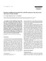
Fig. 1. Lateral radiograph of the stifle showing the relationship
between stifle angle and the cranial cruciate ligament. Metal
landmarks implanted in the cranial cruciate ligament (arrow) are
shown. The cranial cruciate ligament and the fibula cross at a
right angle when the stifle is flexed at 90 degrees. Cr: cranial, Cd:
caudal, F: femur, T: tibia.
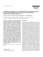
Fig. 2. Photograph of the canine cadaveric stifle joint illustrating
the cranial cruciate ligament. Experimental cranial cruciate
ligament rupture is identified (arrow). L: lateral, M: medial.
pathology [8,11]. However dual-detector helical computed
tomography (CT) is not yet readily available for use in canine
patients.
Recently, the diagnostic utility of single-detector CTA for
identifying ligamentous structures in the normal canine
stifle has been investigated and has been established as a
repeatable imaging protocol [8]. The ligamentous structures
of the normal canine stifle are easily identified using the CTA
protocol described.
The purpose of this study was to optimize the CTA
protocol to obtain sequential true transverse images of
cranial cruciate ligaments and to evaluate the effectiveness
of CTA for detecting partial cranial cruciate ligament
rupture in dogs.
Materials and Methods
Animals
All experimental procedures were approved by the
Institutional Animal Care and Use Committee (Chungbuk
National University, Korea). Four hind limbs obtained from
4 mongrel dogs (body weights 20 to 30 kg) euthanized for
reasons unrelated to this study were used for CTA
investigation. The average age was 16 months. All dogs had
body condition scores (BCS) of 3 on the 5-point BCS system
[5]. Radiographs and synovial fluid examination of the stifle
were performed to confirm the absence of abnormal findings.
The specimens were disarticulated at the hip joint, and all
soft tissues distal to the hip joint were preserved.
CT protocol
Each limb was mounted on a custom-made v-shape
positioner, with the cranial surface of the limb apposed to
the CT couch. The stifle was flexed visually at a 90 degree
angle. All data were collected using a fourth-generation CT
scanner (Picker IQ; Philips Medical Systems, USA). After
acquisition of lateral pilot images, the stifle angle was
readjusted to 90 degrees with the built-in goniometer (Fig.
1). To obtain the true transverse image of the cranial
cruciate ligament, the CT gantry was angled such that the
scanning plane was parallel to the fibula. Two-millimeter
thick, contiguous transverse pre-arthrography CT images
were obtained from just caudal to the femoral epicondyle
to just cranial to the femoral epicondyle. All scans were
performed using a bone algorithm, 85 mA, 130 kVp, and
field of view of 50 mm.
CTA protocol
A 21-gauge needle was directed midway between the
cranial point of the patella and the tibial tuberosity and just
medial to the patella [2]. Digital pressure was applied to the
caudal aspect of the joint opposite the point of entry into the
joint. Iohexol (Omnipaque 300; Nycomed, USA) 150 mg
I/ml was injected into the joint at a volume of 0.3 ml/cm of
the medial to lateral thickness of the joint [2,6]. The joint
was manipulated and massaged to assure even distribution
of the material. The limb was repositioned on the CT couch
as before, and the CT acquisition protocol was repeated.
Surgical procedure
After CTA scans of the intact stifle joint were performed,
the cranial cruciate ligaments were partially transected by
lateral stifle arthrotomy in a routine manner [12]. Partial
transection of the cranial cruciate ligament was performed
locally at the craniomedial band 2 mm distal to the tibial
insertion (Fig. 2). After partial transection was performed,
an extracapsular technique involving lateral imbricating
sutures was used to ensure sealing of the stifle joint.
Residual air within the joint space was removed using the
21-gauge needle. After the procedure was completed,
CTAs were done in the same manner.
Computed tomographic arthrography and partial cranial cruciate ligament 397
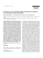
Fig. 3. Two-millimeter sequential transverse computed tomographic arthrography images were scanned parallel to the fibula, femoral
attachment (A), and tibial attachment (I). The cranial cruciate ligament (black arrow) transverse images and caudal cruciate ligamen
t
sagittal images (white arrow) were clearly identified. L: lateral, M: medial.
Image analysis
Using Visus Image Analysis software (Ista-Video Test;
Foresthill Products, USA), the sequential transverse
images of the cranial cruciate ligament were evaluated.
Pre-operative images were compared with post-operative
images in each cadaver.
Results
Nine sequential transverse cranial cruciate ligament
images were obtained (Fig. 3). The initial transverse CTA
image of the intact cranial cruciate ligament at the tibial
attachment revealed a comma shape. The middle stage
revealed a round shape. The final CTA transverse image
obtained at the attachment of the cranial cruciate ligament
to the menisci was eclipse shaped. Total cranial cruciate
ligament slices involved the femoral attachment to the
tibial attachment. The mean number of slices for five
cadavers was 5.7. Five slices were obtained from cadaver
4, while six slices were obtained from the other cadavers.
398 Sungyoung Han et al.
Fig. 4. Comparison of the pre-operative conventional view (A)
and tracing view (C) with the post-operative conventional view
(B) and tracing view (D). The transverse area of the cranial
cruciate ligament image (black arrow) was decreased by 25% on
the post-operative image. The small gas artifact (white arrow)
was considered a normal finding. L: lateral, M: medial.
Tabl e 1 . Transverse area (mm
2
) before and after partial cranial
cruciate ligament rupture
Cadaver
No.
Slice No.
123456
1
2
3
4
Pre
Post
%
Pre
Post
%
Pre
Post
%
Pre
Post
%
0.33
0.34
104
0.54
0.5
91
0.55
0.47
85
0.5
0.46
93
0.48
0.47
99
0.66
0.52
78
0.58
0.56
94
0.56
0.51
90
0.46
0.45
99
0.68
0.62
90
0.68
0.23
*
33
0.55
0.46
*
83
0.57
0.25
*
44
0.71
0.52
*
73
0.7
0.17
*
25
0.5
0.33
*
65
0.56
0.1
*
17
0.74
0.49
*
66
0.8
0.71
88
0.58
0.5
87
0.56
0.3
*
59
0.71
0.64
90
-
-
-
0.66
0.61
93
Pre: pre-operative computed tomographic arthrography, Post:
post-operative computed tomographic arthrography.
*
The slice in
which the partial cranial cruciate ligament rupture was done, % =
post-operative value/pre-operative value ×100.
Images at the same anatomical location were compared
before and after surgery. Defect lesions were identified on
post-surgery CTA images (Fig. 4). Air artifact were also
identified after the procedure was complete.
In the pre-operative period, the cranial cruciate ligament
area range was 0.3∼0.6 mm
2
/kg for cadaver 1, 0.5~0.7
mm
2
/kg for cadaver 2, 0.6∼0.8 mm
2
/kg for cadaver 3, and
0.5∼0.7 mm
2
/kg for cadaver 4. In the post-operative period,
decreases in the area of the cranial cruciate ligament defect
in cadaver 1 were 56% in the fourth slice, 83% in the fifth
slice, and 41% in the sixth slice; in cadaver 2 they were
27% in the fourth slice and 34% in the fifth slice; in cadaver
3 they were 67% in the third slice and 75% in the fourth
slice; in cadaver 4 they were 17% in the third slice and 35%
in the fourth slice (Table 1). The decreases in non-defect
lesion area ranged from -4% to 15%.
In cadaver 2, the area of the lesion in the second slice
decreased by 22%; this finding may have been due to
contrast medium infiltrating the cranial cruciate ligament.
Discussion
We sought to determine the diagnostic utility of single
detector CTA for identifying ligamentous structures in the
normal canine stifle and to establish a repeatable imaging
protocol. The ligamentous structures were easily identified
using the CTA protocol described. Multiplanar reconstructions
were helpful for the evaluation of cranial cruciate
ligaments, medial and lateral menicsi, long digital extensor
tendons, and popliteal tendons.
We tried to orient reconstruction planes to parallel the
axis of the cranial cruciate ligament in order to evaluate its
entire length. The obliquity required to achieve this was
variable within and between dogs. Subtle differences in
limb positioning were not resolved. In addition, there were
limitations in describing the total cranial cruciate ligament
appearance. In an earlier study, the stifle position had no
definite angle and was extended caudally at random. The
CT gantry was angled such that the scanning plane was
parallel to the tibial plateau. For this reason, transverse
cranial cruciate ligament images were obtained atypically
[8]. For successful examination to occur, each patient
needed to be in a fixed position while scanning for constant
images of the cranial cruciate ligament occurred. During
the primary examination, cranial cruciate ligament tension
was observed in various positions. We found that, when the
stifle angle was 90 degrees, the assumption line was
horizontal to the cranial cruciate ligament and vertical to
the fibula. For this reason, the CT gantry was angled such
that the scanning plane was parallel to the fibula. As a
result, we obtained sequential transverse CTA images of
the cranial cruciate ligament.
In the present study, an average of 5.7 slices were taken
from some regular-distant cross sections of whole images
in four cadavers. In the pre-operative stage, the cross
sections of the cranial cruciate ligament progressed from
‘comma’, to ‘round’, to ‘eclipse’, in sequence from the
femoral attachment to the tibial attachment, and the
Computed tomographic arthrography and partial cranial cruciate ligament 399
margination was constantly smooth [4]. However, in the
post-operative period, the comma and round shapes were
maintained at the initial site of femoral attachment, and
afterward the margination became relatively irregular. As a
result, when the area of the two cranial cruciate ligament
pieces were compared, the area in the post-operative period
was smaller than that seen in the pre-operative period at the
same anatomic location. This finding was also noted in the
case of experimental partial ruptures around the tibial
attachment, which we made intentionally.
There are a number of reasons why partial tears were used
as an experimental model. Partial tears in dogs consistently
progress to complete ligament rupture, usually within one
year of the onset of lameness [13]. Furthermore, the definite
diagnosis of partial cranial cruciate ligament tears is more
complicated than the diagnosis of complete tears is. If earlier
surgical procedures prove to retard the progression of
osteoarthritis, it is appropriate to recommend surgery as
soon as possible after the diagnosis of partial tearing has been
confirmed. Thus, partial tear models were used.
In this study, medium to large sized dogs were used for
several reasons. Cruciate ligament disease is particularly
common in large and giant breed dogs, such as the Labrador
Retriever, Rottweiler, and Saint Bernard [7,14], and large
breed dogs are more commonly presented for surgical
management of cranial cruciate ligament rupture. In
addition, the quality of CTA images in large breed dogs is
expected to be better than that seen in small breed dogs [6].
When each experimental partial rupture was performed in
this study, air entered the articular capsule. After the
procedure, air artifact was created near the cranial cruciate
ligament. However, this did not affect the cranial cruciate
ligament.
In all cadavers, the image area for the cranial cruciate
ligaments decreased in partial transection slices. When the
slices from the post-operative period were compared with
those from the pre-operative period in all cadavers, some
decrease in cranial cruciate ligament defect area was seen
in 2 to 3 slices from each cadaver. Considering that the
CTA image thickness is 2 mm, the expected loss would be
between 4 mm and 6 mm in actuality. However, this may
not always be true in clinical practice because we
fashioned a clear transection in the experiment. On
post-operative images, the defects were shown to be
cranial in location. Through CTA image analysis, it was
found that the transverse area of the cranial cruciate
ligament decreased anywhere between 17% and 83%. If
the experimental defect area was not overlooked, the gap
could have been attributed to slight anatomical deviation or
to artifact. These findings support the idea that tears of the
cranial cruciate ligament are visible with the unaided eye
using only CTA, which enables practitioners to make a
standard list for objective examination. Furthermore, our
findings suggest the possibility of establishing a specific
protocol for other ligamentous structures. It is necessary to
do more studies on contrast agent dosing in living subjects,
as well as to better examine the transverse images.
This study showed clear CTA images of acute partial
ruptures of the cranial cruciate ligament. However, most
partial ruptures of the cranial cruciate ligament seen in
clinical practice are chronic, and lesions of the stifle are
effusive or fibrotic. In humans, it is recommended that an
effusive knee be drained prior to the infusion of contrast
medium [4,12]. For this reason, future studies will require
adjustments in the volume or concentration of contrast
medium, as conditions dictate.
References
1. Arnoczky SP, Marshall JL. The cruciate ligaments of the
canine stifle: an anatomical and functional analysis. Am J
Vet Res 1977, 38, 1807-1814.
2. Atilola MA, Pennock PW, Sumner-Smith G. Evaluation
of analytical grade of metrizamide for canine stifle arthrography.
J Am Vet Med Assoc 1984, 185, 436-439.
3. Baird DK, Hathcock JT, Rumph PF, Kincaid SA, Visco
DM. Low-field magnetic resonance imaging of the canine
stifle joint: normal anatomy. Vet Radiol Ultrasound 1998, 2,
87-97.
4. Banfield CM, Morrison WB. Magnetic resonance arthrog-
raphy of the canine stifle joint: technique and applications in
eleven military dogs. Vet Radiol Ultrasound 2000, 41,
200-213.
5. Elliott DA. Disorders of metabolism. In: Nelson RW (ed.).
Small Animal Internal Medicine. p. 818, Mosby, St. Louis,
2003.
6. Gnudi G, Bertoni G. Echographic examination of the stifle
joint affected by cranial cruciate ligament rupture in the dog.
Vet Radiol Ultrasound 2001, 42, 266-270.
7. Hayashi K, Manley PA, Muir P. Cranial cruciate ligament
pathophysiology in dogs with cruciate disease: a review. J
Am Anim Hosp Assoc 2004, 40, 385-390.
8. Samii VF, Dyce J. Computed tomographic arthrography of
the normal canine stifle. Vet Radiol Ultrasound 2004, 45,
402-406.
9. Scavelli TD, Schrader SC, Matthiesen DT, Skorup DE.
Partial rupture of the cranial cruciate ligament of the stifle in
dogs: 25 cases (1982-1988). J Am Vet Med Assoc 1990, 196,
1135-1138.
10. Seong Y, Eom K, Lee H, Lee J, Park J, Lee K, Jang K, Oh
T, Yoon J. Ultrasonographic evaluation of cranial cruciate
ligament rupture via dynamic intra-articular saline injection.
Vet Radiol Ultrasound 2005, 46, 80-82.
11. Vande Berg BC, Lecouvet FE, Poilvache P, Dubuc JE,
Maldague B, Malghem J. Anterior cruciate ligament tears
and associated meniscal lesions: assessment at dual-detector
spiral CT arthrography. Radiology 2002, 223, 403-409.
12. Vande Berg BC, Lecouvet FE, Poilvache P, Maldague B,
Malghem J. Spiral CT arthrography of the knee: technique
and value in the assessment of internal derangement of the
knee. Eur Radiol 2002, 12, 1800-1810.
400 Sungyoung Han et al.
13. Vasseur PB. Stifle joint. In: Slatter D (ed.). Textbook of
Small Animal Surgery. 3rd ed. pp. 2090-2133, Saunders,
Philadelphia, 2003.
14. Whitehair JG, Vasseur PB, Willits NH. Epidemiology of
cranial cruciate ligament rupture in dogs. J Am Vet Med
Assoc 1993, 203, 1016-1019.