Báo cáo khoa học: "Comparative immunohistochemical characterization of canine seminomas and Sertoli cell tumors" ppt
Bạn đang xem bản rút gọn của tài liệu. Xem và tải ngay bản đầy đủ của tài liệu tại đây (4.92 MB, 7 trang )
JOURNAL OF
Veterinary
Science
J. Vet. Sci. (2009), 10(1), 1
7
DOI: 10.4142/jvs.2009.10.1.1
*Corresponding author
Tel: +82-2-450-4153; Fax: +82-2-455-8124
E-mail:
†
First two authors contributed equally to this study.
Comparative immunohistochemical characterization of canine
seminomas and Sertoli cell tumors
Chi-Ho Yu
1,†
, Du-Na Hwang
1,†
, Ji-Young Yhee
1
, Jong-Hyuk Kim
1
, Keum-Soon Im
1
, Whan-Gook Nho
2
,
Young-Soo Lyoo
1
, Jung-Hyang Sur
1,
*
1
Department of Veterinary Pathobiology, Small Animal Tumor Diagnostic Center, College of Veterinary Medicine,
Konkuk University, Seoul 143-701, Korea
2
Department of Animal Science, Korea National Agricultural College, Hwaseong 445-760, Korea
Primary testicular tumors are the most common causes
of cancer in male dogs. Overall, the majority of canine
patients should be cured by testicular surgery. However,
tumor markers are not well-known in veterinary
medicine. We sought to determine using immunohisto-
chemistry whether the combined human testicular tumor
markers (placental alkaline phosphatase, OCT3/4, CD30,
alpha-fetoprotein, inhibin-alpha, vimentin, c-KIT, and
desmin) are expressed in canine seminomas and Sertoli
cell tumors (SCTs). We examined 35 canine testicular
tumors, 20 seminomas and 15 SCTs. c-KIT was expressed
markedly in canine seminomas. Both inhibin-alpha and
vimentin were expressed significantly in canine SCTs. The
results of this study demonstrate differences and
similarities between tumor marker expression of testicular
tumors in dogs and humans. All the main markers in
current routine use are discussed as well as potential
useful markers for benign and malignant tumors, and
tumor progression.
Keywords:
dog, immunohistochemistry, seminoma, Sertoli cell
tumor, tumor markers
Introduction
Testicular tumors arise from germ cells and sex-cord
stromal elements of the testis [22], and are divided into four
general categories: germ cell tumors including seminoma,
teratoma, embryonal carcinoma, and yolk sac carcinoma
arising from the germinal epithelium of the seminiferous
tubules; sex-cord stromal tumors, including Sertoli cell
tumor (SCT) and Leydig (interstitial) cell tumor; mixed
germ cell sex-cord stromal tumors; and primary tumors not
specific to the testis [22,30,32]. The major tumors are
seminoma and SCT, which occur with equal frequency,
showing a prevalence of 0.068-4.6% in mature and old
male dogs [30,31,34]. Seminoma is derived from the germ
cells that constitute the spermatogenic epithelium within
the seminiferous tubules. SCT arises from the supporting
cells within the seminiferous tubules. It is most common in
dogs, but has also been reported in stallions, rams, bulls,
goats, and cats [21,23].
In humans, testicular tumors are the most common
cancers found in men between 15-35-years-of-age, where
90% are germ cell tumors [14,26,32]. Since the rate of
testicular neoplasms is increasing, much research has been
carried out to more accurately diagnose and manage these
patients [2,11]. In particular, many immunohistochemical
markers have been introduced to accurately establish
histological diagnoses and to investigate tumor pathogene-
sis [3,18]. Markers including cytokeratins, c-KIT, CD30,
epithelial membrane antigen, inhibin-alpha, OCT3/4,
placental alkaline phosphatase (PLAP) and alpha
fetoprotein (AFP) are sensitive and specific for the
diagnosis of human testicular tumors [13,17,27,35].
Although tumor markers in canine testicular tumors have
been studied, relatively less is known, which presently
limits their use in diagnosis and evaluation of tumor
pathogenesis.
In the present study, we examined tumor markers
including PLAP, AFP, inhibin-alpha, vimentin, OCT3/4,
CD30, desmin, and c-KIT to determine expression of these
proteins and an appropriate antibody panel. To accomplish
this, we employed immunohistochemical staining of
canine seminomas and SCTs.
2 Chi-Ho Yu et al.
Tabl e 1 . Primary antibodies used for immunohistochemical
staining
Antibody Clone Type Dilution
PLAP PL8/F6 Monoclonal Ready to use
AFP C3 Monoclonal 1:400
Inhibin-α R1 Monoclonal 1:100
Vimentin Vim3B4 Monoclonal 1:400
OCT 3/4 C-10 Monoclonal 1:50
c-KIT C-19 Monoclonal 1:50
Desmin 33 Monoclonal 1:100
CD30 Ber-H2 Polyclonal 1:400
PLAP: placental alkaline phosphatase, AFP: alpha fetoprotein.
Tabl e 2. Frequency of expression of the antigen in canine
seminomas (n = 20) and Sertoli cell tumors (n = 15)*
Antibody
No. immunopositive cases (%)
Seminoma Sertoli cell tumor
PLAP 4 (20%) 6 (40%)
AFP 8 (40%) 8 (53.3%)
Inhibin-α 9 (45%) 14 (93.3%)
Vimentin 5 (25%) 14 (93.3%)
OCT 3/4 0 (0%) 0 (0%)
c-KIT 20 (100%) 10 (66.6%)
Desmin 6 (30%) 5 (33.3%)
CD30 0 (0%) 0 (0%)
*
The result was scored as positive if > 10% of tumor cells showed
the staining. PLAP: placental alkaline phosphatase, AFP: alpha
fetoprotein.
Materials and Methods
Tissues from testicular tumors in dogs
Testicular specimens from 35 male dogs with seminomas
(n = 20) or SCTs (n = 15) were obtained from the files of the
Department of Pathobiology, Small Animal Tumor
Diagnostic Center, Konkuk University, Seoul, Korea. In
addition, two normal testicular samples from neutered
male dogs were used as negative controls. Histopatholo-
gical analyses based on hematoxylin and eosin staining
were performed.
Immunohistochemical staining
Each testis was fixed in 10% neutral buffered formalin.
Blocks that contained testis tumor tissue were embedded in
paraffin wax. Serial 4 μm-thick sections were acquired
from each paraffin-block for immunohistochemical
staining. Monoclonal and polyclonal antibodies (Table 1)
included PLAP (Biogenex, USA), AFP (Biogenex),
inhibin-alpha (Dako, USA), vimentin (Dako), OCT3/4
(Santa Cruz Biotechnology, USA), CD30 (Santa Cruz
Biotechnology), desmin (Biogenex, USA), and c-KIT
(Dako, USA). Sections were deparaffinized in xylene,
rehydrated in a graded ethanol series, treated with 3%
hydrogen peroxide solution for 20 min at room
temperature, and washed three times with phosphate-
buffered saline (PBS; pH 7.4, 137 mM NaCl, 2.7 mM KCl,
10 mM Na
2
HPO
4
, 2 mM KH
2
PO
4
). For inhibin-alpha,
OCT3/4 and CD30, antigen retrieval was performed by
heating slides in Tris-EDTA buffer (pH 9) in a microwave
oven (650 W, high power) for 20 min. After 3 min washes
with PBS, sections to be stained with AFP, vimentin, and
c-KIT antibodies were incubated in a blocking solution of
5% normal goat serum (Vector Laboratories, USA) for 30
min. All primary antibodies were incubated with sections
overnight at 4
o
C. To “visualize” immunolabeling, a
two-step EnVision system (Dako, USA) was applied after
removal of the primary antibody. In this system, EnVision
rabbit/mouse reagent conjugated to horseradish
peroxidase was applied for 40 min at room temperature.
The slides were subsequently washed four times in PBS
and incubated with the supplied substrates until the desired
color intensity developed. The reaction was stopped by
washing in distilled water. Sections were counterstained
with Harris hematoxylin.
Digital image preparation and assessment of
immunohistochemical labeling
Images were acquired using an Olympus BX41
microscope (Olympus, USA) fitted with a Leica DFC 290
digital camera (Leica, Switzerland) and analyzed for
positive signals using Image-Pro Plus software ver. 4.1
(Media Cybernetics, USA). The immunolabeling was
scored as positive if > 10% of tumor cells displayed
staining.
Results
Characteristics of histopathology and immuno-
staining in seminoma and SCTs
The results of immunohistochemical studies are
summarized in Tables 2 and 3. PLAP, AFP, inhibin-alpha,
vimentin, desmin, and c-KIT were expressed in
seminomas and SCTs, while OCT3/4 and CD30 were not
expressed. In normal Leydig cells, inhibin alpha, vimentin,
and desmin antibodies were weakly immunoreactive.
Normal Sertoli cells had focal staining for vimentin and
c-KIT. PLAP, AFP, OCT3/4, and CD30 produced no
immunoreactivity in normal testis (data not shown).
Seminomas consisted of aggregates of germ cells that
Canine seminomas and Sertoli cell tumors 3
Tabl e 3. Immunoreactivity of PLAP, AFP, inhibin-alpha, vimentin,
OCT3/4, CD30, desmin, and c-KIT in normal canine testis (n = 2)*
Antibody
Normal testis
Leydig Sertoli Spermatogenic
cell cell cell
PLAP
AFP
Inhibin-α 1+ 1+
Vimentin 2+ 2+
OCT 3/4
c-KIT 1+
Desmin 1+
CD30
*
= no staining, 1+ = <10% positive cells, 2+ = 10 to 50%
p
ositiv
e
cells, 3+ = >50% positive cells. PLAP: placental alkaline
phosphatase, AFP: alpha fetoprotein.
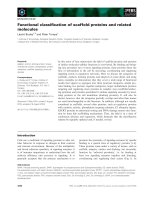
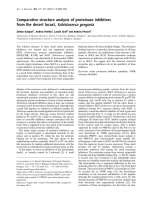
Fig. 1. Immunohistochemical markers in canine seminoma. (A) Seminomas consisted of aggregates of germ cells that filled the affecte
d
seminiferous tubules. The tumor cells were large and polyhedral with vesicular nuclei and prominent nucleoli. H&E stain. (B-F)
Positive signals to tumor cells; (B) alpha-fetoprotein, (C) inhibin-alpha, (D) vimentin, (E) desmin and (F) c-KIT. Immunostain and
counterstain with Harris hematoxylin. Scale bars = A: 350 μm, B-F: 140 μm.
filled the lumens or sheets of the affected seminiferous
tubules. These cells replaced the normal lining of
spermatogenic and Sertoli cells. Large and irregular cellular
aggregates and small clusters of cells were seen to varying
extents. Dense fibrous bands subdivided the tumor into
large discrete nodules. The tumor cells were large and
polyhedral with vesicular nuclei and prominent nucleoli
(Fig. 1A). PLAP staining was moderate-to-diffuse in four
cases. AFP and inhibin-alpha were positive in eight and nine
cases, respectively. AFP was diffusely expressed in tumor
cells (Fig. 1B), while inhibin-alpha demonstrated diffuse
immunoreactivity (Fig. 1C). Vimentin and desmin were
positive in five and six separate cases, respectively.
Expression of vimentin produced strongly positive
immunoreactivity in some tumor cells within some
seminomas (Fig. 1D). Expression of desmin was diffuse and
focal to the cytoplasm of tumor cells (Fig. 1E). No OCT3/4
or CD30 staining was evident in the tumors (data not
shown). C-KIT was strongly positive in all cases (Fig. 1F).
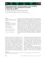
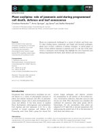
In SCTs, there was abundant fibrous tissue stroma. Cells
within the tumor resembled Sertoli cells that normally
populate the seminiferous tubules, and were arranged into
sheets or tubules separated by fibrous connective tissue.
The tumor cells had small, round or elongated nuclei with
an eosinophilic cytoplasm (Fig. 2A). Of 15 SCT samples,
six cases were positive for PLAP with weak and focal
immunostaining. In eight cases, AFP was expressed in the
cytoplasm of tumor cells (Fig. 2B). Fourteen cases were
positive for inhibin-alpha and vimentin. While the tumor
cells were strongly positive for inhibin-alpha (Fig. 2C),
they were only weakly positive for vimentin (Fig. 2D). All
SCTs were negative for OCT3/4 (data not shown). CD30
was not expressed in tumor cells (data not shown). Focal
immunostaining of desmin was observed in five cases (Fig.
2E). Ten cases were positive for c-KIT, where its
expression was distributed diffusely but strongly in the
cytoplasm of tumor cells (Fig. 2F).
Discussion
Primary testicular tumors are common in mature and old
male dogs [34]. The main types of testicular tumors in dogs
are seminomas and SCTs, which are associated with
4 Chi-Ho Yu et al.
Fig. 2. Immunohistochemical markers in canine Sertoli cell tumors. (A) Cells within the tumor resemble Sertoli cells that normally
p
opulate the seminiferous tubules and are arranged into sheets or tubules separated by fibrous connective tissues. H&E stain. (B-F)
Positive signals to tumor cells; (B) alpha-fetoprotein, (C) inhibin-alpha, (D) vimentin, (E) desmin and (F) c-KIT. Immunostain and
counterstain with Harris hematoxylin. Scale bars = A: 350 μm, B-F: 140 μm.
chryptorchidism. In humans, testicular tumors are the most
common cancers found in men, with an increasing rate of
incidence [26,32]. Substantial research on human
testicular tumors has resulted in more accurate diagnoses
and better management of patients [3,11]. In particular,
many tumor markers have been found for tumor
pathogenesis [1,4,6]. Of these markers, cytokeratins,
c-KIT, CD30, epithelial membrane antigen, inhibin-alpha,
OCT3/4, PLAP, and AFP are widely used because of their
sensitivity and specificity. The usefulness of these as tumor
markers has been assessed in many studies [13,17,27,35].
Canine testicular tumor markers have been studied
[23,28,34]. However, such veterinary tumor markers in
male canine patients still require more study to be useful in
accurate diagnosis. Thus, the purpose of our present study
was to establish the specificity of tumor markers including
PLAP, AFP, inhibin-alpha, vimentin, OCT3/4, CD30,
desmin, and c-KIT in canine seminoma and SCT.
In the current seminoma series, c-KIT was found to be the
most sensitive marker, because tumor cells of all
seminoma cases were homogeneously and strongly
positive for the antibody. c-KIT is a product of the c-KIT
oncogene, which encodes a type III transmembrane
tyrosine kinase receptor that is required in normal
spermatogenesis. High expression of c-KIT is found in
human testicular germ cell tumors, especially in human
seminoma [26]. KIT signal transduction appears to be an
important pathway for carcinogenesis of seminoma [12].
Our result demonstrates that c-KIT is potently expressed
and is potentially a specific tumor marker in canine
seminoma, similar to human tumors. OCT3/4, also known
as otf3 or pou5f1, is a member of the POU family of
transcription factors expressed in pluripotent mouse and
human embryonic stem and germ cells [7,10,29].
Unexpectedly, OCT3/4, which is regarded as a highly
sensitive marker for human seminoma and embryonal
carcinoma [2,33], was not expressed in any canine
seminoma. OCT3/4 may not be a reliably specific tumor
marker in canine seminoma. PLAP, a membrane-bound
enzyme normally synthesized by placental syncytiotro-
phoblasts [4], has been widely applied in the significant
markers of human seminoma. In contrast, PLAP staining
was detected weakly and heterogeneously in canine SCTs.
Although a positive reaction was observed in 20% of cases,
the result was less significant than that observed in human
seminoma PLAP immunostaining. This result might be
related with the classification of testicular tumors
including two types of seminoma. The first type is classical
seminoma (SE) and the second type is spermatocytic
seminoma (SS). Grieco et al. [8] showed that neoplastic
cells are immunoreactive for PLAP in all cases with SE.
Neoplastic cells in SS are essentially negative for PLAP
[8]. Therefore, our result combined with the observations
of Grieco et al. [8] indicate that 20% cases displaying a
positive reaction were likely SE and the 80% cases
displaying negative reaction were SS. Inhibin-alpha, a
subunit of inhibin, is secreted mainly from testicular
Sertoli cells with an additional small contribution from
Leydig cells [4,34]. Although little is known about the cells
secreting inhibin in primary testicular tumors of humans
and older animals [5,9,16], inhibin immunoreactivity has
been biochemically estimated in human and canine
Canine seminomas and Sertoli cell tumors 5
testicular tumors. In the current seminoma series,
inhibin-alpha was observed in the cytoplasm of tumors cell
in 45% of the cases. Vimentin and desmin was expressed in
25% and 30%, respectively, of canine seminoma samples.
As for vimentin and desmin, unlike the diffuse cytoplasmic
staining present in SCTs, immunoreactivity in seminomas
was observed confined to the cytoplasm of the tumor cells.
Vimentin and desmin are known as markers of
mesenchymal origin tumors including connective tissue,
endothelium, hematopoietic cell, and muscle. Because of
the origin of seminoma is germ cells originating from
epithelial tissue, vimentin and desmin are not appropriate
markers. CD30 is a membrane glycoprotein of the tumor
necrosis factor receptor superfamily, which plays a useful
role in identifying primary embryonal carcinoma in
humans [33]. CD30 is also useful for distinguishing
embryonal carcinoma from seminoma [19]. When the
fixation is not adequate, seminoma may be confused with
the solid pattern of embryonal carcinoma [36]. The
distinction between seminoma and embryonal carcinoma
is very important because the treatment differs. Our result
shows that CD30 was not expressed in canine seminoma.
Although there was no case of embryonal carcinoma in this
study, this result suggests that CD30 can be used to
differentiate embryonal carcinoma from seminoma.
These immunohistochemical results demonstrate that
c-KIT is a sensitive marker for seminoma in dogs. In
human seminoma, c-KIT is expressed in seminomas at the
high prevalence rate of 88-100%, making it one of the most
effective immunohistochemical markers [1,18,26].
Although PLAP, AFP, inhibin-alpha, desmin, and vimentin
immunoreactivity were observed in this study, they did not
demonstrate significant results.
The immunoreactivity of SCTs indicates that inhibin-
alpha and vimentin were the most remarkable immuno-
reactivity of all the antibodies used in this study. Inhibin-
alpha and vimentin were expressed with a high prevalence
rate of 93.3% and were diffusely and moderately evident in
the cytoplasm of neoplastic cells. Previous studies did not
detect immunohistochemical expression of inhibin-alpha
in SCTs in dogs [31,34]. In this study, however, this
expression was observed in 93.3% of SCTs. Grootenhuis et
al. [9] showed that peripheral levels of immunodetectable
inhibin in dogs with SCTs are higher than those in normal
dogs. Kawakami et al. [15] also demonstrated that blood
plasma inhibin alpha concentration of dogs with SCT is
higher than normal testis. In human testicular SCT,
Iczkowski et al. [11] reported positive inhibin-alpha
immunostaining in 10 of 11 (91%) testicular SCT. The
results of our study combined with those of Grootenhuis et
al. [9], Kawakami et al. [15] and Iczkowski et al. [11] show
that inhibin-alpha is likely to be the material secreted from
the neoplastic cells of SCTs. In the current series, inhibin
alpha immunostaining of Sertoli cells was evident in
normal testis. The staining intensity in normal Sertoli cells
was consistently lower than in the tumor cells of SCTs.
Sertoli cells are the main source of inhibin production in
the male. This result also suggests that neoplastic Sertoli
cells in SCTs produce abundant inhibin alpha. Vimentin is
a major intermediate filament present in the cytoplasm of
Sertoli cells, and it has been used to identify these cells
[24]. In humans, vimentin immunostaining is positive in
SCTs [20]. Similar to human studies, our canine study
showed that vimentin might be still the intermediate
filament forming the basic structure of the cells of canine
SCTs. PLAP was positive in some cases, although, in
theory, they should not be expressed in SCTs. In humans
PLAP immunostaining is also negative in SCTs [16,20]. To
more precisely determine the expression profile of these
proteins in canine SCTs, further longitudinal studies
should be performed. CD30 was not found in SCTs, which
is consistent with the idea that CD30 is expressed only in
embryonal carcinoma. OCT3/4 was not identified in SCTs.
SCT and seminoma reportedly became OCT3/4-positive
in humans, but such an immunoreaction in neoplastic cells
was not presently demonstrated. Desmin was expressed
focally, and only five cases were positive for the cytoplasm
of tumor cells. Although c-KIT and AFP were expressed
with a diffuse pattern in 10 cases of SCT, they were a
limited marker compared with inhibin-alpha or vimentin.
Based on these results, c-KIT is the most sensitive marker
in canine seminomas, while inhibin-alpha and vimentin are
the most sensitive markers in canine SCTs. Although
OCT3/4 and PLAP are regarded as the most suitable
immunohistochemical markers in human germ cell tumors
including seminoma, these were either not expressed or
only weakly expressed in thecanine seminomas presently
examined. One interpretation of these results might be that
all the antibodies used in this experiment were produced
against human proteins. As a result, these antibodies might
fail to detect canine PLAP or OCT3/4. However, it is also
reasonable to think that OCT3/4 and PLAP are not
effective markers for canine seminoma. Inhibin-alpha and
vimentin were expressed markedly in canine SCTs in a
similar fashion as in human cells. Therefore, these proteins
are specific markers in canine SCTs.
In conclusion, the results of this study demonstrate that
there are differences and similarities between the
expression of testicular tumor markers in dogs and
humans. Further investigation is required to determine
whether expression of c-KIT, inhibin alpha, vimentin,
AFP, and PLAP is related with tumor metastasis or
malignancy, and to elucidate the role of these proteins in
the development of canine testicular tumors.
Acknowledgments
We thank Ms. R-H Jang for her excellent technical
6 Chi-Ho Yu et al.
assistance. This study was supported in part by a grant for
scientific animal research from the Ministry of Agriculture
and Forestry of Korea.
References
1. Biermann K, Klingmüller D, Koch A, Pietsch T, Schorle
H, B
üttner R, Zhou H. Diagnostic value of markers M2A,
OCT3/4, AP-2gamma, PLAP and c-KIT in the detection of
extragonadal seminomas. Histopathology 2006, 49, 290-
297.
2. Cheng L. Establishing a germ cell origin for metastatic
tumors using OCT4 immunohistochemistry. Cancer 2004,
101, 2006-2010.
3. Cheng L, Thomas A, Roth LM, Zheng W, Michael H,
Karim FW. OCT4: a novel biomarker for dysgerminoma of
the ovary. Am J Surg Pathol 2004, 28, 1341-1346.
4. Debora J, Junqi Q, David GB. Immunohistology of the
prostate, bladder, testis and kidney. In: Dabbs DJ (ed.).
Diagnostic Immunohistochemistry. 2nd ed. pp. 75-86,
Churchill Livingstone, New York, 2002.
5. De Jong FH, Grootenhuis AJ, Steenbergen J, van Sluijs
FJ, Foekens JA, ten Kate FJ, Oosterhuis JW, Lamberts
SW, Klijn JG. Inhibin immunoreactivity in gonadal and
non-gonadal tumors. J Steroid Biochem Mol Biol 1990, 37,
863-866.
6. De Vico G, Papparella S, Di Guardo G. Number and size
of silver-stained nucleoli (Ag-NOR clusters) in canine
seminomas: correlation with histological features and
tumour behaviour. J Comp Pathol 1994, 110, 267-273.
7. Goto T, Adjaye J, Rodeck CH, Monk M. Identification of
genes expressed in human primordial germ cells at the time
of entry of the female germ line into meiosis. Mol Hum
Reprod 1999, 5, 851-860.
8. Grieco V, Riccardi E, Rondena M, Ciampi V, Finazzi M.
Classical and spermatocytic seminoma in the dog:
histochemical and immunohistochemical findings. J Comp
Pathol 2007, 137, 41-46
9. Grootenhuis AJ, van Sluijs FJ, Klaij IA, Steenbergen J,
Timmerman MA, Bevers MM, Dieleman SJ, de Jong FH.
Inhibin, gonadotrophins and sex steroids in dogs with Sertoli
cell tumours. J Endocrinol 1990, 127, 235-242
10. Hansis C, Grifo JA, Krey LC. Oct-4 expression in inner
cell mass and trophectoderm of human blastocysts. Mol
Hum Reprod 2000, 6, 999-1004
11. Iczkowski KA, Butler SL. New immunohistochemical
markers in testicular tumors. Anal Quant Cytol Histol 2006,
28, 181-187.
12. Izquierdo MA, Van der Valk P, Van Ark-Otte J, Rubio
G, Germa-Lluch JR, Ueda R, Scheper RJ, Takahashi T,
Giaccone G. Differential expression of the c-kit proto-
oncogene in germ cell tumours. J Pathol 1995, 177, 253-258
13. Jacobsen GK, Jacobsen M. Alpha-fetoprotein (AFP) and
human chorionic gonadotropin (HCG) in testicular germ cell
tumours. A prospective immunohistochemical study. Acta
Pathol Microbiol Immunol Scand [A] 1983, 91, 165-176.
14. Jones TD, Ulbright TM, Eble JN, Baldridge LA, Cheng
L. OCT4 staining in testicular tumors: a sensitive and
specific marker for seminoma and embryonal carcinoma.
Am J Surg Pathol 2004, 28, 935-940.
15. Kawakami E, Hirano T, Hori T, Tsutsui T. Testicular
superoxide dismutase activity, heat shock protein 70
concentration and blood plasma inhibin-alpha concentration
of dogs with a Sertoli cell tumor in a unilateral cryptorchid
testis. J Vet Med Sci 2007, 69, 1259-1262
16. Kommoss F, Oliva E, Bittinger F, Kirkpatrick CJ, Amin
MB, Bhan AK, Young RH, Scully RE. Inhibin-alpha
CD99, HEA125, PLAP, and chromogranin immunoreactivity
in testicular neoplasms and the androgen insensitivity
syndrome. Hum Pathol 2000, 31, 1055-1061
17. Koshida K, Uchibayashi T, Yamamoto H, Hirano K.
Significance of placental alkaline phosphatase (PLAP) in the
monitoring of patients with seminoma. Br J Urol 1996, 77,
138-142.
18. Lau SK, Weiss LM, Chu PG. D2-40 immunohistochemistry
in the differential diagnosis of seminoma and embryonal
carcinoma: a comparative immunohistochemical study with
KIT (CD117) and CD30. Mod Pathol 2007, 20, 320-325.
19. Leroy X, Augusto D, Leteurtre E, Gosselin B. CD30 and
CD117 (c-kit) used in combination are useful for
distinguishing embryonal carcinoma from seminoma. J
Histochem Cytochem 2002, 50, 283-285
20. McCluggage WG, Shanks JH, Whiteside C, Maxwell P,
Banerjee SS, Biggart JD. Immunohistochemical study of
testicular sex cord-stromal tumors, including staining with
anti-inhibin antibody. Am J Surg Pathol 1998, 22, 615-619
21. McEntee K. Scrotum, Spermatic cord and testis proliferative
lesions. In: McEntee K (ed.). Reproductive Pathology of
Domestic Mammals. pp. 279-300, Academic Press, San
Diego, 1990.
22. McLachlan NJ, Kennedy PC. Tumors of the genital system.
In: Meuten DJ (ed.). Tumors in Domestic Animals. 4th ed.
pp. 561-573, Iowa State University Press, Ames, 2002.
23. Miller MA, Hartnett SE, Ramos-Vara JA. Interstitial cell
tumor and Sertoli cell tumor in the testis of a cat. Vet Pathol
2007, 44, 394-397.
24. Mooney EE, Nogales FF, Bergeron C, Tavassoli FA.
Retiform Sertoli-Leydig cell tumours: clinical, morphological
and immunohistochemical findings. Histopathology 2002,
41, 110-117
25. Mostofi FK, Sesterhenn IA. Pathology of germ cell tumors
of testes. Prog Clin Biol Res 1985, 203, 1-34.
26. Nakai Y, Nonomura N, Oka D, Shiba M, Arai Y,
Nakayama M, Inoue H, Nishimura K, Aozasa K, Mizutani
Y, Miki T, Okuyama A. KIT (c-KIT oncogene product)
pathway is constitutively activated in human testicular germ
cell tumors. Biochem Biophys Res Commun 2005, 337,
289-296.
27. Nikolaou M, Valavanis C, Aravantinos G, Fountzilas G,
Tamvakis N, Lekka I, Arapantoni-Dadioti P, Zizi A,
Ghiconti I, Economopoulos T, Pectasides D. KIT
expression in male germ cell tumors. Anticancer Res 2007,
27, 1685-1688.
28. Owston MA, Ramos-Vara JA. Histologic and immuno-
histochemical characterization of a testicular mixed germ
cell sex cord-stromal tumor and a leydig cell tumor in a dog.
Vet Pathol 2007, 44, 936-943.
Canine seminomas and Sertoli cell tumors 7
29. Pera MF, Herszfeld D. Differentiation of human
pluripotent teratocarcinoma stem cells induced by bone
morphogenetic protein-2. Reprod Fertil Dev 1998, 10, 551-
55
30. Peters MA, Teerds KJ, van der Gaag I, de Rooij DG, van
Sluijs FJ. Use of antibodies against LH receptor, 3beta-h
droxysteroid dehydrogenase and vimentin to characterize
different types of testicular tumour in dogs. Reproduction
2001, 121, 287-296.
31. Rajpert-De Meyts E. Recent advances and future directions
in research on testicular germ cell cancer. Int J Androl 2007,
30, 192-197.
32. Rosai J. Rosai and Ackerman's Surgical Pathology. 4th ed.
pp. 1412-1456, Mosby, Edinburgh, 2004.
33. Sung MT, Jones TD, Beck SD, Foster RS, Cheng L. OCT4
is superior to CD30 in the diagnosis of metastatic embryonal
carcinomas after chemotherapy. Hum Pathol 2006, 37,
662-667.
34. Taniyama H, Hirayama K, Nakada K, Numagami K,
Yaosaka N, Kagawa Y, Izumisawa Y, Nakade T, Tanaka
Y, Watanabe G, Taya K. Immunohistochemical detection
of inhibin-alpha, -betaB, and -betaA chains and 3beta-
ydroxysteroid dehydrogenase in canine testicular tumors
and normal testes. Vet Pathol 2001, 38, 661-666.
35. Teng LH, Lu DH, Xu QZ, Fu YJ, Yang H, He ZL.
Expression and diagnostic significance of OCT4, CD117
and CD30 in germ cell tumors. Zhonghua Bing Li Xue Za
Zhi 2005, 34, 711-715.
36. Ulbright TM, Amin MB, Young RH. Tumors of the Testis,
Adnexa, Spermatic Cord, and Scrotum. 3rd ed. pp. 59-85,
Armed Forces Institute of Pathology, Washington DC, 1999.