Báo cáo khoa học: " A biosensor assay for the detection of Mycobacterium avium subsp. paratuberculosis in fecal samples" potx
Bạn đang xem bản rút gọn của tài liệu. Xem và tải ngay bản đầy đủ của tài liệu tại đây (587.01 KB, 8 trang )
JOURNAL OF
Veterinary
Science
J. Vet. Sci. (2009), 10(1), 35
42
DOI: 10.4142/jvs.2009.10.1.35
*Corresponding author
Tel: +1-607-253-3675; Fax: +1-607-253-3083
E-mail:
A biosensor assay for the detection of Mycobacterium avium subsp.
paratuberculosis in fecal samples
Vijayarani Kumanan
1
, Sam R. Nugen
2
, Antje J. Baeumner
2
, Yung-Fu Chang
1,
*
1
Animal Health Diagnostic Center, Department of Population Medicine and Diagnostic Sciences, College of Veterinary
Medicine, and
2
Department of Biological and Environmental Engineering, Cornell University, Ithaca, NY, USA
A simple, membrane-strip-based lateral-flow (LF)
biosensor assay and a high-throughput microtiter plate assay
have been combined with a reverse transcriptase
polymerase chain reaction (RT-PCR) for the detection of a
small number (ten) of viable Mycobacterium (M.) avium
subsp. paratuberculosis (MAP) cells in fecal samples. The
assays are based on the identification of the RNA of the
IS900 element of MAP. For the assay, RNA was extracted
from fecal samples spiked with a known quantity of (10
1
to
10
6
) MAP cells and amplified using RT-PCR and identified
by the LF biosensor and the microtiter plate assay. While the
LF biosensor assay requires only 30 min of assay time, the
overall process took 10 h for the detection of 10 viable cells.
The assays are based on an oligonucleotide sandwich
hybridization assay format and use either a membrane flow
through system with an immobilized DNA probe that
hybridizes with the target sequence or a microtiter plate
well. Signal amplification is provided when the target
sequence hybridizes to a second DNA probe that has been
coupled to liposomes encapsulating the dye, sulforhodamine
B. The dye in the liposomes provides a signal that can be
read visually, quantified with a hand-held reflectometer, or
with a fluorescence reader. Specificity analysis of the assays
revealed no cross reactivity with other mycobacteria, such
as M. avium complex, M. ulcerans, M. marium, M. kansasii,
M. abscessus, M. asiaticum, M. phlei, M. fortuitum, M.
scrofulaceum, M. intracellulare, M. smegmatis, and M. bovis.
The overall assay for the detection of live MAP organisms is
comparatively less expensive and quick, especially in
comparison to standard MAP detection using a culture
method requiring 6-8 weeks of incubation time, and is
significantly less expensive than real-time PCR.
Keywords:
feces, lateral flow biosensor assay, liposomes,
Mycobacterium avium subsp. paratuberculosis, RT-PCR
Introduction
Mycobacterium avium subsp. paratuberculosis (MAP) is
the causative agent of Johne’s disease (JD), a chronic
intestinal granulamatous infection affecting domestic and
wild ruminants [7,11,15,32]. Although cattle are usually
infected early in life, clinical signs do not develop until 2-4
years of age, which makes early diagnosis of this infection
a difficult task. JD is considered to be an economically
important disease and accounts for an annual loss of $220
million to the US dairy industry [25]. The proposed, but
poorly defined association of MAP with Crohn’s disease in
human beings, is also of concern [13,18,23,24]. The in vivo
diagnosis of MAP infections is quite challenging and
difficult in the pre-clinical stages since the majority of
infected animals do not show symptoms of the disease.
Although the isolation and identification of MAP is the
most definitive test for diagnosis, it is time-consuming and
labor-intensive, requiring 8-12 weeks. Contamination is an
added problem when MAP is cultured from fecal samples.
Although, PCR for IS900 sequences is of diagnostic value,
at times PCR leads to false positive amplification due to the
presence of environmental bacteria with similar sequences
[10]. Novel sequences recently identified in the genome of
MAP appear specific and may also be used in nucleic acid-
based diagnostic tests [6,16]. Real time PCR-based assays,
which involve high equipment costs and trained personnel,
can be used only under well-established laboratory
conditions and serological tests may lack sensitivity [8].
Most diagnostic laboratories continue to use traditional
culture methods; few laboratories use molecular methods
along with culture methods [14,21,26,30,31]. Development
of bioanalytical systems, such as biosensors coupled with
a reverse transcriptase PCR to achieve low limits of
detection, will be useful in the rapid and accurate detection
of MAP.
Biosensors based on nucleic acid hybridization and
liposome signal amplification have been shown to be very
useful in developing rapid, inexpensive, and easy-to-
36 Vijayarani Kumanan et al.
handle systems for the detection and quantification of RNA
molecules [1,2,4,5]. A biosensor is a lateral flow assay that
provides visual or reflectance data within about 20 min of
overall assay time [3]. A biosensor uses a membrane flow-
through system with an immobilized DNA probe that
hybridizes with the target. Signal amplification is provided
when the target sequence hybridizes to a second DNA probe
coupled to liposomes encapsulating the dye, sulforhodamine
B (SRB). The amount of liposomes captured in the detection
zone can be either read visually or quantified with a hand-
held reflectometer.
For MAP diagnosis, the IS900 gene, with 15-20 copies
[27], has been routinely used in PCR-based detection
systems. However, in the past, IS900 primers have also
amplified IS900-like PCR products, probably from
environmental mycobacteria, and resulting in false
positive results [10]. Despite this possibility, IS900 gene
amplification should still serve as a good indicator when
coupled to a high-specificity hybridization reaction, as
proposed here. Apart from IS900, other novel sequences,
such as ISMav2 [26] and ISMap02 [20,27], could also be
potential candidates in PCR-based assays. In the current
study, the development of a rapid biosensor assay for the
detection of live MAP organisms employing IS900 gene
sequences is described. This is the first time the biosensor
assay for MAP has been demonstrated.
Materials and Methods
Bacterial strain and growth
Mycobacterium avium subsp. paratuberculosis-66115-98,
a clinical isolate available from the Department of
Population Medicine and Diagnostic Sciences at Cornell
University, was grown in 7H9 medium, supplemented with
10% oleic acid-albumin-dextrose-catalase (Becton, Dickinson
and Company, USA) and Mycobactin J (Allied Monitor,
USA). The cultures were grown at 37
o
C for 8 weeks and used
in this study.
RNA extraction
MAP cultures were centrifuged at 12,000 rpm for 10 min.
One ml of Trizol was added to the pellet, and the mixture was
passed through the syringe and needle (22 gauge) several
times. The mixture was kept at room temperature for 5 min.
Two hundred μl of chloroform was added and mixed
vigorously for 15 sec and incubated at room temperature for
3 min. The mixture was spun in a microcentrifuge at 12,000
rpm for 15 min at 4
o
C. The supernatant was transferred to
a fresh microcentrifuge tube and an equal volume of 70%
alcohol was added at room temperature. The mixture was
transferred to the minispin column of a RNeasy kit (Qiagen,
USA) and RNA was isolated following the manufacturer’s
protocol. The isolated RNA samples were treated with 10
U/μl of RNase-free DNase I (Qiagen, USA) at 37
o
C for 10
min, followed by heat inactivation at 95
o
C for 5 min, and then
chilled on ice.
Estimation of cell quantity by optical density
MAP organisms were quantified by measuring the optical
density at 550 nm as described earlier [17]. An optical
density of 0.25 at 550 nm was equivalent to approximately
10
8
organisms per ml.
Quantitation of cell number
The organisms were harvested by centrifugation, diluted
in phosphate buffered saline (PBS; NaCl, 0.8%; KCl,
0.02%; Na
2
HPO
4
, 0.115%; and KH
2
PO
4
, 0.02% [pH 7.2])
containing 0.05% Tween-80, loaded on the platform of an
improved Neubauer haemocytometer chamber, and visually
counted.
Preparation of spiked fecal samples
Fecal samples were collected from healthy animals for
initial standardization. Ten-fold serial dilutions of viable
MAP organisms were prepared from a stock suspension of
10
8
organisms. Aliquots of each bacterial dilution (900 μl)
were added to 100 mg of feces to yield bacterial numbers
between 10
1
and 10
6
. For samples from infected animals,
25-50 gm of fecal samples were collected from 8 calves
challenged with 10
7
MAP cells/animal in milk replacer for
7 consecutive days. One hundred mg of fecal samples
collected 2, 4, 6, 8, and 10 days after challenge were used
for RNA isolation.
RNA extraction from spiked fecal samples
RNA was extracted from spiked fecal samples containing
10
1
to 10
6
organisms using Trizol (Invitrogen, USA) and
the extracted RNA was resuspended in 10 μl RNase-free
water. The isolated RNA samples were treated with 10
U/μl of RNase-free DNase I (Qiagen, USA) at 37
o
C for 10
min, followed by heat inactivation at 95
o
C for 5 min, and
then chilled on ice.
Reverse-Transcriptase PCR
RNA isolated from spiked fecal samples was amplified
using a one-step RT-PCR kit (Qiagen, USA). IS900 primers
were used for amplification. The RT-PCR products were
electrophoresed and checked on a 1% agarose gel containing
5 μg of ethidium bromide. The amplified products were
used in the biosensor assay.
Preparation of membranes
Polyethersulfone membranes (Pall, USA) were cut into
4.5 mm × 7.5 cm strips. Streptavidin was diluted in 0.4 M
NaHCO
3
/Na
2
CO
3
buffer (pH 9.0) containing 5% methanol
in a final concentration of 20 pmol/μl. Streptavidin was
spotted on the membrane strips using a Camag Linomat IV
TLC sample applicator (Camag Scientific, USA) and
A biosensor assay for the detection of Mycobacterium avium subsp. paratuberculosis in fecal samples 37
Function Sequence 5’-3’ Length Location in IS900
Forward primer
Reverse primer
Capture probe
Reporter probe
ACCGTGCGCCCGGGAATATA
GGAGTTGATTGCGGCGGTGA
TTGGCCGATGGAGGCGAGGT
*
GATCGACCTCAACGCCGG
†
20 nt
20 nt
20 nt
18 nt
482-501
358-377
383-402
412-429
*
The capture probe is biotinylated at the 5’ end.
†
The reporter probe had a 20 base oligonucleotide tag (gggggtgggggtgggggtgg) at the 3’ end.
Table 1. Details of the IS900 gene (Accession No. X16293) probes and primers used
incubated for 20 min at room temperature. The membranes
were dried for an additional 1.5 h in a vacuum oven (-15
psi) at 55
o
C. Subsequently the membranes were incubated
in a blocking solution of 0.5% polyvinylpyrrolidone,
0.015% casein in Tris-buffered saline (TBS, 20 mmol/l
Tris; 150 mmol/l NaCl; and 0.01% NaN
3
[pH 7.5]) for 30
min. Following this, the membranes were dried in a
vacuum oven (-15 psi) at 30
o
C for 3 h, and stored in
vacuum-sealed bags at 4
o
C until used.
Preparation of liposomes
A slightly modified protocol [3] of the reverse phase
evaporation method [28] was used for the preparation of
liposomes. Briefly, 40.3 μmol dipalmitoyl phosphatidyl
choline, 21 μmol dipalmitoyl phosphatidyl glycerol, and
51.7 μmol cholesterol were dissolved in a mixture of
chloroform, methanol, and isopropyl ether (30 ml : 5 ml :
30 ml) by sonication using a round bottom flask in a water
bath at 45
o
C. Subsequently, 50 μl of cholesterol-tagged
reporter probe (corresponding to 0.013 mol%) was added
to the mixture and sonicated in a 45
o
C water bath. To the
lipid mixture, a total of 4 ml of 150 mM SRB in 0.02 mol/l
phosphate buffer (pH 7.5; 516 mmol/kg) was added and
sonicated for 5 min. The organic solvents were evaporated
in a rotary evaporator so that the liposomes formed
spontaneously, entrapping SRB. The liposomes were
extruded 11 times through 2 μm and 0.6 μm filters using a
mini- extruder and polycarbonate filters (Avanti Polar
Lipids, USA) to obtain a uniform particle size. Liposomes
were purified from the free dye by gel filtration using a
Sephadex G50 column, followed by dialysis against 0.01
mol/l PBS (pH 7.0) containing sucrose to increase the
osmolarity to 590 mmol/l. Purified liposomes were stored
at 4
o
C until used.
Primers and probes
The details of primers and probes used in this study are
presented in Table 1. The capture and reporter probes used
in this study were prepared synthetically. A synthetic target
with the following sequence was used to optimize the assay
conditions, which has been found to be useful in previous
RNA biosensor assay developments. This sequence is
essentially made up of sequences antisense to the capture
(bold and italics) and reporter (bold and underlined) probes
plus additional sequences at the 5’ and 3’ ends homologous
to the IS900 sequence, as follows: (5’CGATCAGCAAC
GCGGCGCCGCCGGCGTTGAGGTCGATC
GCCCAC
GTGACCTCGCCTCCATCGGCCAACGTCGTCACCG
CCGCAATCA 3’).
Lateral flow biosensor assay
The assay was performed by mixing 1.5 μl (1.5 μg) of the
target sequence (RT-PCR product), 0.5 μl of forward
primer (1 μM), 0.5 μl of reverse primer (1 μM), 1 μl of
capture probe (1 pmol), 1 μl of reporter probe (2 pmol), and
4 μl of master mix (20% formamide, 4× sodium saline
citrate [SSC], 0.4% Ficoll type 400, and 0.4 M sucrose) in
a microcentrifuge tube. The mixture was denatured at 95
o
C
for 5 min, annealed at 60
o
C for 1 min, and transferred to a
glass tube. To this mixture, 2 μl of liposomes (tagged with
the reporter probe) was added and incubated at 60
o
C for 20
min. After incubation, the membrane strip (with 20 pmol of
streptavidin) was inserted into the glass tube, and the
hybridization mixture was allowed to migrate up the strip.
Subsequently, 35 μl of running buffer (40% formamide, ×8
SSC [1.35 M sodium chloride, 0.135 M sodium citrate, and
0.01% sodium azide {pH 7.0}], 0.2% Ficoll, and 0.2 M
sucrose) was added to the glass tube to flush the solution up
the membrane. After 8-10 min, when all of the running
buffer had run the length of the strip, the signal at the
capture zone was analyzed with the BR-10 reflectometer
(ESECO Speedmaster, USA). The reflectometer measures
the reflectance of light at a wavelength of 560 nm, which is
close to the maximum absorbance of the SRB that is
encapsulated within the liposomes.
Microtiter assay
Reacti-Bind Neutravidin-linked microtiter plates were
obtained from Pierce Biotechnology (USA). The plates
were washed twice with 200 μl of wash buffer (PBS
containing 0.05% [v/v] Tween-20 and 0.01% bovine serum
albumin), and once with 200 μl of PBS. To each well, 100
μl of biotinylated capture probe (0.1 μM in 50 mM
potassium phosphate buffer [pH 7.5] containing 1 mM
EDTA) was added and incubated for 30 min at room
temperature. Unbound capture probe was removed and the
38 Vijayarani Kumanan et al.
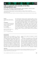
Fig. 1. Dose-response curve of the optimized lateral flow
biosensor assay using quantified synthetic DNA target sequence.
The intensity of the signals increased as the concentration of the
target sample increased. Assays were run in triplicate. The value
for the negative control was 1.04 ± 3.
Fig. 2. Biosensor assay done with the RT-PCR product of RN
A
isolated from fecal samples spiked with 10
1
to 10
6
MAP
organisms. Three strips were used for each dilution (10
1
to 10
6
) o
f
sample. One strip each for positive control (PC) and negative
control (NC) were used. Positive signals are seen at the capture
zone even with the RT- PCR product of RNA extracted from feca
l
samples containing 10 organisms of MAP. RFR: reflectomete
r
reading, CFU: colony forming units.
wells were washed thoroughly with 200 μl of wash buffer,
followed by 200 μl of hybridization buffer (4× SSC, 20%
formamide, 0.2% Ficoll, and 0.2 M sucrose). The target
(RT-PCR product) and the reporter probe (0.2 μM in 50
mM potassium phosphate buffer [pH 7.5] containing 1 mM
EDTA) were diluted in hybridization buffer, and denatured
at 95
o
C for 5 min. To this mixture, 3 μl of liposomes for
each well was added and incubated at 60
o
C for 20 min. One
hundred μl of this mixture was added to each well and
incubated at 60
o
C for 30 min. The plates were washed
twice with 200 μl PBS-sucrose buffer and 50 μl of 30 mM
OG was added. After a 5 min incubation period, the
fluorescence of the bound liposomes was measured at
λ
ex
=
540/35 nm and
λ
em
= 590/25 nm.
Results
Optimization and development of a lateral-flow
biosensor assay based on a synthetic IS900 sequence
The lateral-flow biosensor assay was developed and
optimized using universal membranes, liposomes, and
specific capture and reporter probes for IS900.
Initially, a synthetic DNA target was used to optimize the
assay to assess the signal-to-noise ratios with a relatively
large dynamic range and the highest signal obtainable. The
standard lateral-flow biosensor assay was run in triplicate
with 1 μl of synthetic DNA with 8 different concentrations
ranging from 1-1,000 fmol μ/l. The limit of detection was
determined using the signal obtained for the negative
control plus three times the standard deviation at that point.
The data showed that the limit of detection was as low as 1
fmol of the synthetic target sequence per assay with a
dynamic range from 1-1,000 fmol (Fig. 1). The negative
control contained water instead of target sequence and had
a value of 1.04 ± 3.
Lateral-flow biosensor assay with the RT-PCR
product of the IS900 gene
After optimization the lateral-flow biosensor assay with
the synthetic IS900 sequence, the assay was performed
with the RT-PCR product of the IS900 gene sequence. The
RT-PCR product of RNA isolated from cultured MAP was
used for optimization of the assay. In order to allow the
capture and reporter probes to hybridize with the double-
stranded DNA target sequence, denaturing, and hybridization
conditions were optimized. For final assays, the target
(RT-PCR product), probes, and primers were denatured at
95
o
C for 5 min, annealed at 60
o
C for 1 min, and hybridized
with liposomes. Annealing at 60
o
C was done to prevent the
re-association of thermally-denatured double-stranded
DNA strands.
Lateral-flow biosensor assay with the RT-PCR product
of the RNA extracted from spiked fecal samples
Positive signals were noticed at the capture zone, even with
the RT-PCR product of RNA extracted from fecal samples
containing only 10 organisms of MAP per 100 mg of feces
(Fig. 2). We also performed the assay with a limited number
of fecal samples collected from 2, 4, 6, 8, and 10 days from
calves orally challenged with MAP. Fecal samples collected
2 and 4 days after challenge gave positive signals in the
biosensor assay, which concurred with the MAP culture
A biosensor assay for the detection of Mycobacterium avium subsp. paratuberculosis in fecal samples 39
Fig. 3. Effect of synthetic DNA target concentration (0-1,000
nM) on the fluorescence signal assessed by microtiter plate assay.
Each point is the average of triplicate determinations at each o
f
the target concentrations tested and the error bars represent one
standard deviation. A detection limit of 0.1 nM was obtained
based on the value of the lowest concentration tested to be above
the value of the negative control plus three times the standard
deviation of the negative control.
Fig. 4. Effect of RT-PCR products of RNA extracted from spike
d
fecal samples (containing 10
1
to 10
6
organisms) on the fluorescenc
e
signal assessed by microtiter plate assay. Each point is the averag
e
of 3 determinations with error bars representing one standar
d
deviation. The detection limit was found to be as low as 10 CFU
based on the value of the lowest CFU tested to be above the valu
e
of the negative control plus three times the standard deviation o
f
the negative control.
Organisms ATCC # Origin
Expected
result
Reflectometer
reading
N
egative control
M
. avium subsp.
paratuberculosis
M
. ulcerans
M
. marium
M
. kansasii
M
. abscessus
M
. avium
M
. phlei
M
. fortuitum
subsp. fortuitum
M
. scrofulaceum
M
. intracellulare
M
. smegmatis
M
. bovis
Staphylococcus
aureus
1943
297
12478
19977
2576
11758
6841
19981
13950
19420
19210
66115-98
(Cattle)
UN
UN
Human
UN
UN
Hay/grass
Human
Human
Human
Human
Bovine
Dog
Negative
Positive
Negative
Negative
Negative
Negative
Negative
Negative
Negative
Negative
Negative
Negative
Negative
Negative
0
52
2
0
0
3
7
0
0
5
3
0
4
1
Table 2. Specificity of biosensor assay to Mycobacterium (M.)
avium subsp. paratuberculosis
results. The MAP culture results of the positive fecal samples
had 8 and 5 colony forming units (CFU), respectively, 2 and
4 days post-challenge. However, fecal samples collected 6,
8, and 10 days after challenge were found to be negative by
both the biosensor assay and MAP culture studies. The
coefficient of variance ranged from 0.59-3.7 for the different
levels of MAP organisms in the spiked fecal samples tested
by the biosensor assay.
Microtiter plate assay with the RT-PCR product of
RNA extracted from spiked fecal samples
For optimization, the biotinylated capture probe was
immobilized to the microtiter plates coated with neutravidin
and the synthetic single-stranded DNA target for IS900
was allowed to hybridize prior to the addition of the
reporter probe and the SRB encapsulating liposomes. The
assay was run in triplicate with 1 μl of synthetic DNA target
at 7 different concentrations ranging from 0.001-1,000 nM.
In order to decrease the limit of detection, liposomes were
lysed with a detergent, releasing the otherwise self-
quenched SRB dye and detected using fluorescence (Fig.
3). A limit of detection of 0.1 nM was obtained calculating
the lowest concentration detected that is above a value of
the negative control plus three times the standard deviation
of the negative control.
The lateral-flow biosensor assay was compared with the
microtiter plate assay employing the same probe and target
sequences for the detection of RNA extracted from fecal
samples. The microtiter plate assay was performed with
the RT-PCR product of the IS900 gene after denaturation at
95
o
C for 5 min and hybridized at 60
o
C. Positive signals
were obtained when 1.5 μl of target was used in the assay.
The detection limit was found to be as low as 10 CFU when
RT-PCR product of RNA extracted from fecal samples
spiked with 10
1
to 10
6
organisms (Fig. 4). This was the
same limit of detection obtained for the simple LF assay.
40 Vijayarani Kumanan et al.
Specificity of the assay
The specificity of the lateral-flow biosensor assay was
evaluated with samples from closely related mycobacteria
for false positive reactions. These mycobacteria were
cultured under optimal conditions and the RNA extracted
was used in the RT-PCR reactions. No false positive signals
were detected for any of the mycobacteria tested (Table 2).
Discussion
The majority of the diagnostic tests available for MAP
detection is based upon the amplification of insertion
sequences (IS elements). In this study, we used the IS900 gene
because of its uniqueness in the MAP genome [9,22,29] and
its comparably high copy number. Diagnostic tests based on
IS900 elements have a high level of sensitivity because of
the copy number [27]. In this study, we developed lateral flow
and a microtiter assays. In the microtiter assay, the detection
of the amplified target sequence is achieved through surfactant-
induced liposome lysis and release of encapsulated dye
molecules with subsequent fluorescent detection [12].
Although the hybridization of the probes with the target is
usually done at 41
o
C [3] in the case of single-stranded RNA
sequences, the hybridization was optimized at 60
o
C to suit
the high G + C content of the MAP genome.
Generally, milk and feces are considered to be the most
suitable clinical specimens for the diagnosis of JD.
However, because of the presence of large amounts of fat
and calcium ions, milk is regarded as a difficult specimen
for the detection of MAP organisms [19]. Hence, we used
fecal samples in this assay. The lateral flow biosensor assay
was performed with the RT-PCR product of RNA extracted
from spiked fecal samples containing 10
1
to 10
6
organisms
in order to assess the sensitivity of the assay.
The results of our study indicated that the lateral flow
biosensor assay was effective, even in the detection of 10
MAP organisms in the spiked fecal samples. Apart from
the spiked fecal samples, we also tested fecal samples from
experimentally infected animals, wherein fecal samples
collected 2 and 4 days post-challenge gave positive results
by the lateral flow biosensor assay. Shedding of MAP in
feces has been reported to be inconsistent after challenge,
at least during early stages. Moreover, there could be
colonization of the organisms in the intestines which could
have resulted in the non-detection of MAP at 6, 8, and 10
days post-challenge. However, 2 and 4 days post-challenge
samples were also positive by MAP culture results with 8
and 5 CFU, respectively, which in turn indicated the ability
of this method in detecting low levels of MAP organisms.
Moreover, with the use of the RT-PCR product, in general
only viable organisms present in the feces will be detected
which provides an excellent tool for diagnosis. The existing
cultural and serological methods accurately predict MAP
infections during clinical stages when most animals shed
large numbers of organisms, compared to subclinical
stages when fecal shedding occurs at low levels with lesser
frequencies. The present study with detection limits as low
as 10 organisms is well-suited for the present day diagnostic
requirements of JD. These results indicated that this assay
is highly sensitive and could be used to detect animals in
the early stage of infection with very low MAP shedding.
In addition to the rapid lateral flow assay that is suitable
for low-sample numbers, a microtiter plate assay was
developed for the detection of the RT-PCR product of RNA
extracted from spiked fecal samples. Comparison of the
lateral flow biosensor assay with the microtiter plate assay
indicated that the detection limit of both assays were
similar (10 CFU). With no false positive signals with the
closely related mycobacteria tested in this study, this assay
was considered to have excellent specificity.
In conclusion, the results of our study indicated that the
IS900 gene sequence-based lateral flow biosensor assay
developed is sensitive and specific for the detection MAP
organisms in fecal samples. The assay was found to be
effective in detecting as few as 10 organisms per 100 mg of
feces. This assay will be useful in identifying animals in
their early clinical stage, shedding low numbers of MAP in
their feces, which can allow their quick removal from the
rest of the herd, thereby avoiding further environmental
contamination. Although one would expect a perfect dose
response in the results between 10 and 10
6
organisms, the
results were not as expected, which could possibly be due
to the presence of PCR inhibitors in the fecal samples. This
assay is comparatively cheaper and does not require costly
equipments in comparison to real-time PCR or PCR
coupled with Southern blotting. In this assay, reverse
transcription PCR is being used instead of regular PCR
which will help in detecting live MAP organisms.
Moreover, the results can be obtained in a shorter time, in
contrast to MAP culture techniques which take at least 6-8
weeks. Therefore, the present work was carried out with an
idea of developing bioanalytical systems that are simple
and yet highly sensitive. With the availability of small,
easy-to-carry thermal cyclers, this assay could be developed
as a portable assay which may cater to the needs of first
responder emergency teams and clinicians in the field.
Acknowledgments
This research was supported, in part, by the Cornell
University Agricultural Experiment Station federal formula
funds Project No. NYC-478462 received from the
Cooperative State Research, Education and Extension
Service of the U.S. Department of Agriculture, the Animal
Health Diagnostic Center technique development fund, and
the New York State Science and Technology Foundation
(CAT).
A biosensor assay for the detection of Mycobacterium avium subsp. paratuberculosis in fecal samples 41
References
1. Ahn-Yoon S, DeCory TR, Baeumner AJ, Durst RA.
Ganglioside-liposome immunoassay for the ultrasensitive
detection of cholera toxin. Anal Chem 2003, 75, 2256-2261.
2. Baeumner AJ. Biosensors for environmental pollutants and
food contaminants. Anal Bioanal Chem 2003, 377, 434-445.
3. Baeumner AJ, Jones C, Wong CY, Price A. A generic
sandwich-type biosensor with nanomolar detection limits.
Anal Bioanal Chem 2004, 378, 1587-1593.
4. Baeumner AJ, Leonard B, McElwee J, Montagna RA. A
rapid biosensor for viable B. anthracis spores. Anal Bioanal
Chem 2004, 380, 15-23.
5. Baeumner AJ, Pretz J, Fang S. A universal nucleic acid
sequence biosensor with nanomolar detection limits. Anal
Chem 2004, 76, 888-894.
6. Bannantine JP, Barletta RG, Stabel JR, Paustian ML,
Kapur V. Application of the genome sequence to address
concerns that Mycobacterium avium subspecies paratuberculosis
might be a foodborne pathogen. Foodborne Pathog Dis 2004,
1, 3-15.
7. Beard PM, Rhind SM, Buxton D, Daniels MJ, Henderson
D, Pirie A, Rudge K, Greig A, Hutchings MR, Stevenson
K, Sharp JM. Natural paratuberculosis infection in rabbits
in Scotland. J Comp Pathol 2001, 124, 290-299.
8. Clarke CJ, Patterson IA, Armstrong KE, Low JC.
Comparison of the absorbed ELISA and agar gel
immunodiffusion test with clinicopathological findings in
ovine clinical paratuberculosis. Vet Rec 1996, 139, 618-621.
9. Collins DM, Gabric DM, De Lisle GW. Identification of a
repetitive DNA sequence specific to Mycobacterium
paratuberculosis. FEMS Microbiol Lett 1989, 51, 175-178.
10. Cousins DV, Whittington R, Marsh I, Masters A, Evans
RJ, Kluver P. Mycobacteria distinct from Mycobacterium
avium subsp. paratuberculosis isolated from the faeces of
ruminants possess IS900-like sequences detectable IS900
polymerase chain reaction: implications for diagnosis. Mol
Cell Probes 1999, 13, 431-442.
11. Dukes TW, Glover GJ, Brooks BW, Duncan JR,
Swendrowski M. Paratuberculosis in saiga antelope (Saiga
tatarica) and experimental transmission to domestic sheep. J
Wildl Dis 1992, 28, 161-170.
12. Edwards KA, Baeumner AJ. Optimization of DNA-tagged
liposomes for use in microtiter plate analyses. Anal Bioanal
Chem 2006, 386, 1613-1623.
13. El-Zaatari FA, Osato MS, Graham DY. Etiology of Crohn's
disease: the role of Mycobacterium avium paratuberculosis.
Trends Mol Med 2001, 7, 247-252.
14. Ellingson JL, Koziczkowski JJ, Anderson JL. Comparison
of PCR prescreening to two cultivation procedures with PCR
confirmation for detection of Mycobacterium avium subsp.
paratuberculosis in U.S. Department of Agriculture fecal
check test samples. J Food Prot 2004, 67, 2310-2314.
15. Greig A, Stevenson K, Henderson D, Perez V, Hughes V,
Pavlik I, Hines ME 2nd, McKendrick I, Sharp JM.
Epidemiological study of paratuberculosis in wild rabbits in
Scotland. J Clin Microbiol 1999, 37, 1746-1751.
16. Herthnek D, B
ölske G. New PCR systems to confirm
real-time PCR detection of Mycobacterium avium subsp.
paratuberculosis. BMC Microbiol 2006, 6, 87.
17. Hughes VM, Stevenson K, Sharp JM. Improved preparation
of high molecular weight DNA for pulsed-field gel
electrophoresis from mycobacteria. J Microbiol Methods
2001, 44, 209-215.
18. Hulten K, El-Zimaity HM, Karttunen TJ, Almashhrawi
A, Schwartz MR, Graham DY, El-Zaatari FA. Detection
of Mycobacterium avium subspecies paratuberculosis in
Crohn's diseased tissues by in situ hybridization. Am J
Gastroenterol 2001, 96, 1529-1535.
19. Khare S, Ficht TA, Santos RL, Romano J, Ficht AR,
Zhang S, Grant IR, Libal M, Hunter D, Adams LG. Rapid
and sensitive detection of Mycobacterium avium subsp.
paratuberculosis in bovine milk and feces by a combination
of immunomagnetic bead separation-conventional PCR and
real-time PCR. J Clin Microbiol 2004, 42, 1075-1081.
20. Li L, Bannantine JP, Zhang Q, Amonsin A, May BJ, Alt
D, Banerji N, Kanjilal S, Kapur V. The complete genome
sequence of Mycobacterium avium subspecies paratuberculosis.
Proc Natl Acad Sci USA 2005, 102, 12344-12349.
21. Marsh I, Whittington R, Cousins D. PCR-restriction
endonuclease analysis for identification and strain typing of
Mycobacterium avium subsp. paratuberculosis and
Mycobacterium avium subsp. avium based on polymorphisms
in IS1311. Mol Cell Probes 1999, 13, 115-126.
22. Moss MT, Green EP, Tizard ML, Malik ZP, Hermon-Taylor
J. Specific detection of Mycobacterium paratuberculosis by
DNA hybridisation with a fragment of the insertion element
IS900. Gut 1991, 32, 395-398.
23. Naser SA, Ghobrial G, Romero C, Valentine JF. Culture
of Mycobacterium avium subspecies paratuberculosis from
the blood of patients with Crohn's disease. Lancet 2004, 364,
1039-1044.
24. Naser SA, Hulten K, Shafran I, Graham DY, El-Zaatari
FA. Specific seroreactivity of Crohn's disease patients against
p35 and p36 antigens of M. avium subsp. paratuberculosis.
Vet Microbiol 2000, 77, 497-504.
25. Ott SL, Wells SJ, Wagner BA. Herd-level economic losses
associated with Johne's disease on US dairy operations. Prev
Vet Med 1999, 40, 179-192.
26. Shin SJ, Chang YF, Huang C, Zhu J, Huang L, Yoo HS,
Shin KS, Stehman S, Shin SJ, Torres A. Development of a
polymerase chain reaction test to confirm Mycobacterium
avium subsp. paratuberculosis in culture. J Vet Diagn Invest
2004, 16, 116-120.
27. Stabel JR, Bannantine JP. Development of a nested PCR
method targeting a unique multicopy element, ISMap02, for
detection of Mycobacterium avium subsp. paratuberculosis
in fecal samples. J Clin Microbiol 2005, 43, 4744-4750.
28. Turnbull PC. Definitive identification of Bacillus anthracis-
a review. J Appl Microbiol 1999, 87, 237-240.
29. Vary PH, Andersen PR, Green E, Hermon-Taylor J,
McFadden JJ. Use of highly specific DNA probes and the
polymerase chain reaction to detect Mycobacterium
paratuberculosis in Johne's disease. J Clin Microbiol 1990,
28, 933-937.
30. Whitlock RH, Wells SJ, Sweeney RW, Van Tiem J.
ELISA and fecal culture for paratuberculosis (Johne's
disease): sensitivity and specificity of each method. Vet
42 Vijayarani Kumanan et al.
Microbiol 2000, 77, 387-398.
31. Whittington RJ, Marsh I, Turner MJ, McAllister S, Choy
E, Eamens GJ, Marshall DJ, Ottaway S. Rapid detection of
Mycobacterium paratuberculosis in clinical samples from
ruminants and in spiked environmental samples by modified
BACTEC 12B radiometric culture and direct confirmation by
IS900 PCR. J Clin Microbiol 1998, 36, 701-707.
32. Whittington RJ, Marsh IB, Whitlock, RH. Typing of IS
1311 polymorphisms confirms that bison (Bison bison) with
paratuberculosis in Montana are infected with a strain of
Mycobacterium avium subsp. paratuberculosis distinct from
that occurring in cattle and other domesticated livestock.
Mol Cell Probes 2001, 15, 139-145.