Báo cáo khoa học: " A suspected case of Lyme borreliosis in a hunting dog in Korea" pps
Bạn đang xem bản rút gọn của tài liệu. Xem và tải ngay bản đầy đủ của tài liệu tại đây (1.01 MB, 3 trang )
JOURNAL OF
Veterinary
Science
Case Report
J. Vet. Sci. (2009), 10(1), 89
91
DOI: 10.4142/jvs.2009.10.1.89
*Corresponding author
Tel: +82-2-880-1267; Fax: +82-2-888-0659
E-mail:
A suspected case of Lyme borreliosis in a hunting dog in Korea
Ul Soo Choi
1
, Hyun Wook Kim
2
, Sung Eun You
2
, Hee Jeong Youn
3,
*
1
Department of Veterinary Clinical Pathology, College of Veterinary Medicine, Chonbuk National University, Jeonju
561-756, Korea
2
Haemaru Referral Animal Hospital, Sungnam 463-050, Korea
3
KRF Priority Zoonotic Disease Research Institute, College of Veterinary Medicine, Seoul National University, Seoul
151-742, Korea
A two-year-old male Pointer had been presented with
anorexia, cachexia, and weight loss of 10-day duration. Upon
physical examination, fever, lethargy, superficial lymph node
enlargement, and tick infestation were noted. The only
abnormality in CBC and serum chemistry analyses was mild
hyperglobulinemia. Spleen was enlarged by radiography, and
the lymph nodes showed neutrophilic lymphadenitis by
cytological examination. A polymerase chain reaction test for
babesiosis and commercial ELISA tests for Ehrlichia canis,
heartworm, and Lyme disease was negative except for Lyme
disease, which was verified by both an IFA-IgG test and a
quantitative C
6
assay. Doxycycline was administered for 2
weeks and the recovery was uneventful. Post-treatment C
6
titer decreased to within normal limits.
Keywords:
dog, fever, hyperglobulinemia, Lyme disease
Lyme disease is a tick-borne disease caused by Borrelia
(B.) burgdorferi sensu lato. It affects humans and dogs
worldwide, and has been reported in North America,
Europe, Asia, Australia, South America, and Africa [3].
Vectors of B. burgdorferi sensu lato include various species
of hard ticks of the Ixodes spp
In Korea, B. afzelii, B. garinii, and B. valaisiana have
been identified in ticks and several clinical cases of Lyme
disease in humans have been reported [1,2,4,11]. However,
there are no known occurrences in dogs. We report a
possible case of Lyme borreliosis in a hunting dog evaluated
by a commercial C
6
ELISA kit, IFA, and a laboratory
quantitative C
6
assay.
A 2-year-old male Pointer dog was presented to Haemaru
Referral Animal Hospital with signs of anorexia, cachexia,
and weight loss of a 10-day duration. The body condition
score decreased from 3/5 to 1/5 at presentation. The dog
was born and raised in Kwangju, Kyonggi-do. It had been
used in hunting, and was exposed to ticks. The hunting area
covered Bongwha and Youngju, Kyongsangnam-do. Upon
physical examination, a mild fever (39.7
o
C), lethargy, and
enlargement of both superficial cervical lymph nodes were
noted. Initial screening tests include CBC, serum chemistry,
cytology of fine needle aspirates of enlarged lymph nodes,
and abdominal radiography. Blood works and serum
chemistry results were all within normal limits, except for
mild hyperglobulinemia (4.6 g/dl, reference range 2.5-4.5
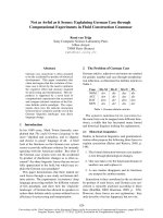
g/dl) (Table 1). The radiography showed that the spleen was
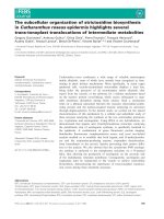
enlarged (Fig. 1). Smears of lymph node aspirates revealed
a predominant population of small lymphocytes with a
lesser number of medium to large lymphocytes, plasma
cells, neutrophils, and macrophages, consistent with
neutrophilic lymphadenitis (Fig. 2). Based on this finding,
increased serum globulin was considered to be associated
with chronic inflammation, and lymphoproliferative neoplasia
was ruled out. On the day of the first visit, infectious
diseases were screened using a commercial ELISA kit
(SNAP 3Dx; IDEXX, USA) for a heartworm (Ehrlichia
canis) antigen, and a Lyme antibody (C
6
antibody). All the
tests were negative except for Lyme disease. On the same
day, EDTA whole blood was also submitted to the College
of Veterinary Medicine, Chonbuk National University, for
PCR testing for E. canis and Babesia (the results were
negative for both). To confirm the positive results of the
ELISA kit for Lyme disease, the rest of the patient’s serum
was submitted to an IDEXX Reference Laboratory (USA)
for a C
6
titer assay and to Antech Diagnostics (USA) for a
Lyme IgG assay. The C
6
serum titer was 30 U/ml (positive
≥ 30), and the IgG titer was 1 : 256 (negative < 1 : 64).
These results were highly suggestive of active borreliosis.
Treatment with doxycycline (10 mg/kg s.i.d, PO; Myung
In Pharm, Korea) and supportive drugs (vitamin C, thiamine,
serratiopeptidase and famotidine) was administered for 2
weeks. The dog responded well to treatment and the clinical
90 Ul Soo Choi et al.
CBC results Serum chemistry
Name Reference ranges Result Unit Name Reference ranges Result Unit
WBC
RBC
HGB
HCT
MCV
MCHC
MCH
PLT
6.0-17.0
5.5-8.5
12-18
37-58
66-77
32-36
19.5-24.8
200-500
16.6
6.08
14.1
40.8
67.1
34.6
23.2
423
10
3
/μl
10
6
/μl
g/dl
%
Fl
g/dl
10
3
/μl
BUN
Creatinine
Phosphorous
Ca
Total protein
Albumin
Globulin
ALT
ALP
Total
bilirubin
Cholesterol
Amylase
Glucose
Na
K
Cl
7.0-27.0
0.5-1.8
2.5-6.8
7.9-12.0
5.2-8.2
2.3-4.0
2.5-4.5
10-100
23-212
0.0-0.9
110-320
500-1,500
74-143
144-160
3.5-5.8
109-122
11
0.5
4.9
10.2
7.3
2.7
4.6
35
105
0.3
160
791
110
145
3.8
114
mg/dl
mg/dl
mg/dl
mg/dl
g/dl
g/dl
g/dl
U/l
U/l
mg/dl
mg/dl
U/l
mg/dl
mmol/l
mmol/l
mmol/l
WBC: white blood cell, RBC: red blood cell, HGB: hemoglobin, HCT: hematocrit, MCV: mean corpuscular volume, MCHC: mean
corpuscular hemoglobin concentration, MCH: mean corpuscular hemoglobin, PLT: platelet, BUN: blood urea nitrogen, ALT: alanine
transaminase, ALP: alkaline phosphatase.
Tabl e 1 . CBC and serum chemistry results
Fig. 1. Radiograph of the spleen. The abdomen was decreased
by
lack of fat. In the lateral view, soft tissue density structure
(arrows) was identified in the ventral region of the middle
abdomen, and this structure was considered to be splenomegaly
based on the position and shape.
Fig. 2. Microphotograph of the enlarged superficial cervical
lymph node. Note the mixed population of lymphocytes,
neutrophils, and plasma cells with a predominance of small
lymphocytes. Some of the cells were lysed in the
b
ackground.
Wright stain, ×400.
signs disappeared. 3 weeks after the initiation of treatment,
the lymph nodes had returned to normal size and serum
globulin levels had decreased to 2.7 g/dl. Post-treatment C
6
level was 10 U/ml.
C
6
peptide is derived from the VlsE antigen, B. burgdorferi
surface protein expressed when B. burgdorferi is transmitted
to the dog but not expressed in the tick, in tissue culture, or
in Lyme vaccines [5,7,8,10]. The sensitivities and
specificities of this peptide-based ELISA are reportedly
equivalent or superior to those of western blot assays alone
or a combination of whole-cell based ELISA and western
A suspected case of Lyme borreliosis in a hunting dog in Korea 91
blot analysis [6,7,9]. The C
6
antibody titer has been found
to decrease post-treatment because production of the
antibody to the C
6
peptide may depend on the presence of
a viable organism. For this reason, post treatment C
6
levels
may be used as an indicator of therapy outcome [6,7,10].
The patient in this report was presumptively diagnosed
with Lyme borreliosis based on an increased C
6
antibody
titer. This diagnosis was confirmed by both a commercial
Lyme antibody test kit and a laboratory quantitative C
6
assay
(IDEXX Reference Laboratory, USA) along with an
increased IgG titer by IFA. Normal lymph nodes, resolution
of the anorexia and fever, and decreased C
6
antibody titer
post treatment also supported our diagnosis. In a previous
report, the median percent decline in C
6
level relative to
pretreatment values at 6 and 12 months were found to be
68.0% and 83.3%, respectively [6]. However, in this case,
a PCR test using peripheral blood was unsuccessful. The
negative PCR test may be attributed to the fact that it was
done using a peripheral blood sample. Connective tissue,
synovia, or skin samples near the tick bite are preferred for
PCR because Borrelia organisms rarely spread
hematogenously. The organism can also be isolated from
the skin area for an extended period. In this case, there was
tick infestation when it came to the animal hospital, so if a
skin sample from near the tick bite site was used for PCR,
positive results would have been possible. A skin biopsy
sample from as close as possible to the tick-bite should be
submitted for a reliable PCR test [3,12].
To the best of our knowledge, canine Lyme borreliosis has
never been reported in Korea. This disease might be
underestimated in dogs because B. burgdorferi sensu lato
has been isolated in ticks and wild rodents in Korea [1,4,11].
Patients could also easily be overlooked due to good responses
to therapy without a proper diagnosis. If a symptomatic dog
has outdoor activity and a history of tick infestation, Lyme
disease should be included in the differential diagnosis.
Acknowledgment
This work was supported by Korean Research Foundation
grant (KRF-005-E00078).
References
1. Chae JS, Yu do H, Shringi S, Klein TA, Kim HC, Chong
ST, Lee IY, Foley J. Microbial pathogens in ticks, rodents
and a shrew in northern Gyeonggi-do near the DMZ, Korea.
J Vet Sci 2008, 9, 285-293.
2. Choi YJ, Han SH, Park JM, Lee KM, Lee EM, Lee SH,
Song HJ, Koh YS, Lee KW, Jang WJ, Park KH. First
molecular detection of Borrelia afzelii in clinical samples in
Korea. Microbiol Immunol 2007, 51, 1201-1207.
3. Hartmann K, Greene CE. Diseases caused by systemic
bacterial infections. In: Ettinger SJ, Feldman EC (eds.).
Textbook of Veterinary Internal Medicine: Diseases of the
Dog and Cat. 6th ed. pp. 619-625, Elsevier, St. Louis, 2005.
4. Lee SH, Kim BJ, Kim JH, Park KH, Kim SJ, Kook YH.
Differentiation of Borrelia burgdorferi sensu lato on the
basis of RNA polymerase gene (rpoB) sequences. J Clin
Microbiol 2000, 38, 2557-2562.
5. Levy S, O'Connor TP, Hanscom JL, Shields P. Utility of
an in-office C
6
ELISA test kit for determination of infection
status of dogs naturally exposed to Borrelia burgdorferi. Vet
Ther 2002, 3, 308-315.
6. Levy SA, O'Connor TP, Hanscom JL, Shields P, Lorentzen
L, Dimarco AA. Quantitative measurement of C
6
antibody
following antibiotic treatment of Borrelia burgdorferi antibody-
positive nonclinical dogs. Clin Vaccine Immunol 2008, 15,
115-119.
7. Liang FT, Jacobson RH, Straubinger RK, Grooters A,
Philipp MT. Characterization of a Borrelia burgdorferi
VlsE invariable region useful in canine Lyme disease
serodiagnosis by enzyme-linked immunosorbent assay. J
Clin Microbiol 2000, 38, 4160-4166.
8. Littman MP, Goldstein RE, Labato MA, Lappin MR,
Moore GE. ACVIM small animal consensus statement on
Lyme disease in dogs: diagnosis, treatment, and prevention.
J Vet Intern Med 2006, 20, 422-434.
9. Mogilyansky E, Loa CC, Adelson ME, Mordechai E,
Tilton RC. Comparison of Western immunoblotting and the
C
6
Lyme antibody test for laboratory detection of Lyme
disease. Clin Diagn Lab Immunol 2004, 11, 924-929.
10. O'Connor TP, Esty KJ, Hanscom JL, Shields P, Philipp
MT. Dogs vaccinated with common Lyme disease vaccines
do not respond to IR6, the conserved immunodominant
region of the VlsE surface protein of Borrelia burgdorferi.
Clin Diagn Lab Immunol 2004, 11, 458-462.
11. Park KH, Chang WH, Schwan TG. Identification and
characterization of Lyme disease spirochetes, Borrelia
burgdorferi sensu lato, isolated in Korea. J Clin Microbiol
1993, 31, 1831-1837.
12. Speck S, Reiner B, Streich WJ, Reusch C, Wittenbrink
MM. Canine borreliosis: a laboratory diagnostic trial. Vet
Microbiol 2007, 120, 132-141.