Báo cáo khoa học: " Porcine abortion outbreak associated with Toxoplasma gondii in Jeju Island, Korea" pot
Bạn đang xem bản rút gọn của tài liệu. Xem và tải ngay bản đầy đủ của tài liệu tại đây (2.85 MB, 5 trang )
JOURNAL OF
Veterinary
Science
J. Vet. Sci. (2009), 10(2), 147
151
DOI: 10.4142/jvs.2009.10.2.147
*Corresponding author
Tel: +82-64-754-3387; Fax: +82-64-702-9920
E-mail:
Porcine abortion outbreak associated with Toxoplasma gondii in Jeju
Island, Korea
Jae-Hoon Kim
1,
*
, Kyung-Il Kang
2
, Wan-Cheul Kang
3
, Hyun-Joo Sohn
4
, Young-Hwa Jean
4
, Bong Kyun Park
5
,
Yongbaek Kim
6
, Dae-Yong Kim
5
1
College of Veterinary Medicine, Jeju National University, Jeju 690-756, Korea
2
Department of Veterinary Pathology, College of Veterinary Medicine, University of Georgia, Athens, GA 30602-7388, USA
3
Jeju Veterinary Research Institute, Jeju 690-962, Korea
4
National Veterinary Research and Quarantine Service, Anyang 430-824, Korea
5
College of Veterinary Medicine, Seoul National University, Seoul 151-742, Korea
6
College of Veterinary Medicine, North Carolina State University, Raleigh, NC 27606, USA
This report deals with the acute onset of an abortion
outbreak and high sow mortality in one pig herd consisted of
1,200 pigs and 120 sows on Jeju Island, Korea. Affected
pregnant sows showed clinical signs, including high fever,
gradual anorexia, vomiting, depression, recumbency,
prostration, abortion, and a few deaths. Four dead sows, five
aborted fetuses from the same litter, and 17 sera collected
from sows infected or normal were submitted to the Pathology
Division of the National Veterinary Research and Quarantine
Service for diagnostic investigation. Grossly, hepatomegaly
and splenomegaly were observed in sows. Multiple necrotic
foci were scattered in the lungs, liver, spleen, and lymph nodes.
Microscopically, multifocal necrotizing lesions and protozoan
tachyzoites were present in the lesions. Tachyzoites of
Toxoplasma (T.) gondii were detected immunohistochemically.
Latex agglutination showed that the sera of 7 of 17 (41.2%)
sows were positive for antibody to T. gondii. The disease
outbreak in this herd was diagnosed as epizootic toxoplasmosis.
To our knowledge, this is the first report of porcine
toxoplasmosis with a high abortion rate and sow mortality in
Korea.
Keywords:
abortion, pig, sow mortality, tachyzoite, Toxoplasma
gondii
Introduction
Toxoplasmosis is caused by infection with Toxoplasma
(T.) gondii, a coccidian parasite that can infect humans and
animals [2,6,15]. Postnatally, animals and humans
generally become infected after ingesting food and water
contaminated with sporulated oocysts or by consuming
raw or undercooked meat containing tissue cysts [2,7].
Although toxoplasmosis is generally asymptomatic, primary
infections in pregnant women and animals may cause
abortions, fetal abnormalities, or perinatal death [2,6].
Most T. gondii infections in pigs are subclinical [2], and
transplacental infections are less common than post-natal
infections [2]. Although abortions related to T. gondii are
uncommon, they may occur in sows infected during
pregnancy [2]. Pigs infected transplacentally may be born
premature, dead, or weak, or they may die soon after birth
[15].
In Korea, T. gondii infections have been reported in
humans [20] and many domestic animals, including cats
[5], dogs [9], and pigs [19]. To our knowledge, however,
there have been no reports of abortions in animals related
to this parasite in Korea. Herein we describe an abortion
outbreak due to T. gondii infection in sows.
Materials and Methods
Case histories
During September 2002, an outbreak of abortion, lasting
10 days, was observed in one pig herd in Jeju Island, Korea.
The index herd was a 120-mixed breed-sow feeder pig
producer herd located in Hallym County, in the western part
of Jeju Island. This pig farm was isolated from other pig
farms and the herd was housed in 2 separate pens. Weaned
piglets were housed in the first pen. The second pen was
divided into 4 rooms, which housed delivered sows, pregnant
sows, fattening pigs, and grower pigs in rooms 1∼4,
respectively. The herd was maintained using a continuous
flow system and had been routinely vaccinated for Japanese
encephalitis virus, porcine parvovirus, and a few bacterial
148 Jae-Hoon Kim et al.
respiratory and enteric pathogens such as Bordetella
bronchiseptica, Pasteurella multocida, Erysipelothrix
rhusiopathiae, and Escherichia coli. One month before the
disease outbreak, the source of commercial feed was changed.
Water supply was from a private well. Three dogs were
also present at the farm. There were five pig farms located
within a 100 m radius.
At the time of the outbreak, the second room of the second
pen housed 84 pregnant sows. Two weeks after changing
the feed, 10 sows, especially first parity sows, exhibited
poor appetite and vomiting. An abnormal stink was present
in the new feed without unusual gross appearance at that
time. Among the clinical signs observed in the 37 affected
pregnant sows were fever (temperature of > 37
o
C), gradual
anorexia, vomiting, depression, recumbency, prostration,
and abortion. Sixteen sows died within 7 days of the initial
manifestation of symptoms. Abortion usually occurred 3-5
days after the onset of clinical signs, and at any stage of
gestation. The abortion rate was high (44%), and the
mortality rate of the sows was 19%. Intensive antibiotics
including neomycin, gentamicin and cefazolin and
symptomatic therapies had no effect.
Necropsy and histopathology
Two dead sows, five aborted fetuses from the same litter,
and sera collected from 12 sows that had aborted and from
5 normal sows were submitted to the Pathology Division of
the National Veterinary Research and Quarantine Service
(NVRQS) for diagnostic investigation. Later additional
internal tissues from two aborted sows obtained in the
middle of culling were also submitted to the NVRQS.
Grossly, the four sows had cutaneous cyanosis in the ears,
snout, and ventral abdomen. The submandibular and
mediastinal lymph nodes were enlarged and bright red in
color. The lungs did not fully collapse and exhibited pinpoint
yellowish white foci throughout the whole lobes. Mild to
moderate hepatomegaly and splenomegaly were also observed.
Pale, white, dry miliary foci were scattered in the liver,
spleen, and lymph nodes. The length from crown to rump
of the fetuses ranged from 22 to 24 cm. No significant gross
abnormalities were noted in any of the aborted fetuses.
Tissue samples from the lungs, heart, liver, kidneys, spleen,
stomach, intestine, lymph nodes, and brain of sows and
fetuses were fixed in 10% phosphate-buffered formalin,
dehydrated, embedded in paraffin, sectioned at 4 μm, and
stained with hematoxylin and eosin for light microscopic
examination.
Immunohistochemistry
Immunohistochemical assays for T. gondii were performed
on the replicated paraffin sections of the lungs, liver and
lymph nodes, as described previously [9,19]. Sections
were mounted on Probe-On slides (Fisher, USA) and
incubated with the primary antibody, unlabelled rabbit
polyclonal antibody (Elite, USA) against T. gondii. The
presence of antigen was determined using standard avidin-
biotin-peroxidase complex (ABC) methods, according to
the manufacturer’s protocol (Elite, USA), with 3, 3-
diaminobenzidine (Elite, USA) as the chromogen. Control
procedures included omission of the primary antibody and
substitution of an isotype-matched irrelevant antibody.
Polymerase chain reaction (PCR) and virus isolation
Samples were assayed for the presence of classical swine
fever (CSF) virus, Aujeszky’s disease (AD), porcine
reproductive and respiratory syndrome virus (PRRSV),
and porcine circovirus type 2 (PCV-2) using PCR, as
described previously [10,17]. Reverse transcriptase-PCR
for CSF virus was conducted on tissue homogenates from
the sow to amplify the 5’ untranslated region and E2
envelope glycoprotein gene [1]. Virus was isolated to
exclude infection with AD infection.
Fluorescent antibody test and bacterial culture
Indirect fluorescent antibody (FA) tests for CSF, AD,
PRRSV, and PCV-2 were performed on the replicated cryo-
sections of the tonsils and lungs as described previously [8,
10]. Cryo-sections were fixed in cold acetone for 5 min and
incubated with primary monoclonal or polyclonal antibody
against each pathogen, followed by fluorescein isothiocyanate-
conjugated secondary antibody (Dakocytomation, Denmark)
directed against the primary antibody.
Aseptically collected tissue samples from the lungs, liver,
spleen, lymph nodes, and intestine were cultured on blood
agar, MacConkey agar (BBL, USA), and Chocolate agar
(Hanil Komed, Korea) at 37
o
C under aerobic and anaerobic
conditions.
Serological tests
Antibody titers to T. gondii were determined using a
commercial latex agglutination (LA) test kit (Eiken Chemical,
Japan) [11]. The reactions were performed in 96-well U-
bottom polystyrene microplates (BD Biosciences, USA) at
two-fold dilutions. To each well 25 μl of T. gondii antigen-
coated latex particles suspension was added, followed by
incubation overnight at room temperature. An agglutination
titer ≥ 1:32 was considered positive. The serum antibody
titers to CSF virus and AD virus were also screened by
ELISA methods as described previously [17]. An indirect
FA test to determine the antibody titers for Neospora (N.)
caninum was performed as previously described [11].
Results
Histopathology and immunohistochemistry
Microscopically, moderate multifocal necrotizing
pneumonia with intralesional protozoan tachyzoites was
present in the lungs of sows. Mild perivascular or
Porcine abortion associated with Toxoplasmosis 149
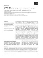
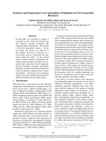
Fig. 2. Cerebrum of aborted sow, showing focal necrotic encephalitis.
H&E. ×200.
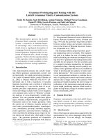
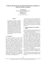
Fig. 3. Lung of an aborted porcine fetus, showing severe pulmonar
y
necrosis. H&E, ×200. Insert: Note brown tachyzoites of Tox opl as m
a
g
ondii in alveolar macrophages. ABC stain, ×400.
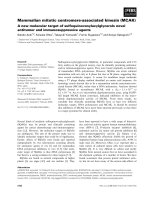
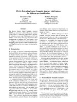
Fig. 1. Liver of aborted sow, showing multifocal necrotic hepatitis.
H&E, ×200. Insert: Note brown tachyzoites of Tox oplasma
g
ondi
i
in sinusoid and Kupffer cell. ABC stain, ×400.
peribronchiolar lymphoid cuffings associated with
mycoplasmal pneumonia were observed in two sows. Severe
multifocal necrotizing granulomatous cholangiohepatitis,
characterized by the infiltration of lymphocytes, plasma
cells, macrophages and a few neutrophils and protozoan
tachyzoites, were observed in the livers (Fig. 1). The lymph
nodes presented with severe lymphadenitis, with focal to
diffuse necrosis and protozoan tachyzoites, and the spleens
showed moderate congestion, lymphoid depletion and
multifocal necrosis. Lymphohistiocytic meningoencephalitis
with disseminated malacic foci and perivascular cuffing was
observed in the brains (Fig. 2).
Three of five aborted fetuses showed multifocal necrotic
lesions and intralesional or adjacent protozoan tachyzoites
in the lungs (Fig. 3), liver, lymph nodes, and brain. In
addition, the brains showed necrotic encephalitis, gliosis,
and perivascular cuffing.
The inflamed organs of both sows and fetuses contained
numerous T. gondii tachyzoites, present as fine brownish
granules in the necrotic areas of parenchymas and in the
cytoplasm of infiltrated macrophages (Fig. 1, 3 Insert).
PCR, FA, virus and bacteria isolation
Sows and fetuses were negative for CSF, AD, PRRSV,
and PCV-2 by PCR, virus isolation and FA tests. No
specific bacterial pathogens were isolated, except for
Pasteurella multocida, which was isolated from one sow
with pneumonic consolidation.
Serological tests
At the time of submission, 7 of 17 (41.2%) selected sows
were positive for T. gondii by an LA test and were judged
to have been exposed to the parasite. LA titers of
seropositive sows ranged from 1 : 32 (4), 1 : 64 (2), to 1 :
128 (1), respectively. All seropositive sows experienced
recent abortions. The five normal sows without abortion
were seronegative for T. gondii. Only one sow in the herd
was serologically positive for CSF virus by ELISA,
whereas none was serologically positive for AD virus and
N. caninum.
Discussion
Clinical signs and pathological changes may be non-
specific, requiring full panels of microbiological and
serological tests to confirm the exact etiology. Diagnostic
tests were able to rule out major endemic swine infectious
diseases in Korea, including CSF, AD, PRRS, and PCV-2.
Histopathologic examination of the major parenchymal
organs of sows and aborted fetuses revealed many
necrotic lesions associated with protozoan tachyzoites.
Using T. gondii specific antibody, the presence of T. gondii
tachyzoites was demonstrated in the formalin-fixed tissues
from sows and fetuses. In addition, serological tests
revealed high antibody titers to T. gondii in aborted sows
from this herd. Based on these clinical signs, histopathology,
immunohistochemistry and microbiology, the herd was
150 Jae-Hoon Kim et al.
diagnosed with toxoplasmosis. As toxoplasmosis is zoonotic,
the remaining 68 pregnant sows present in the same room
were culled 2 weeks after the outbreak of this disease.
Although clinical toxoplasmosis has been reported in
young piglets, little was known of abortions associated
with T. gondii and rates of congenital infection in pigs [2].
Epidemiologically, porcine toxoplasmosis has been
classified into sporadic neonatal, postnatal, and epizootic
infection [2]. Sporadic fatal toxoplasmosis in piglets has
been reported in several countries, including the USA,
Japan, and Korea. Although many infected piglets were
born dead or sick, or became sick within 3 months of birth,
others remained clinically normal.
Epizootic outbreaks of toxoplasmosis have been reported
in Italy [4], Singapore [13], and Taiwan [18]. In Italy, there
were 4 simultaneous outbreaks of toxoplasmosis with high
mortality in different pig herds in 2 different provinces
over a 1 month period [4]. The morbidity rate was as high
as 60%, and the mortality rate ranged from 10% to 42% in
fattening pigs weighing 60 to 180 kg. In Taiwan, 51 of 66
pregnant gilts infected with T. gondii on a single farm
aborted within 2 months [18]. Most fetuses were stillborn,
some were mummified, and few were born alive but died
within a few days. In Singapore, a large outbreak of
toxoplasmosis was observed in a herd containing 540 pigs
[13]. Many pigs in different age groups, ranging from
piglets to sows, became sick and died. The overall
morbidity rate was 35.7% and the overall mortality rate
was 11.8%.
In this study, higher abortion rates, up to 44%, were
observed and unusually high sow mortality rates, up to
19%, were primarily associated with toxoplasmosis over a
very short period of time. All clinical signs were restricted
to pregnant sows, especially those at any stages of gestation.
Although the sample size was relatively small, the high
seroprevalence in aborted sows indicated an active
toxoplasmosis infection in this particular herd. Transplacental
infection of T. gondii were also observed. In sum, most
clinical aspects and laboratory results in this herd were
very similar to epizootic toxoplasmosis reported in other
countries.
At the initial outbreak, toxoplasmosis was not suspected.
Therefore, a thorough epidemiological survey was not
conducted to determine the origin of T. gondii. In addition,
the feed could not be investigated due to a disagreement
with the farmer. All remaining pregnant sows were
destroyed 2 weeks after the disease outbreak. Hence, the
precise origin of T. gondii remains unknown. Studies
aiming to clarify the sources of pig infection for T. gondii
have suggested that ingestion of oocysts in contaminated
feed, water, soil, and living animals were the main sources
of infection [2,14]. In some countries, eating infected
rodents has been regarded as a source of infection [16].
Cannibalism has been shown experimentally to be another
possible route of infection [2]. However, most studies have
suggested that oocysts shed by cats are the most common
source. Cats may excrete millions of oocysts after
ingesting only one bradyzoite or one tissue cyst, and many
tissue cysts may be present in one infected mouse [3,6].
Although oocysts are shed only for a short period (1∼2
weeks) in the life of a cat, the enormous numbers shed
assure widespread contamination of the environment [6].
According to the system used to manage this herd, the
pigs in individual rooms of the second pen were fed
different feed. As they drank the same water from a private
well and new animals had not been introduced into the herd
in the period leading up to the disease outbreak, a change of
feed source was the only variation within this herd.
Because the disease outbreak in this herd was restricted to
pregnant sows housed in the second room, and there was no
recurrence of infection after the other pregnant sows were
destroyed, the source of the parasite may have been the
feed source or an animal contaminated with oocysts.
Recently, the density of stray cats has been gradually
increased in Jeju Island. Many wild rodents and stray cats
were freely introduced into the old-fashioned small farms
in Jeju. Although the feed for the sows were not able to be
investigated, it is assumed that the feed, which had an
abnormal stink, might have been contaminated in some
way with feces contained T. gondii oocysts from wild
animals, such as stray cats. According to a previous study,
the seropositive rate of T. gondii in the residents of Jeju
Island is relatively high compared to that of the past 30
years in Korea [20]. This suggested that incomplete
cooked porcine meat and mammals such as pigs and deer
may acts as reservoir hosts for T. gondii on Jeju Island. To
prevent T. gondii infection in pigs, pig farms should be
treated periodically with rodenticides, and no cats and wild
rodents should be allowed to enter their living quarters.
Moreover, animal feed should be carefully stored to
prevent contamination by cats.
Acknowledgments
This work was supported by a grant (Code #20070401
034009) from the BioGreen 21 Program run by the Rural
Development Administration of Korea.
References
1. Cha SH, Choi EJ, Park JH, Yoon SR, Kwon JH, Yoon
KJ, Song JY. Phylogenetic characterization of classical
swine fever viruses isolated in Korea between 1988 and
2003. Virus Res 2007, 126, 256-261.
2. Dubey JP. A review of toxoplasmosis in pigs. Vet Parasitol
1986, 19, 181-223.
3. Dubey JP. Oocyst shedding by cats fed isolated bradyzoites
and comparison of infectivity of bradyzoites of the VEG
strain Toxoplasma gondii to cats and mice. J Parasitol 2001,
Porcine abortion associated with Toxoplasmosis 151
87, 215-219.
4. Gelmetti D, Sironi G, Finazzi M, Gelmini L, Rosignoli C,
Cordioli P, Lavazza A. Diagnostic investigations of
toxoplasmosis in four swine herds. J Vet Diagn Invest 1999,
11, 87-90.
5. Han DU, Lee CG, Kang MI, Jang H, Kim HS, Kim HJ,
Wee SH. Serological studies on Toxoplasma gondii,
hantavirus and some rickettsial pathogens in stray cats in
Korea. Korean J Vet Public Health 1999, 23, 301-310.
6. Hill D, Dubey JP. Toxoplasma gondii: transmission,
diagnosis and prevention. Clin Microbiol Infect 2002, 8,
634-640.
7. Hill DE, Chirukandoth S, Dubey JP, Lunney JK, Gamble
HR. Comparison of detection methods for Toxoplasma
gondii in naturally and experimentally infected swine. Vet
Parasitol 2006, 141, 9-17.
8. Hwang EK, Kim JH, Kim BH, Park CK, Choi SH.
Infectious agents associated with swine abortions and
stillbirths in Korea. RDA J Vet Sci 1998, 40, 48-53.
9. Kang HW, Kang SC, Yang HS, Bae JH, Kim JH. Co-
infection of canine distemper virus and Toxoplasma gondii
in a dog. J Vet Clin 2004, 21, 80-82.
10. Kim JH, Hwang EK, Kim YJ, Sohn HJ. Pathologic studies
in piglets naturally infected with porcine reproductive and
respiratory syndrome virus. Korean J Vet Pathol 1997, 1,
125-133.
11. Kim JH, Lee JK, Hwang EK, Kim DY. Prevalence of
antibodies to Neospora caninum in Korean native beef cattle.
J Vet Med Sci 2002, 64, 941-943.
12. Kim JH, Roh IS, Sohn HJ, Jean YH, Hwang EK, Yoon
KJ. Porcine circovirus infection in weaned pigs with
postweaning multisystemic wasting syndrome in Korea.
Korean J Vet Res 2003, 43, 463-469.
13. Koh JGW, Loh H, Teng MF, Cheok WC. Toxoplasmosis
in a pig herd. Singapore Vet J 1978, 2, 17-22.
14. Lehmann T, Graham DH, Dahl E, Sreekumar C, Launer F,
Corn JL, Gamble HR, Dubey JP. Transmission dynamics of
Toxoplasma gondii on a pig farm. Infect Genetic Evol 2003, 3,
135-141.
15. Lindsay DS, Blagburn BL, Dubey JP. Coccidia and other
protozoa. In: Straw BE, D’Allaire S, Mengeling WL, Taylor
DI (eds.). Diseases of Swine. 8th ed. pp. 661-664. Iowa State
University Press, Ames, 1999.
16. Lubroth JS, Dreesen DW, Ridenhour RA. The role of
rodents and other wildlife in the epidemiology of swine
toxoplasmosis. Prev Vet Med 1983, 1, 169-178.
17. Lyoo YS, Park CK, Chang CH. Diagnostic Manual for
Animal Diseases. pp. 3-39, LeeKong World, Seoul, 1997.
18. Pan IC, Young SS, Wang CT, Yeh YC, Pan IJ, Chen HC.
Toxoplasmosis in domestic animals: abortion and stillbirth
in asymptomatic carrier gilts. Bull Inst Zool Acad Sin 1962,
1, 89-100.
19. Roh IS, Han JH, Kim JH, Ahn BW. Toxoplasmosis in
piglets. Korean J Vet Res 1997, 37, 817-823.
20. Yang HJ, Jin KN, Park YK, Hong SC, Bae JM, Lee SH,
Choi HS, Hwang HS, Chung YB, Lee NS, Nam HW.
Seroprevalence of toxoplasmosis in the residents of Cheju
island, Korea. Korean J Parasitol 2000, 38, 91-93.