Báo cáo khoa học: "Effects of mosapride on motility of the small intestine and caecum in normal horses after jejunocaecostomy" potx
Bạn đang xem bản rút gọn của tài liệu. Xem và tải ngay bản đầy đủ của tài liệu tại đây (413.22 KB, 4 trang )
JOURNAL OF
Veterinary
Science
J. Vet. Sci. (2009), 10(2), 157
160
DOI: 10.4142/jvs.2009.10.2.157
*Corresponding author
Tel: +81-155-49-5378; Fax: +81-155-49-5378
E-mail:
Effects of mosapride on motility of the small intestine and caecum in
normal horses after jejunocaecostomy
Kouichi Okamura
1
, Naoki Sasaki
2,
*
, Takuya Kikuchi
2
, Aya Murata
2
, Inhyung Lee
3
, Haruo Yamada
2
, Hisashi
Inokuma
2
1
The United Graduate School of Veterinary Sciences, Gifu University, 1-1 Yanago, Gifu-shi, 501-1193, Japan
2
Department of Veterinary Clinical Science, Obihiro University of Agriculture & Veterinary Medicine, Inada, Obihiro,
Hokkaido, 080-8555, Japan
3
Department of Veterinary Surgery and Anesthesiology, College of Veterinary Medicine, Seoul National University, Seoul
151-742, Korea
The purpose of the present study was to evaluate the
prokinetic effects of mosapride with non-invasive assessment
of myoelectrical activity in the small intestine and caecum of
healthy horses after jejunocaecostomy. Six horses underwent
celiotomy and jejunocaecostomy, and were treated with
mosapride (treated group) at 1.5 mg/kg per osos once daily
for 5 days after surgery. The other six horses did not receive
treatment and were used as controls (non-treated group).
The electrointestinography (EIG) maximum amplitude was
used to measure intestinal motility. Motility significantly
decreased following surgery. In the treated group, the EIG
maximum amplitude of the small intestine was significantly
higher than in the controls from day 6
∼
31 after treatment.
These findings clearly indicate that mosapride could overcome
the decline of intestinal motility after jejunocaecostomy in
normal horses.
Keywords:
horse, ileus, mosapride, small intestine
Introduction
Jejunocaecostomy is performed in horses if the distal ileum
requires resection due to strangulating obstruction and/or
stenosis or bypass [6-8]. However, after jejunocaecostomy,
the horses experienced reduced intestinal motility with
postoperative ileus, resulting in death [2,5,12]. From rodent
studies, it is apparent that at least three major mechanisms
are involved in manipulation-induced postoperative ileus
(neurogenic, inflammatory, and pharmacologic) [1].
Medical treatment with prokinetic agents is known to be
effective for gastrointestinal motility dysfunctions, such as
postoperative ileus [3,14,15].
Mosapride selectively acts on the 5-hydroxytryptamine 4
(5HT
4
) receptor, thereby increasing neuronal release of
acetylcholine in the digestive tract and promoting
gastrointestinal motility in dogs [17,18]. Although it has
been reported that mosapride promotes motility in the small
intestine and caecum of horses [13], no study has investigated
the effects of mosapride on reduced motility of the small
intestine and caecum using electrointestonography (EIG) in
horses after celiotomy and jejunocaecostomy. Therefore,
the purpose of the present study was to evaluate the effects
of mosapride on motility of the small intestine and caecum
in horses after jejunocaecostomy using EIG.
Materials and Methods
Experimental animals
Twelve healthy thoroughbred horses (5 stallions, 4 mares,
and 3 geldings) were used in the present study. Six horses
(5.0 ± 3.2 years old; 484 ± 32 kg) were treated with mosapride
(treated group) after jejunocaecostomy, and the other six
horses (6.8 ± 4.0 years old; 504 ± 41 kg) were used as
controls (non-treated group). The horses were fed an ordinary
two-meal diet per day (0.9 kg oats, 0.3 kg bran, and 3.5 kg
dried grass per meal) with unrestricted water intake. Housing
and care of the horses and conduct of the study were in
accordance with a protocol approved by the Obihiro
University Institutional Animal Care and Use Committee.
Surgical procedure
All foods and water were withheld for 12 h before the
surgery. All horses were intravenously pre-medicated with
4 μg/kg of medetomidine (Domitor; Orion, Japan), and
anesthesia was intravenously induced 5 min later with 0.03
mg/kg of diazepam (10 mg, Horizon; Yamanouchi
158 Kouichi Okamura et al.
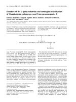
Fig. 1. Electrointestinography (EIG) electrode position. A: Small
intestine, B: Ceacum, ●: EIG mini-amplifier, ○: EIG in differen
t
electrodes.
Pharmaceutical, Japan) and 2.2 mg/kg of ketamine
(Veterinary Ketalar 50; Sankyo Yell Yakuhin, Japan).
Guaifenesin (25∼50 mg/kg; Kyoto Pharmaceutical
Industries, Japan) was infused rapidly until the horse became
ataxic. The trachea was then intubated and the horse was
held on a surgical table. Anesthesia was maintained by
inhalation of halothane and oxygen. At the beginning of
surgery, 20 mg/kg of cefalotin sodium (Coaxin; Tobishi
Pharmaceutical Industries, Japan) and 1.1 mg/kg of flunixin
meglumine (Banamine; Dainippon Sumitomo Pharmaceutical,
Japan) were administered to each horse intravenously.
The surgical area was routinely prepared for aseptic
surgery in dorsal recumbency and a ventral midline celiotomy
was performed. The caecum was exteriorized and the apex
was pulled caudally to expose the dorsal taenital band. The
surgical method of jejunocaecostomy used in the present
study was essentially the same as performing hand suturing
as used by Donawick et al. [4]. The ileum and jejunum
were resected approximately 50 and 200 cm proximal to
the ileocaecal junction, respectively.
After the surgical intervention, 20 mg/kg of cefalotin
sodium and 1.1 mg/kg of flunixin meglumine were
intravenously administered twice a day at 9 : 00 and 18 : 00
for 5 days, and 10,000 ml of lactated Ringer’s solution
(Solulact; Terumo, Japan) was intravenously administered
twice a day for 4 days. Incisions were treated as necessary.
Water was offered ad libitum starting 13 h after the surgery,
and the horses were fed 0.7 kg of alfalfa softened by hot
water 47 h after operation. The quantity of feed was then
gradually increased over a period of 5 days until the
amount of their meal diet reached its former level.
Treatment with mosapride
Mosapride (Gasmotin; Dainippon Sumitomo Pharmaceutical,
Japan) at a dose of 1.5 mg/kg was administered in 1,000 ml
distilled water via a nasogastric tubation once daily at 9:00
am for 5 consecutive days.
Evaluation of intestinal motility
EIGs of the small intestine and caecum were performed
on conscious horses at rest in a stall. After clipping the hair
over the paralumbar fossa on the left and right sides of the
abdomen, the skin was washed and EIG electrodes were
installed via surface electrodes (Vitrode M-150 Disposable
Electrodes; Nihon Kohden, Japan) at three sites: the front
edge of the tuber coxae (EIG mini-amplifier), the intersection
of the horizontal line extending from the tuber coxae and
the rear edge of the last rib (noninductive electrodes), and
the apex of an inverted regular triangle formed by placing
the other two electrodes on the other apexes (EIG mini-
amplifier; Fig. 1). At a sampling rate of 1 Hz, the frequency
was measured within the range of 1.6∼12 cycles per min.
An electrogastrographic (EGG) recorder (Nipro EG; A&D,
Japan) and a digitrapper EGG system were used to measure
the percutaneous potential of the small intestine and caecum
[10]. The system was attached to the trunk by means of a
saddle, a girth, and a saddlecloth.
Preoperative EIGs were recorded for 24 h before the
surgical operation after horses were fed an ordinary diet.
Postoperative EIG data were collected immediately
following recovery from general anesthesia, continued to
be recorded for 10 consecutive days, and were thereafter
collected on days 17, 24, and 31 for 24 h each day.
For EIG analysis, a running spectrum method with fast
Fourier transform was used. The waveform was divided
into 1-min intervals. The relative value of maximum
amplitude (μv) of the wave form at 10 min per h was
calculated. Preoperative data were regarded as 100% of
relative values.
Statistical analysis
Data were indicated as the mean ± SD. Two-way repeated
measure ANOVA was used to determine significant
differences between the treated group and the control.
Significant differences were evaluated by a post-hoc test
(Fisher’s PLSD). The significance level was set at p <
0.05.
Results
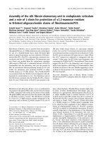
In the control group, the EIG maximum amplitudes of the
small intestine and caecum on postoperative day 1 were
significantly lower than those recorded before the surgical
operation (44.2 ± 19.1% vs. 100.0 ± 0.0% for the small
intestine and 45.7 ± 19.3% vs. 100.0 ± 0.0% for the
caecum, Figs. 2A and B). The EIG maximum amplitudes
of the small intestine and caecum tended to increase from
postoperative days 2∼10; however, they stayed significantly
lower than those before the surgical operation (p < 0.05).
EIG maximum amplitudes from postoperative days 10∼31
continued to increase reaching preoperative levels.
In mosapride-treated horses, the EIG maximum amplitudes
of the small intestine were significantly higher than those
in the control group from postoperative days 6∼31 (Fig.
2A). Although the EIG maximum amplitudes of the
caecum in mosapride-treated horses tended to be higher
Effects of mosapride on motility of the small intestine and caecum in normal horses after jejunocaecostomy 159
Fig. 2. Electrointestinography maximum amplitude of the small intestine (A) and caecum (B). Preoperative value was taken as 100%
and each value was shown as mean ± SD, [○: treated group (N = 6), △: control (N = 6)].
a,b
Significant differences (p < 0.05) compare
d
with each preoperative value of the treated group (A) and the control group (B).
*
,†
Significant differences (p < 0.05, p < 0.01) compared
between the treated group and the control group on the same day.
than those in the control group, the difference was not
statistically significant (Fig. 2B).
Discussion
Mosapride selectively acts on 5-HT
4
receptors and increases
the level of acetylcholine released from cholinergic nerve
endings in the digestive tract [17,18]. Acetylcholine binds to
muscarinic receptors on the smooth muscle and induces
contractions. Generally, mosapride is administrated three
times a day in humans. However, it has been reported that
administration of 1.5 mg/kg once a day significantly
enhances small intestinal and caecal motility in horses
[13]. Therefore, in the present study, mosapride was
administered at 1.5 mg/kg once a day for 5 consecutive
days.
In mosapride-treated horses, the parameters measured
returned to their preoperative levels earlier compared to the
control group. These findings indicate that mosapride
effectively improves a decline in intestinal motility by
increasing both peristalsis and the number of intestinal
contractions [11]. In addition, the EIG maximum amplitudes
from days 2∼31 were significantly higher in mosapride-
treated horses than in the control group. It is considered
that the EIG maximum amplitude reflects contractility of
smooth muscle [9]. Therefore, it is believed that mosapride
enhances contractile motility and thus alleviates decline in
intestinal motility.
In the caecum, no significant difference between the two
groups was observed in the EIG maximum amplitude.
Therefore, we conclude that mosapride acts more effectively
on the small intestine than on the caecum in healthy horses.
It has been reported that the distribution of 5HT
4
receptor
differs depending on gastrointestinal sites and species. In
horses, it is known that 5HT
4
receptors are widely
distributed in the ileum and pelvic flexure [16]. Therefore,
it is suggested that the difference in the distribution of the
5HT
4
receptor is at the base of different effects of
mosapride in the small intestine and caecum.
In this study, we demonstrated that administration of
mosapride is an effective treatment for the decline of intestinal
motility in the period following jejunocaecostomy in healthy
horses. Mosapride appeared to have more of an effect on
motility in the small intestine than in the caecum. It is
therefore concluded that administration of mosapride may
improve the decline of intestinal motility after surgery, but
further studies are needed to understand the mechanism by
which this drug may act as a prokinetic and to determine
whether the potential beneficial effects on intestinal
motility occur in clinical colic cases.
References
1. Bauer AJ, Boeckxstaens GE. Mechanisms of postoperative
ileus. Neurogastroenterol Motil 2004, 16 (Suppl 2), 54-60.
2. Cohen ND, Lester GD, Sanchez LC, Merritt AM,
Roussel AJ Jr. Evaluation of risk factors associated with
development of postoperative ileus in horses. J Am Vet Med
Assoc 2004, 225, 1070-1078.
3. Dart AJ, Hodgson DR. Role of prokinetic drugs for
treatment of postoperative ileus in the horse. Aust Vet J
1998, 76, 25-31.
4. Donawick WJ, Christie BA, Stewart JV. Resection of
diseased ileum in the horse. J Am Vet Med Assoc 1971, 159,
1146-1149.
5. Embertson RM, Colahan PT, Brown MP, Peyton LC,
Schneider RK, Granstedt ME. Ileal impaction in the horse.
J Am Vet Med Assoc 1985, 186, 570-572.
6. Ford TS, Freeman DE, Ross MW, Richardson DW,
160 Kouichi Okamura et al.
Martin BB, Madison JB. Ileocecal intussusception in
horses: 26 cases (1981-1988). J Am Vet Med Assoc 1990,
196, 121- 126.
7. Frankeny RL, Wilson DA, Messer NT, Campbell-Beggs
C. Jejunal intussusception: a complication of functional end-
to-end stapled anastomoses in two ponies. Vet Surg 1995,
24, 515-517.
8. Freeman DE. Surgery of the small intestine. Vet Clin North
Am Equine Pract 1997, 13, 261-301.
9. Kajimoto T. Relation between electrogastrography and
gastric electromyogram, mechanical activity. J Smooth
Muscle Res 1995, 31, 93-107.
10. Sasaki N, Lee I, Ayukawa Y, Yamada H. Clinical
applications of electrointestinography in the horse. J Equine
Sci 2004, 15, 85-92.
11. Sasaki N, Mizuno Y, Yoshihara T. The application of
electrocecography for evaluation of cecum motility in
horses. J Vet Med Sci 1998, 60, 1221-1226.
12. Sasaki N, Murata A, Lee I, Yamada H. Evaluation of
equine cecal motility by ausculation, ultrasonography and
electrointestinography after jejunocecostomy. Res Vet Sci
2008, 84, 305-310.
13. Sasaki N, Okamura K, Yamada H. Effects of mosapride, a
5-hydroxytryptamine 4 receptor agonist, on electrical
activity of the small intestine and cecum in horses. Am J Vet
Res 2005, 66, 1321-1323.
14. Smith CL, Dowling BA, Dart AJ. Recent advances in
equine abdominal surgery. Vet J 2005, 170, 41-51.
15. Van Hoogmoed LM, Nieto JE, Snyder JR, Harmon FA.
Survey of prokinetic use in horses with gastrointestinal
injury. Vet Surg 2004, 33, 279-285.
16. Weiss R, Abel D, Scholtysik G, Straub R, Mevissen M.
5-Hydroxytryptamine mediated contractions in isolated
preparations of equine ileum and pelvic flexure:
pharmacological characterization of a new 5-HT(4) agonist.
J Vet Pharmacol Ther 2002, 25, 49-58.
17. Yoshida N, Ito T, Karasawa T, Itoh Z. AS-4370, a new
gastrokinetic agent, enhances upper gastrointestinal motor
activity in conscious dogs. J Pharmacol Exp Ther 1991, 257,
781-787.
18. Yoshida N, Omoya H, Oka M, Furukawa K, Ito T,
Karasawa T. AS-4370, a novel gastrokinetic agent free of
dopamine D2 receptor antagonist properties. Arch Int
Pharmacodyn Ther 1989, 300, 51-67.