Báo cáo khoa học: "The effect of doxycycline on canine hip osteoarthritis: design of a 6- months clinical trial" pps
Bạn đang xem bản rút gọn của tài liệu. Xem và tải ngay bản đầy đủ của tài liệu tại đây (376.55 KB, 9 trang )
JOURNAL OF
Veterinary
Science
J. Vet. Sci. (2009), 10(3), 239
247
DOI: 10.4142/jvs.2009.10.3.239
*Corresponding author
Tel: +66-53-948046; Fax: +66-53-274710
E-mail:
The effect of doxycycline on canine hip osteoarthritis: design of a 6-
months clinical trial
Korakot Nganvongpanit
1,3,
*
, Peraphan Pothacharoen
3
, Niyada Suwankong
1,2
, Siriwan Ong-Chai
1,3
, Prachya
Kongtawelert
1,3
1
Bone and Joint Research Laboratory, Department of Veterinary Biosciences and Public Health, and
2
Department of Small
Animal Clinic, Faculty of Veterinary Medicine, Chiang Mai University, Chiang Mai 50100, Thailand
3
Thailand Excellence Center for Tissue Engineering, Department of Biochemistry, Faculty of Medicine, Chiang Mai
University, Chiang Mai 50000, Thailand
Twenty-five dogs were included in a randomized, double-
blind trial to assess the efficacy of doxycycline (DOX) orally
administered twice a day at 4 mg/kg/day (n = 12) for the
treatment of osteoarthritis of the hip. Chondroitin sulfate
(CS; 525 mg/day) was used as a positive control (n = 13).
Dogs were re-examined monthly for 6 months after initiation
of treatment. The assessment protocol included clinical
score, radiographic findings and serum osteoarthritis
biomarkers. Dogs treated with DOX showed statistically
significant improvements (p
<
0.05) in lameness, joint
mobility, pain on palpation, weight-bearing and overall
score at 2, 6, 4, 4 and 4 months, respectively, after treatment.
Biomarker levels of CS-WF6 epitope and hyaluronan were
significantly increased and decreased (p
<
0.05) at 2 and 3
months after treatment compared to pretreatment. These
results showed that DOX had a positive therapeutic effect in
dogs with osteoarthritis.
Keywords:
biomarkers, canine, chondroitin sulfate, doxycycline,
osteoarthritis
Introduction
Osteoarthritis (OA) is a chronic, disabling condition for
which there are no cure and few useful treatments [9].
Clinical features include joint pain, instability, limitation
of motion and function impairment. The pathogenesis of
OA, albeit not yet well understood, is often linked to joint
injury, biochemical alterations and ageing [20]. Pharmacological
treatment alternatives for OA can be divided into two
groups: symptom-modifying and disease-modifying drugs
[7]. Symptom-modifying drugs are at present the prescription
of choice for patients with OA. Drugs in this group are
analgesics and non-steroidal anti-inflammatory drugs,
which are effective in relieving the symptoms of OA [8].
There has recently been a lot of debate about some
biological agents that are thought to have both symptom-
modifying and disease-modifying properties [8,11].
Most of the compounds suggested as disease- modifying
drugs are physiological molecules contained in articular
tissues such as glucosamine sulfate and chondroitin sulfate
(CS) [13]. A tetracycline antibiotic, doxycycline (DOX),
has been successfully used to treat a wide-range of bacterial
infections. In addition to its effects as an antibiotic, laboratory
studies with animals and with human tissue have shown that
doxycycline can inhibit the degradation of cartilage in a way
that could be useful for the treatment of OA [24-26,38]. DOX,
reportedly limits cartilage degradation and significantly
ameliorates the degenerative changes that occur in OA
joints [24-26,38]. Specifically, DOX orally administered at
low dosages appeared to reduce the rate and extent of joint
pathology in a canine model of OA [19,36,37]. However,
most of the papers published about DOX and joint
pathology were done in vitro [1,2,25,38]. Therefore, this
study aims to investigate whether the long- term use of DOX
can favorably modify the progression of OA in dogs.
Moreover, to confirm preclinical data suggesting that DOX
can slow the progression of OA. The experimental design
was developed according to potential clinical use, with
clinical score, radiographic findings and serum osteoarthritis
biomarkers as primary outcome measures.
Materials and Methods
Animals
Twenty-five client-owned dogs were included in this
study, 12 males and 13 females aged between 1∼7 (4.21 ±
1.63) years old. Twenty dogs were Golden retrievers and 5
240 Korakot Nganvongpanit et al.
Tabl e 2 . Radiographic scoring system for assessing dogs with
osteoarthritis
Grade Radiographic evaluation
0 Normal Not affected
1 Mild Doubtful narrowing of joint space and
possible osteophytic lipping
2 Moderate Definite osteophytes and possible
narrowing of joint space
3 Severe Moderate multiple osteophytes, definite
narrowing of joints space, some sclerosis and
possible deformity of bone contour
4 Very severe Large osteophytes, marked narrowing of
joint space, severe sclerosis and definite
deformity of bone contour
Tabl e 1 . Clinical scoring system for assessing dogs with osteoarthritis
Criterion Grade Clinical evaluation
Lameness 1 Walk normally
2 Slightly lame when walking
3 Moderately lame when walking
4 Severely lame when walking
5 Reluctant to rise and will not
walk more than five paces
Joint mobility 1 Full range of motion
2 Mild limitation (10∼20%) in
range of motion; no crepitus
3 Mild limitation (10∼20%) in
range of motion; crepitus
4 Moderate limitation (20∼50%) in
range of motion; ± crepitus
5 Severe limitation (>50%) in
range of motion; ± crepitus
Pain on 1 None
palpation 2 Mild signs; dog turns head
in recognition
3 Moderate signs; dog pulls limb away
4 Severe signs; dog vocalizes or
becomes aggressive
5 Dog will not allow palpation
Weight bearing 1 Equal on all limbs standing and walking
2 Normal standing; favors affected limb
when walking
3 Partial weight-bearing standing and walking
4 Partial weight-bearing standing;
non-weight-bearing walking
5 Non-weight-bearing standing and walking
Overall score 1 Not affected
of clinical 2 Mildly affected
condition 3 Moderately affected
4 Severely affected
5 Very severely affected
dogs were Labrador retrievers. Informed owner consent
was obtained and the trial protocol was approved by the
Faculty of Veterinary Medicine, Chiang Mai University’s
Ethics Committee, Chiang Mai, Thailand.
Inclusion/exclusion criteria
Golden and Labrador retriever dogs with clinical signs of
chronic lameness, stiffness and joint pain and radiological
evidence of OA of the hip were considered eligible for this
study. Animals which were pregnant, receiving medication,
or had hepatic, cardiovascular, gastrointestinal and
neurological disease, were excluded. Dogs with lameness
due to lumbosacral instability, infection, immune disease
and fractures and dogs which previously received drug or
dietary supplement for OA treatment were also excluded.
Pretreatment evaluation
Dogs were clinically examined and blood samples were
collected for baseline hematology, blood chemistry and
biomarker for OA. Radiographs of hip joints were
interpreted by two veterinarians.
Treatment protocol
The dogs were randomly assigned to two treatment
groups. The first group (DOX group) received doxycycline
(2 mg/kg body weight twice daily; Osoth Inter Laboratories,
Thailand) [19], the second group (CS group) served as
control group and received chondroitin sulfate (Fortiflex,
525 mg/dog daily; Virbac, USA). Animal were re-assessed
monthly for clinical evaluation and blood collection, while
radiographs were taken every 2 months. Treatment was
stopped on the end of the 6th month.
Assessment protocol
Two veterinarians recorded the severity of the clinical
signs at each monthly visit using an ordinal scoring system
(Table 1) [10] and all veterinarians scored blind to the
group classification. The radiographs of hip joints were
taken every 2 months (3 times per animal) and were
interpreted by the 2 veterinarians using the Takahashi
scoring system (Table 2) [28]. Three milliliters of blood
was collected monthly from the cephalic vein to assess the
levels of OA biomarkers [16,17,22,23].
Clinical score
Efficacy of the treatment was determined by the mean of
a clinical scoring system [10] that assessed the animal
specific lameness, joint mobility, pain on palpation,
weight-bearing and overall score of clinical condition. The
dogs had to walk and trot 6 meters 3 times for the evaluation
The effect of doxycycline for canine osteoarthritis treatment 241
Tabl e 3. Sex, age and
b
ody weight distribution for all 25 dogs
which completed the trial
Group Total
Gender
Age (months) Weight (kg)
Male Female
Doxycycline 12 5 7 52.25 ± 20.25 30.17 ± 5.13
Chondroitin 13 7 6 49.33 ± 19.58 28.42 ± 5.28
sulfate
Age and weight data are expressed as mean ± SD; neither were
significantly different between the two groups (p > 0.05).
Tabl e 4 . Comparison of pre-treatment clinical and radiographic
scores for doxycycline (DOX) and chondroitin sulfate (CS) groups
Parameter
Group
p-value
DOX CS
Lameness 3.92 ± 0.76 3.67 ± 0.89 0.44
Joint mobility 3.08 ± 0.86 3.00 ± 0.60 0.80
Pain on palpation 2.31 ± 0.48 2.33 ± 0.49 0.90
Weight bearing 3.69 ± 0.75 3.67 ± 0.78 0.93
Overall score 3.46 ± 0.78 3.58 ± 0.90 0.72
Radiography score 2.46 ± 0.66 2.67 ± 0.78 0.48
The data are expressed as mean ± SD which were not significantly
different between two group.
of lameness by 2 veterinarians, following palpation on hip
joint for joint mobility and pain evaluation. The palpation
was performed by 2 veterinarians, 30 min apart.
Radiographs
Structural joint changes were assessed on serial radiographs
performed according to the standardized technique
recommended by Takahashi [28]. Radiographs were taken
for each animal at enrollment and at 3 and 6 months after
treatment by the same technician using a usual X-ray
machine (Kelex, Thailand). Ventrodorsal radiographs
were obtained with the dog’s hip and the leg in full
extension position. Repositioning of the dog for subsequent
radiographs were guided by the original film and the same
radiographic setting (i.e. kilovolts, milliamperes and
milliseconds) were used. All radiographs in a dog set (3
films) were interpreted for all evaluations concomitantly
by 2 veterinarians using the criteria in Table 2.
Hematology and biochemistry
Blood samples were analyzed for complete blood counts,
including hematocrit, hemoglobin level, red blood cell
count and white blood cell count and the platelet count.
Two mililiters of serum were analyzed for aspartate
aminotransferase, alanine aminotranferase, blood urea
nitrogen and creatinine.
Biomarker assay
The biomarker assays were enzyme-linked immunoassays
(ELISA) as previously described [16-18,22,23]. This study
used 2 biomarkers; CS-WF6 epitope and hyaluronan (HA).
Competitive immunoassay using monoclonal antibody
WF6
A mouse monoclonal antibody WF6 was raised against a
shark cartilage aggrecan preparation and a quantitative
ELISA for the epitope recognized by monoclonal antibody
WF6 was modified from a previous study [16,22]. The
antibody was specific for intact CS chains and showed no
interaction with other sulfated glycosaminoglycans,
hyaluronan or other polyanions, such as DNA, RNA or
dextran sulfate. The standard used in the assay was shark
cartilage aggrecan (A1 fraction) (Sigma-Aldrich, USA) at
concentrations of 19∼10,000 ng/mL in 6% Bovine serum
albumin (BSA) in Tris Incubation (TI) buffer (0.1 M Tris
HCl, pH 7.4 containing 0.15 M sodium chloride, 0.1%
Tween 20 and 0.1% BSA). Diluted human serum samples
(1 : 5 in 6% BSA-TI) were added to 1.5 mL plastic tubes
containing an equal volume of WF6 (cell culture
supernatant, 1 : 200 dilution in TI buffer). They were
incubated at 37
o
C for 1 h, and then added to the microtiter
plate, which was pre-coated with shark aggrecan (A1
fraction). Non-specific protein binding was blocked with
BSA. The plates were then incubated at 37°C for 1 h, and
the wells were then washed and peroxidase-conjugated
anti-mouse IgM antibody (1 : 2,000) was added (100 mL/
well; in TI buffer). The bound conjugate was detected by
adding ortho-phenylenediamine (o-PD) substrate (100
mL/well in 0.05 M citrate buffer, pH 5.0). The reaction was
stopped after 10 min with 50 mL/well of 4 M sulfuric acid,
and absorbance was determined using a microplate reader
(Titertek multiscan Mcc/340; ICN-Flow, USA) at 492/690
nm. The concentration of WF6 epitope in supernatant
samples was calculated by reference to a standard curve.
ELISA-based assay for HA using biotinylated HA-
binding proteins (HABPs)
Human serum samples or standard HA (HealonR;
Pharmacia Pharmaceutical AB, Sweden) at various
concentrations [19∼10,000 ng/mL in 6% bovine serum
albumin (BSA)-phosphate buffer saline (PBS) pH 7.4]
were added to 1.5 mL plastic tubes containing biotinylated
HABPs prepared as described above (1 : 200 in 0.05 M
Tris-HCl buffer, pH 8.6). The tubes were incubated at room
temperature for 1 h, and then samples were added to the
microplate, which was precoated with umbilical cord HA
(100 mL/well of 10 mg/mL) and blocked with 1% BSA
242 Korakot Nganvongpanit et al.
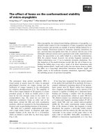
Fig. 1. Mean scores for lameness, joint mobility, pain on palpation, weight bearing and overall. Black = DOX group; white = CS group.
*Values were significantly different compare to month 0 within the groups (p < 0.05).
†
Values were significantly different
b
etween
groups within the month (p < 0.05).
(150 mL/well). The plate was then incubated at room
temperature for 1 h. The wells were then washed and
peroxidase-conjugated antibiotin antibody (1 : 2,000
dilution; Zymed, USA), 100 mL/well in PBS, was added.
The plate was incubated at room temperature for another
hour. The detection of conjugated antibody was with o-PD
substrate and plate reading was carried out as described
above. The concentration of HA in samples was calculated
from the standard curve [16,23].
Data collection and statistics
The results of CS and HA analyses are presented as mean ±
SD. The non-parametric 2-sample Mann-Whitney procedure
was used to test for differences between the DOX and CS
groups. The radiograph and clinical sign scores were
calculated as mean ± SD. The non-parametric 2-sample
The effect of doxycycline for canine osteoarthritis treatment 243
Fig. 2. Mean radiography scores. Black = DOX group; white =
CS group. Vertical bar means a standard deviation.
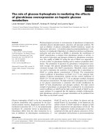
Fig. 3. Mean of relative change (%) of serum CS-WF6 epitope and hyaluronan (HA). Black = DOX group; white = CS group. *Values
were significantly different compare to month 0 within the groups (p < 0.05).
†
Values were significantly different between groups
within the month (p < 0.05).
Mann-Whitney procedure was used to test for differences
between the DOX and CS groups in the same month and
between before and after treatment. The relative data was
analyzed using the Statistical Analysis System version 8.0
(SAS Institute, USA) software package. p ≤ 0.05 was
considered to be significant.
Results
Thirty dogs were enrolled in the trial but 5 were
withdrawn due the failure to attend an assessment
appointment (2 dogs in the DOX group, 1 in the CS group)
and death from a car accident (1 dog in the DOX group and
1 in the CS group). Table 3 shows the summary of age, sex
and body weight data of 25 dogs completing the trial to the
6th month. All dogs enrolled in the trial had hemogram and
biochemical profile results within reference range throughout
the trial (6 months). Comparisons of pre-treatment disease
score found no significant difference (p > 0.05) between
the DOX and CS groups (Table 4).
Fig. 1 presents the 5 clinical score data before treatment
(month 0) and at one month intervals until 6 months. The
lameness score in the DOX group showed significant
improvements (p < 0.05) at th e 2nd month, while in the
CS group it showed significant improvements (p < 0.05)
in the first month. Joint mobility score in the DOX and CS
groups showed significant improvements (p < 0.05) at the
6th and 4th months, respectively. Pain at palpation, weight
bearing and the overall score in the DOX group showed
significant improvements (p < 0.05) at the 4th month, but
in the CS group it showed significant improvements (p <
0.05) earlier, in the 2nd month of treatment. Lameness,
joint mobility, pain of palpation and overall score in the CS
group was significant better (p > 0.05) than in the DOX
group. The weight bearing score in the CS group was
significant better (p > 0.05) than in the DOX group from
the first month.
Radiography scores are shown in Fig. 2. Those scores
were not significantly difference between treatment groups
(p > 0.05). When comparing with the pretreatment scores,
there also was no significant difference (p < 0.05).
The results of serum biomarkers for OA are shown in Fig.
3. The level of CS-WF6 epitope in the DOX and CS groups
were significantly higher than before treatment. Differences
between the 2 groups were significant in the first and
second months after treatment. The level of HA in the
DOX and CS groups were significantly higher than before
treatment after 3 and 1 months of treatment, respectively.
Discussion
The results of this study showed that dogs with OA had
244 Korakot Nganvongpanit et al.
significant improvements in score for clinical evaluation
and biomarker levels when treated with oral DOX.
However, all of these effects occurred slower when
compared to CS.
Disease-modifying or symptomatic slow-acting drugs are
more interesting because some have been shown to be
effective in improving symptoms and in reducing OA
cartilage degradation with a reasonable safety profile
[8,11]. These drugs have shown onset of efficacy and a
prolonged residual effect once treatment is stopped [7]. As
this study was a clinical trial, CS was chosen as positive
control in order to address ethical responsibilities and the
welfare of the participant dogs. According to previous
studies, CS has been shown to reduce pro-inflammatory
factors, modify the cellular death process and improve the
anabolism/catabolism balance of extracellular cartilage
matrix [12,13]. At the same time it has proven to have a
positive effect on the pathological process involving the
synovial tissue and subchondral bone. These mechanisms
could, account for the beneficial results observed in some
clinical trials [12,13,31].
The recognized limitation of this study was the lack of an
objective assessment of the joint. It was not possible to
perform ground force reaction measurements as was done
in the trials of Hazewinkel [6], Moreau [14] and Vasseur
[32] as this was a multicenter trial. Subjective assessment
of weight bearing by 2 blinded veterinarians was used
instead. Our study found that DOX had a slower effect on
clinical improvements compare to CS. The overall score
was improved 3 months after treatment, while CS had a
significant effect 2 months after treatment. Moreover, we
found that DOX did not improve the measures of pain as
well as CS. Radiographic findings did not show any
significant changes. In agreement with a published study
[4], DOX did not significantly prevent the onset of
progressive joint space narrowing (JSN) in the contralateral
knee, and did not improve measures of pain or function of
the OA knee.
The morphological changes in OA include alterations in
the cartilage, subchondral bone, and synovial membrane
[9,20]. Current knowledge points to an important
involvement of the matrix metalloproteases (MMPs) class
in the OA process [20]. Collagenase-3 (MMP-13) was
demonstrated to play a major role in cartilage degeneration.
It is also suggested that another enzyme, aggrecanase-2, or
ADAMTS-5 plays a predominant role in the proteolysis of
OA cartilage aggrecan [5,27].
It is recognized that MMPs play a role in the pathologic
breakdown of the joint extracellular matrix in OA. It is
known that low-dose regimens of a tetracycline analogue,
namely DOX can inhibit some MMPs, hence reducing the
extracellular matrix breakdown [1,2]. A recent study
examined the effects of DOX on knee OA progression [4].
The primary outcome measure was JSN in the medial
tibiofemoral compartment. Obese females with a unilateral
OA knee were randomly assigned to receive 30 months of
treatment with DOX or placebo. The loss of joint space
width in the index knee in the DOX group was less than in
the placebo group. This study showed that DOX can reduce
the progression of established OA in this patient
population. It provides the first proof of concept of the
effectiveness of anti-MMP strategies for developing
disease-modifying drugs.
Inhibition of the MMPs superfamily is a very logical
objective in OA. Moreover, tetracyclines inhibit collagenase
levels and nitric oxide production in vitro, thereby
decreasing chondrocyte MMPs activity and increasing
proteoglycan synthesis attenuating OA in animal models
[1]. In agreement with a previous study, Pardy [19] showed
DOX treatment conserved bone strain energy density at 72
weeks. Doxycycline had little effect on the degradation of
superficial osseous tissue at 36 week after anterior cruciate
ligament transection (ACLT); by 72 weeks, DOX in ACLT
canine model limited subchondral bone loss within the first
3 mm of periarticular bone with established OA.
Significant bone loss occurred in the deeper trabecular
bone for all groups. Substantial architectural adaptation
within deeper trabecular bone accompanied changes in
mechanics in early and established OA. In 1991, Yu was
done in vitro studies, and indicated that levels of neutral
MMPs in OA cartilage are elevated and that doxycycline
inhibits collagenolytic and gelatinolytic activity in extracts
of OA cartilage [38]. However, before tetracycline and its
analogues, or even MMP inhibitors, can be considered to
be an effective treatment in preventing knee OA
progression, further investigations are needed.
A novel monoclonal antibody CS-WF6, which
recognizes a native epitope in CS chain [23], was elevated
after treatment. The finding of elevated levels of serum
CS-WF6 epitope after treated with both DOX and CS
reflector to alteration the metabolism of the cartilage. In
chronic OA, the level of CS-WF6 epitope is higher than
normal because the native CS chain in cartilage was
degraded and release into the blood system [16,17,23]. The
elevation of CS-WF6 epitope in this study was shown both
drugs induced the synthesis of CS-chain in cartilage lead to
have more proportion of CS in the cartilage. This new CSs
were source of the degradation process in OA joint which
made a CS-WF6 epitope up-regulation. We found DOX
had a slower effect on cartilage metabolism than CS. In this
study, the level of CS-WF6 epitope was found to be
significantly elevated after 1 and 2 months in CS and DOX
groups, respectively.
In an inflammatory rat model of arthritis, it was
demonstrated that serum HA levels correlated with the
degree of synovitis and clinical arthritis [3]. HA plays the
key role in immobilizing aggrecans in articular cartilage;
this balances the tension and compressive resilience in the
The effect of doxycycline for canine osteoarthritis treatment 245
collagen network by its osmotic properties. Also, the HA
levels were related to joint inflammation in humans [21].
Serum HA has been studied as a biomarker of disease
progression, since significantly increased levels were
reported in cases of rheumatoid arthritis and progressive
osteoarthritis, compared to the normal population
[3,21,23]. In our study, the HA levels were significantly
decreased after 1 and 2 months in CS and DOX groups,
respectively. This means that both DOX and CS decreased
the level of inflammation in the joint. Compared between 2
groups, CS reduce inflammation significantly faster than
DOX.
The one importance issue in using DOX as disease-
modifying drug in OA is the microbial resistance. This trial
did not study the effect of using a chronic, sub-antimicrobial
dose of DOX on microbial resistance. However, many
studies had proved that using sub-antimicrobial dose DOX
(20 mg twice a day) had no effect on the microbial
resistance [29,35]. Microbial studies have documented
the lack of any antimicrobial effect on the normal flora,
periodontal and/or opportunistic pathogens, or change in
antibiotic susceptibilities following the use of a sub-
antimicrobial dose of DOX up to 9 months in double-
blinded, placebo controlled, multicentered studies [29].
These studies examined the effect, or lack of effect, of
sub-antimicrobial dose DOX on the sub-gingival flora
[29,30,35] and on antibiotic resistances within this flora
[29] in a periodontitis population. Likewise, there was no
detectible effect of a 9-month regimen of sub-antimicrobial
dose of DOX on the intestinal flora of a periodontitis
population cross-sectionally, relative to placebo control, or
longitudinally within the sub-antimicrobial dose DOX
treatment group [33]. In 2007 Walker and colleague [34]
had reported that there was no evidence that exposure to
DOX, 20 mg twice a day, resulted in cross- or multiantibiotic
resistance. No evidence was present that the use of a
sub-antimicrobial dose of DOX for a period of 24 months
in a population of periodontally diseased osteopenic
women exerted any detectible effect on the microbial flora
as determined by total anaerobic counts and total counts for
actinomyces and streptococci. There was no evidence that
a sub-antimicrobial dose of DOX resulted in the
colonization or overgrowth by periodontal and/or
opportunistic pathogens. In our trial, we used 4 mg/kg
DOX daily, which is a sub-antimicrobial dose (10 mg/kg
daily) [15], related to all publications which mention above
that possible is using 4 mg/kg DOX daily has no effect on
the microbial resistance. However, all studies on microbial
resistance had been done in humans not canine, so to fulfill
this hypothesis, the sub-antimicrobial dose effect on
microbial resistance needs to be done in canines in the
future.
OA is, by far, the most common type of arthritis in human
and animals encountered worldwide, yet the development
of effective disease-modifying treatments has lagged
behind that of other arthritides. Current challenges that
need to be met are an ideal pharmacy by using novel
knowledge of the biochemistry, molecular biology and
imaging findings that stop progression of disease and
recovery of the cartilage function. This study showed one
of the drugs which can be used as an OA disease-modifying
drug, even though the efficacy was not as great as the
positive control. However, comparing the cost-benefit of
DOX, we believe that DOX will be the disease-modifying
drugs of choice for treated OA in dogs. Indeed, the results
of the present study suggest that using DOX 4 mg/kg daily
for 6 months had no effect on the liver and kidney
functions. This drug can improve the clinical signs of OA
in a dog within 4 months. Moreover, we showed orally
administered DOX can alter the anabolism of the articular
cartilage. This information may prove useful for using
DOX as disease-modifying drug in clinical practice.
Acknowledgments
The authors would like to express their gratitude and
thanks to all veterinarians and technicians at the Bone and
Joint Research Laboratory, and the Small Animal Hospital,
Faculty of Veterinary Medicine, Chiang Mai University for
their kind support. This project was supported by The 2007
Young Researcher Grant, Chiang Mai University, Thailand
and The National Research Council of Thailand (Research
program of drug, chemical, medical material and
equipment).
References
1. Amin AR, Attur MG, Thakker GD, Patel PD, Vyas PR,
Patel RN, Patel IR, Abramson SB. A novel mechanism of
action of tetracyclines: effects on nitric oxide synthases.
Proc Natl Acad Sci USA 1996, 93, 14014-14019.
2. Attur MG, Patel RN, Patel PD, Abramson SB, Amin AR.
Tetracycline up-regulates COX-2 expression and prostaglandin
E2 production independent of its effect on nitric oxide. J
Immunol 1999, 162, 3160-3167.
3. Bj
örk J, Kleinau S, Tengblad A, Smedegård G. Elevated
levels of serum hyaluronate and correlation with disease
activity in experimental models of arthritis. Arthritis Rheum
1989, 32, 306-311.
4. Brandt KD, Mazzuca SA, Katz BP, Lane KA,
Buckwalter KA, Yocum DE, Wolfe F, Schnitzer TJ,
Moreland LW, Manzi S, Bradley JD, Sharma L, Oddis
CV, Hugenberg ST, Heck LW. Effects of doxycycline on
progression of osteoarthritis: Results of a randomized,
placebo-controlled, double-blind trial. Arthritis Rheum
2005, 52, 2015-2025.
5. Glasson SS, Askew R, Sheppard B, Carito B, Blanchet T,
Ma HL, Flannery CR, Peluso D, Kanki K, Yang Z,
Majumdar MK, Morris EA. Deletion of active ADAMTS5
prevents cartilage degradation in a murine model of
246 Korakot Nganvongpanit et al.
osteoarthritis. Nature 2005, 434, 644-648.
6. Hazewinkel HA, van den Brom WE, Theyse LF,
Pollmeier M, Hanson PD. Comparison of the effects of
firocoxib, carprofen and vedaprofen in a sodium urate
crystal induced synovitis model of arthritis in dogs. Res Vet
Sci 2008, 84, 74-79.
7. Lequesne M, Brandt K, Bellamy N, Moskowitz R,
Menkes CJ, Pelletier JP, Altman R. Guidelines for testing
slow acting drugs in osteoarthritis. J Rheumatol Suppl 1994,
41, 65-71.
8. Maddison JE, Johnston KJ. Nonsteroidal anti-inflammatory
drugs and chondroprotective agents. In: Maddison JE, Page
SW, Church D (eds.). Small Animal Clinical Pharmacology.
pp. 251-269, Saunders, London, 2002.
9. Martel-Pelletier J, Lajeunesse D, Fahmi H, Tardif G,
Pelletier JP. New thoughts on the pathophysiology of
osteoarthritis: One more step toward new therapeutic
targets. Curr Rheumatol Rep 2006, 8, 30-36.
10. McCarthy G, O’donovan J, Jones B, McAllister H, Seed
M, Mooney C. Randomised double-blind, positive-
controlled trial to assess the efficacy of glucosamine/
chondroitin sulfate for the treatment of dogs with
osteoarthritis. Vet J 2007, 174, 54-61.
11. McNamara PS, Johnston SA, Todhunter RJ. Slow-
acting, disease-modifying osteoarthritis agents. Vet Clin
North Am Small Anim Pract 1997, 27, 863-881.
12. Michel BA, Stucki G, Frey D, De Vathaire F, Vignon E,
Bruehlmann P, Uebelhart D. Chondroitins 4 and 6 sulfate
in osteoarthritis of the knee: a randomized, controlled trial.
Arthritis Rheum 2005, 52, 779-786.
13. Monfort J, Pelletier JP, Garcia-Giralt N, Martel-
Pelletier J. Biochemical basis of the effect of chondroitin
sulphate on osteoarthritis articular tissues. Ann Rheum Dis
2008, 67, 735-740.
14. Moreau M, Dupuis J, Bonneau NH, Desnoyers M.
Clinical evaluation of a nutraceutical, carprofen and
meloxicam for the treatment of dogs with osteoarthritis. Vet
Rec 2003, 152, 323-329.
15. Morgan RV. Handbook of Small Animal Practice. 5th ed. p.
1351, Saunders, St. Louis, 2008.
16. Nganvongpanit K, Itthiarbha A, Ong-Chai S, Kongtawelert
P. Evaluation of serum chondroitin sulfate and hyaluronan:
biomarkers for osteoarthritis in canine hip dysplasia. J Vet
Sci 2008, 9, 317-325.
17. Nganvongpanit K, Suwankong N, Jitpean S, Ong-Chai S.
The changes of serum chondroitin sulfate in the induced
osteoarthritic dogs after chitosan polysulfate administration.
J Thai Vet Pract 2005, 17, 27-39.
18. Paimela L, Heiskanen A, Kurki P, Helve T, Leirisalo-
Repo M.
Serum hyaluronate level as a predictor of
radiologic progression in early rheumatoid arthritis. Arthritis
Rheum 1991, 34, 815-821.
19. Pardy CK, Matyas JR, Zernicke RF. Doxycycline effects
on mechanical and morphometrical properties of early- and
late-stage osteoarthritic bone following anterior cruciate
ligament injury. J Appl Physiol 2004, 97, 1254-1260.
20. Pelletier JP, Martel-Pelletier J, Raynauld JP. Most recent
developments in strategies to reduce the progression of
structural changes in osteoarthritis: today and tomorrow.
Arthritis Res Ther 2006, 8, 206.
21. Poole AR, Witter J, Roberts N, Piccolo F, Brandt R,
Paquin J, Baron M. Inflammation and cartilage metabolism
in rheumatoid arthritis. Studies of the blood markers
hyaluronic acid, orosomucoid, and keratan sulfate. Arthritis
Rheum 1990, 33, 790-799.
22. Pothacharoen P, Siriaunkgul S, Ong-Chai S,
Supabandhu J, Kumja P, Wanaphirak C, Sugahara K,
Hardingham T, Kongtawelert P. Raised serum chondroitin
sulfate epitope level in ovarian epithelial cancer. J Biochem
2006, 140, 517-524.
23. Pothacharoen P, Teekachunhatean S, Louthrenoo W,
Yingsung W, Ong-Chai S, Hardingham T, Kongtawelert
P. Raised chondroitin sulfate epitopes and hyaluronan in
serum from rheumatoid arthritis and osteoarthritis patients.
Osteoarthritis Cartilage 2006, 14, 299-301.
24. Ryan ME, Greenwald RA, Golub LM. Potential of
tetracyclines to modify cartilage breakdown in osteoarthritis.
Curr Opin Rheumatol 1996, 8, 238-247.
25. Smith GN Jr, Brandt KD, Hasty KA. Activation of
recombinant human neutrophil procollagenase in the
presence of doxycycline results in fragmentation of the
enzyme and loss of enzyme activity. Arthritis Rheum 1996,
39, 235-244.
26. Smith GN Jr, Yu LP Jr, Brandt KD, Capello WN. Oral
administration of doxycycline reduces collagenase and
gelatinase activities in extracts of human osteoarthritic
cartilage. J Rheumatol 1998, 25, 532-535.
27. Stanton H, Rogerson FM, East CJ, Golub SB, Lawlor
KE, Meeker CT, Little CB, Last K, Farmer PJ, Campbell
IK, Fourie AM, Fosang AJ. ADAMTS5 is the major
aggrecanase in mouse cartilage in vivo and in vitro. Nature
2005, 434, 648-652.
28. Takahashi M, Naito K, Abe M, Sawada T, Nagano A.
Relationship between radiographic grading of osteoarthritis
and the biochemical markers for arthritis in knee
osteoarthritis. Arthritis Res Ther 2004, 6, R208-212.
29. Thomas J, Walker C, Bradshaw M. Long-term use of
subantimicrobial dose doxycycline does not lead to changes
in antimicrobial susceptibility. J Periodontol 2000, 71
,
1472-1483.
30. Thomas JG, Metheny RJ, Karakiozis JM, Wetzel JM,
Crout RJ. Long-term sub-antimicrobial doxycycline
(Periostat) as adjunctive management in adult periodontitis:
Effects on subgingival bacterial population dynamics. Adv
Dent Res 1998, 12, 32-39.
31. Uebelhart D, Malaise M, Marcolongo R, de Vathaire F,
Piperno M, Mailleux E, Fioravanti A, Matoso L, Vignon
E. Intermittent treatment of knee osteoarthritis with oral
chondroitin sulfate: a one-year, randomized, double-blind,
multicenter study versus placebo. Osteoarthritis Cartilage
2004, 12, 269-276.
32. Vasseur PB, Johnson AL, Budsberg SC, Lincoln JD,
Toombs JP, Whitehair JG, Lentz EL. Randomized,
controlled trial of the efficacy of carprofen, a nonsteroidal
anti-inflammatory drug, in the treatment of osteoarthritis in
dogs. J Am Vet Med Assoc 1995, 206, 807-811.
33. Walker C, Preshaw PM, Novak J, Hefti AF, Bradshaw
M, Powala C. Long-term treatment with sub-antimicrobial
The effect of doxycycline for canine osteoarthritis treatment 247
dose doxycycline has no antibacterial effect on intestinal
flora. J Clin Periodontol 2005, 32, 1163-1169.
34. Walker C, Puumala S, Golub LM, Stoner JA, Reinhardt
RA, Lee HM, Payne JB. Subantimicrobial Dose Doxycycline
Effects on Osteopenic Bone Loss: Microbiologic Results. J
Periodontol 2007, 78, 1590-1601.
35. Walker C, Thomas J, Nang
ó
S, Lennon J, Wetzel J,
Powala C. Long-term treatment with subantimicrobial dose
doxycycline exerts no antibacterial effect on the subgingival
microflora associated with adult periodontitis. J Periodontol
2000, 71, 1465-1471.
36. Yu LP Jr, Burr DB, Brandt KD, O’Connor BL, Rubinow
A, Albrecht M. Effects of oral doxycycline administration
on histomorphometry and dynamics of subchondral bone in
a canine model of osteoarthritis. J Rheumatol 1996, 23,
137-142.
37. Yu LP Jr, Smith GN Jr, Brandt KD, Myers SL,
O’Connor BL, Brandt DA. Reduction of the severity of
canine osteoarthritis by prophylactic treatment with oral
doxycycline. Arthritis Rheum 1992, 35, 1150-1159.
38. Yu LP Jr, Smith GN Jr, Hasty KA, Brandt KD.
Doxycycline inhibits type XI collagenolytic activity of
extracts from human osteoarthritic cartilage and of
gelatinase. J Rheumatol 1991, 18, 1450-1452.