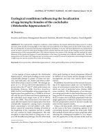
Báo cáo lâm nghiệp: "Chemical composition of the periderm in relation to in situ water absorption rates of oak, beech and spruce fine roots" doc
Bạn đang xem bản rút gọn của tài liệu. Xem và tải ngay bản đầy đủ của tài liệu tại đây (2.44 MB, 10 trang )
763
Ann. For. Sci. 60 (2003) 763–772
© INRA, EDP Sciences, 2004
DOI: 10.1051/forest:2003071
Original article
Chemical composition of the periderm in relation to in situ water
absorption rates of oak, beech and spruce fine roots
Christoph LEUSCHNER
a
*, Heinz CONERS
a
, Regina ICKE
b
, Klaus HARTMANN
c
, N. Dominique EFFINGER
d
,
Lukas SCHREIBER
c
a
Abt. Ökologie und Ökosystemforschung, Albrecht-von-Haller-Institut für Pflanzenwissenschaften, Universität Göttingen,
Untere Karspüle 2, 37073 Göttingen, Germany
b
Abt. Ökologie, Fachbereich 19, Universität Gh Kassel, Heinrich-Plett-Strasse 40, 34132 Kassel, Germany
c
Institut für Botanik, Ökophysiologie der Pflanzen, Universität Bonn, Kirschallee 1, 53115 Bonn, Germany
d
Lehrstuhl Botanik II, Universität Würzburg, Julius-von-Sachs-Platz 3, 97082 Würzburg, Germany
(Received 18 March 2002; accepted 11 September 2002)
Abstract – The water absorption by terminal branch roots of mature oak, beech and spruce trees was measured in situ with miniature sap flow
gauges for 11 consecutive days and related to the suberin and lignin content of the fine root periderm. All fine roots contained a well-developed
periderm, whereas no primary white roots were present. Mean root water uptake decreased in the sequence beech - spruce - oak. Oak roots
contained twice as much suberin and a thicker periderm than beech, and had smaller mean water uptake rates (201 vs. 508 g m
–2
root surface d
–1
).
However, spruce with 2 to 7 times smaller suberin contents had lower uptake rates (346 g m
2
d
–1
) than beech with more suberin. We conclude
that the relationship between periderm chemistry and water absorption is only weak in the three species. Other factors such as hydraulic
resistances in the soil-root interface, or the size of water potential gradients may be more influential in regulating root water uptake.
lignin / miniature sap flow gauge / root hydraulic conductivity / root surface area / suberin
Résumé – Relation entre la composition chimique du périderme et le taux d’absorption d’eau, in situ, des petites racines de chêne, hêtre
et épicéa. On a mesuré, in situ, avec des sondes miniaturisées de flux de sève, pendant 11 jours consécutifs, l’absorption d’eau par les extrêmités
des racines de sujets adultes de chêne, hêtre et épicéa. Celle-ci a été ensuite mise en relation avec le contenu en subérine du périderme des petites
racines. Toutes ces petites racines présentaient un périderme bien développé, sans racines primaires blanches. Les différentes espèces se classent
pour le prélèvement d’eau moyen dans l’ordre décroissant suivant : hêtre, épicéa, chêne. Les racines de chêne contenaient deux fois plus de
subérine et comportaient un périderme plus épais que le hêtre. Leur prélèvement d’eau était en moyenne plus faible (201 contre 508 g m
–2
de
surface racinaire et par jour). Cependant, l’épicéa, avec un contenu de subérine 2 à 7 fois inférieur, présentait un taux de prélèvement plus faible
(346 g m
2
et par jour) que le hêtre qui a pourtant plus de subérine. Nous en concluons que la relation entre la composition chimique du périderme
et l’absorption d’eau n’est que faible pour les trois espèces. D’autres facteurs, tels que la résistance hydraulique à l’interface sol/racines, ou
l’importance des gradients de potentiels hydriques, pourraient jouer un rôle plus important pour la régulation du prélèvement d’eau.
lignine / sonde miniaturisée de flux de sève / conductivité hydraulique des racines / surface racinaire / subérine
1. INTRODUCTION
Water flow along the soil-plant-atmosphere continuum
(SPAC) crosses two major plant-environment interfaces, the
root surface where plant water uptake occurs, and the leaf mes-
ophyll surface where transpiration takes place. Despite its
importance in the SPAC, relatively little is known about the
factors and processes that govern root water uptake. Major
advances in our understanding of water uptake by plant roots
have been made by introducing pressure probe techniques
which allow the measurement of root radial hydraulic conduc-
tivity (Lpr) in excised roots (root pressure probe), or cell
hydraulic conductivity (Lp) in selected root cells (cell pressure
probe) under defined conditions in the laboratory [40, 41]. By
applying these techniques to root systems of various herba-
ceous and woody plant species, it has been shown that the
radial hydraulic conductivity of a root may vary considerably
in response to external (e.g. soil moisture, temperature or
anoxia) or internal factors (e.g. plant water and nutrient status),
but may also change with root development and age [7, 30, 41,
* Corresponding author:
764 C. Leuschner et al.
42]. Moreover, comparison among different plant species
revealed large differences in Lpr that partly seem to be spe-
cies-specific. According to root pressure probe data, the Lpr of
roots of woody species was smaller by an order of magnitude
than that of herbaceous species [41]. Laboratory studies with
young excised root systems indicate that different tree species
may differ significantly in root Lpr as well: Norway spruce
(Picea abies Karst.), sessile oak (Quercus petraea (Matt.)
Liebl.) and European beech (Fagus sylvatica L.) differed at
least fourfold in Lpr with spruce having the highest and beech
the lowest conductivity [34, 43, 44].
A negative relationship between root Lpr and the amount of
suberin in the apoplastic barriers of a root has been found in
several investigations with herbaceous roots [11, 38, 52]. Sim-
ilarly, the large difference in Lpr between herbaceous and
woody roots was attributed to a higher degree of suberisation
in woody roots [41]. We would expect that the observed dif-
ferences in Lpr among spruce, oak and beech roots are a conse-
quence of differences in peridermal suberin content in these
species. In the root systems of adult trees of these species,
white growing roots represent only a low percentage of the
total root surface area. The vast majority of fine roots refers to
mature suberised roots with the periderm representing the
main apoplastic barrier [27]. This situation contrasts with that
in most herbaceous roots where endodermis and exodermis
play this role.
Pressure probe studies with excised root systems or
selected root cells can yield valuable insight into the hydraulic
and osmotic properties of root systems but these techniques do
not provide sufficient information for predicting in situ water
absorption rates of roots in the soil. This is because root
hydraulic conductivity is only one factor among others (e.g.
hydraulic conductivity of the root-soil interface, conductivity
of mycorrhizal hyphae, soil-to-root water potential gradient)
which control water flow from the soil into the root. For tech-
nical reasons, it has been difficult to quantify water uptake
rates of roots in undisturbed soil and, thus, to extrapolate data
on root hydraulic conductivity to in situ root water uptake
rates. Therefore, the question as to whether differences in root
anatomy, chemistry and hydraulics will lead to substantial dif-
ferences in water uptake rates in a shared soil volume, or
whether uptake rates among co-existing plant species are more
or less similar, still remains open. There is the possibility that
species-specific differences in root hydraulics are simply lost
at the level of root water uptake under field conditions if other
influential factors are equally or even more important than
Lpr. Experimental data on root water absorption, which are
needed to solve this problem, are virtually non-existent.
The recently developed miniature sap flow technique pro-
vides a welcome opportunity to study tree root water uptake in
the soil under in situ conditions [9]. For the first time, a
method allows to measure water absorption of tree terminal
branch roots in the field without disturbing soil structure, soil
moisture and mycorrhizal infection of root tips. In this study,
the miniature sap flow technique in combination with root sur-
face area determination was used to compare water absorption
per root surface area in three co-existing temperate tree spe-
cies in a mixed stand. In Central Europe, sessile oak, European
beech and Norway spruce have been found to differ in the sen-
sitivity of their leaf water status and growth to soil drought
with oak being the least sensitive and spruce the most sensitive
species [2, 12, 23, 48]. Consequently, spruce is restricted to
sites with moderate to high rainfall (> 650 mm) but is absent
from regions with low precipitation and/or sandy soils where,
in many cases, oak dominates over both spruce and beech [12].
We compare the water absorption rates of terminal branch
roots (diameter: 3–4 mm) of co-existing oak, beech and spruce
trees measured in situ and relate them to the contents of
suberin and lignin in the root periderm. This approach contrasts
with earlier laboratory studies on the relationship of radial
hydraulic conductivity and the chemical composition of apo-
plastic transport barriers in plant roots in two ways: (a) chem-
ical data are expressed in relation to root surface area instead
of root mass, and (b) water absorption instead of hydraulic
conductivity is measured. The following hypotheses are
tested: (i) species-specific differences in water absorption
rates are related to the suberin and lignin contents of the root
periderm, and (ii) suberin and lignin content and, thus, water
absorption, are a function of periderm thickness.
2. MATERIALS AND METHODS
2.1. Study site
Field measurements of root water uptake and sampling of root bio-
mass were conducted in an old-growth mixed beech/oak/spruce stand
in the vicinity of Unterlüss in the southern part of the Lüneburger
Heide (Lower Saxony, Germany, 52° 45’ N, 10° 30’ E). The study
plot is located in close proximity to forest site no. OB5 where root
system structure and root functioning have been studied in mature
beech and oak trees by [5, 16, 22–24]. The plot consists of 90- to 100-
yr-old beech, 180- to 200-yr-old oak, and 80- to 100-yr-old spruce
trees at similar stem densities (total number of trees per hectare: ca.
250) that form a closed canopy of 28–32 m in height. Shrub and her-
baceous layers are missing.
Located in the diluvial lowlands of NW Germany on Saalian melt
water sands (115 m asl), this site is characterised by soil profiles
(spodo-dystric cambisols) with thick organic layers (mean depth of
the entire organic profile, i.e. Of+Oh horizons, is 72 mm). The
organic profile is highly acidic with pH values of 3.0 and 2.6 (in KCl)
and Ca
2+
/H
+
quotients of 0.2 in the equilibrium soil solution of the
upper (Of) and lower organic horizons (Oh), respectively. Measure-
ments using the in situ-soil incubation method [33] showed that about
85% of the profile total of net nitrogen mineralisation is supplied by
these organic horizons, which are much more important for plant
nutrition than the mineral soil [24].
The climate is humid sub-oceanic (annual means: 8.0 °C,
800 mm). The ground water table is far below the rooting horizon.
Periods of low rainfall in summer irregularly lead to substantial water
shortage in the sandy mineral soil and in the forest floor. Gravimetric
monitoring of soil water content (θ) in the densely rooted organic Of
and Oh layers on the forest floor showed that θ may be reduced to less
than 10 vol% during summer which corresponds to soil matric poten-
tials < –1.5 to –2.0 MPa in this substrate [22]. In such periods,
drought-induced fine root mortality can affect the root systems of
beech (but not of oak) in the superficial organic horizons [16, 23].
2.2. Root sampling and anatomical investigation
For investigating root anatomy and peridermal chemistry, we
extracted 11 branch root systems per species in a 3 × 3 m plot bor-
dered by an oak, a beech and a spruce tree separated by about 10 m.
Root water absorption 765
The stem diameters of the trees were representative of the respective
tree species in the stand. The 11 roots were sampled in direct proxim-
ity of those roots that were used for root sap flow measurements (see
below). We applied compressed air (0.2–0.4 MPa) to completely
expose the appending root systems without damaging fine rootlets
and peridermal surfaces. The sampled fine root systems had a length
of 0.5 to 0.9 m from the cut to the terminal tip, and were highly
branched with a large number of ectomycorrhizal root tips. The root
material was sealed in plastic bags and transported to the laboratory.
Five roots were analysed for their diameter/distance relationship,
three were used for anatomical investigation, and another three for
chemical analysis. The relationship between root diameter and dis-
tance from the terminal root tip was measured with a caliper rule at
20 mm intervals. For anatomical investigation, three branch roots per
species were transferred to ethanol (70%), dehydrated in a sequence
of ethanol/water mixtures, and infiltrated with a solution of Technovit
plus hardener (Fa. Kulzer, Frankfurt, Germany). The solid samples
were cut with a microtom (Leitz, Wetzlar, Germany) at about 6–7,
70–100, 200–300, 400–500, and 600–800 mm from the terminal root
tip (corresponding to root diameters of 0.25, 0.50, 1.0, 2.0 and
3.0 mm, respectively). The transverse cross-sections were stained
with Toluidin blue and Sudan III, and investigated under a micro-
scope (100–1000x) for the following anatomical parameters that may
characterise the radial water water flow path in a root: root diameter
(mean of largest and smallest diagonal), average periderm thickness,
average number of peridermal cell layers, and presence/absence of an
endodermis. In most cross-sections investigated, we were able to
identify annual growth rings based on the vessel structure in the
xylem as well, a parameter used to estimate the minimum age of a
root segment.
2.3. Isolation of cell walls from roots
Three terminal branch roots per species were used for chemical
analysis. Because fresh root material is needed for cell wall analysis,
we did not investigate the instrumented roots but extracted branch
roots in direct vicinity of those roots that were used for sap flow
measurement. The root material was immediately transported to the
laboratory and fractionated into the diameter classes < 0.5 mm, 0.5–
1.0 mm, and 1.0–2.0 mm. The root surface area of all samples was
determined with a WinRhizo (Régent, Quebec, Canada) image anal-
ysis unit for relating the suberin and lignin content to peridermal sur-
face area. Cell walls of the root segments in the three diameter classes
were isolated enzymatically in a manner similar to a method
described previously by [39]. Briefly, the freshly harvested root parts
were incubated in an enzymatic buffer solution (10
–2
mol L
–1
NaAc
at pH 4.50, 25 °C) containing 0.25% (w/v) cellulase (Onozuka R-10,
Serva, Heidelberg, Germany) and 0.25% (w/v) pectinase (Macero-
zyme R-10, Serva). Peridermal cell walls which resisted the enzy-
matic attack were separated mechanically under a binocular micro-
scope from the lignified stele using two precision forceps after
approximately three weeks of maceration. The heavily lignified cen-
tral cylinder was not subjected to further analysis. Isolated cell wall
material was washed twice with borate buffer (10
–2
mol L
–1
Na
2
B
4
O
7
, pH 9) and deionized water, dried and stored over phospho-
rus pentoxide for further use.
2.4. Depolymerization and analyses of suberin
and lignin content in isolated peridermal cell walls
Prior to the chemical depolymerization procedures, suberin and
lignin were thoroughly extracted at 60 °C for 12 h using chloroform/
methanol (1:1 v/v) and dried again. The dry material was subjected to
specific chemical degradation methods depolymerizing either suberin
or lignin as described in detail by [49–51]. Chloroform/methanol
extracts were used for analysis after solvent evaporation without fur-
ther purification. After transesterification of the remaining cell wall
material, suberin was analysed with methanol borontrifluoride
(MeOH/BF
3
; Fluka) according to [20]. Thioacidolysis was used for
the detection of lignin [21]. Three replicate samples per root fraction
were investigated.
2.5. In situ-measurement of root water absorption
In the past few years, considerable progress has been made in
measuring water flux in tree roots under in situ conditions in undis-
turbed soil (e.g., [15, 17, 18, 25]). In this study, the recently intro-
duced miniature sap flow technique [9, 37] was used to measure
water flow in coarse roots (3–4 mm in diameter) of oak, beech and
spruce, and to relate it to the surface of the distal branch root system
in order to obtain water absorption rates per root surface area. Details
on gauge design, operation, and calibration are given in [9]. Briefly,
segments of intact, 3 to 4 mm roots of mature trees are uncovered by
pressurised air (0.2–0.4 MPa) to mount sap flow gauges that are
heated continuously by a film resistance heater with 0.04 to 0.07 W.
The gauge design is in accordance with [35, 36]; it applies the heat
balance equation to small-diameter roots [37]. The dissipation of heat
in distal and radial direction along the root is monitored at time inter-
vals of 15 s with two sets of thermocouples and a thermopile. Axial
water flow in the root is calculated for 15-min averages by solving the
heat balance equation for the portion of heat transported with mass
flow in axial direction. By cutting the root segment under water and
measuring water uptake volumetrically, the gauge data can easily be
calibrated by an independent method [9].
In contrast to earlier attempts to measure root sap flow in coarse
and large roots with diameters > 10 mm, the miniature sap flow tech-
nique allows the investigation of roots that are small enough to be
extracted quantitatively with all appending terminal branch roots
after measurement. The appending root systems were extracted with
pressurised air (0.2–0.4 MPa) and sealed in plastic bags prior to trans-
port to the laboratory. The samples were soaked in demineralised
water, and soil residues were removed using a 0.25 mm wire mesh.
Live (biomass) and dead root sections (necromass) were separated
under the dissecting microscope using the degree of cohesion of stele
and periderm, root elasticity, and colour. A dark periderm and stele,
or a white, but non-turgid, stele and periderm, or the complete loss of
the stele were used as indicators of root death. These criteria had been
established in 20 root samples that were stained with triphenyltetra-
zolium chloride (TTC) according to the procedure described by [19]
and sorted into live and dead fractions according to the presence of
the red stain (reduced TTC). To distinguish the three tree species, dif-
ferences in colour, periderm surface structure and ramification were
used [16]. The root surface area of the samples (biomass only) was
determined visually with a WinRhizo image analysis unit. Measured
root sap flow was then related to fine root surface area (units: g m
–2
d
–1
or mol m
–2
s
–1
).
Four to five oak, beech and spruce branch roots each (diameter 3–
4 mm) were selected for study. The 13 studied roots (length to termi-
nal tip: 500 to 900 mm) penetrated the organic Of and Oh horizons
with a multitude of branch roots. The roots co-existed in the 3 × 3m
plot bordered by an oak, a beech and a spruce tree separated by about
10 m. After measurement the roots were traced to these donor trees.
A soil coring study in a nearby plot had shown that the fine root sys-
tems of the three tree species intermingled completely in this mixed
stand [23]; this allows the conclusion that all 13 roots were extracting
water in soil of similar soil moisture status. Sap flow measurements
were conducted on 11 consecutive days during the summer of 1999
(August 29–September 8). This period was selected for being typical
for mid-summer atmospheric and soil moisture conditions. Extensive
rainfall during mid of August had saturated the topsoil to soil mois-
ture contents of 23 to 30 vol%. During the measuring period, a high
766 C. Leuschner et al.
atmospheric water demand prevailed on bright or partly overcast
days, which led to a soil moisture reduction to 17 vol% on September 8.
Thus, a natural soil drying cycle with moderate drought effects in
early September was included in the study. Detailed results of these
flux measurements are presented in [11].
2.6. In situ-estimation of root hydraulic conductivity Lp
Root hydraulic conductivity Lp can be obtained from J
s
, the volu-
metric flux density across the root surface (in m
3
m
–2
s
–1
) and the
water potential gradient Ψ
surface
– Ψ
xylem
(in Pa) by equation (1)
Lp = J
s
/ (Ψ
surface
– Ψ
xylem
)(1)
if variations in membrane permeability to solutes are neglected. Flux
density J
s
equals root water absorption (J
v
, in m
3
s
–1
) divided by root
surface area A
r
(in m
2
). We attempted to obtain Lp for the tree branch
roots under in situ conditions in the undisturbed rhizosphere. We cal-
culated Lp by recording surface-related water absorption with minia-
ture sap flow gauges, and by measuring the corresponding water
potential gradient between root xylem and soil with a pressure cham-
ber and tensiometers. Root water absorption on a surface area basis
was recorded at 15-min intervals for several hours. Five terminal
branch roots (length about 100 mm, diameter c.1 mm) were carefully
uncovered with pressurised air without damage to root surfaces, cut
and the pressure potential of the root xylem immediately measured
with a pressure chamber (PMS, Corvallis, Oregon, USA) [28]. The
chamber measurements were conducted in a similar manner as done
with leaves and completed within 1 min after the cut to minimise
errors due to water loss. Three tensiometers with ceramic cups of
20 × 50 mm being equipped with pressure transducers recorded the soil
matric potential in close proximity of the studied branch roots to obtain
a crude estimate of the water potential at the root surface (Ψ
surface
).
The tensiometer data were taken every 15 min and averaged over the
three instruments. The potential gradient Ψ
surface
– Ψ
xylem
was calcu-
lated as the difference between pressure chamber and tensiometer
readings assuming that the osmotic potential of the soil can be
neglected in the very poor sandy soils of this site. We investigated
4 to 5 branch roots per species in the mineral topsoil (0 to 100 mm)
on 4 days between June 24 and September 9, 1999.
2.7. Statistical analysis
We used Scheffé’s multiple comparison procedure to test for sig-
nificant differences among the three species with respect to root water
absorption rates, root anatomical properties, and suberin and lignin
contents in the periderm. Scheffé’s test was also applied for compar-
ing root diameter classes for their suberin and lignin contents in the
periderm.
3. RESULTS
3.1. Root anatomy
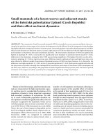
All oak, beech and spruce branch roots which were inves-
tigated for anatomy showed the mature second stage of tree
root development. A thin but clearly differentiated periderm
was already present at a distance of about 5 mm from the ter-
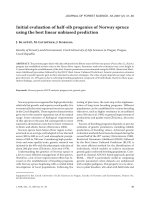
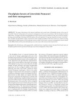
minal root tip. As an example for the three species, Figure 1
shows cross-sections of beech branch roots at two distances
from the root tip. The primary stage of root development with
stele, endodermis, cortex and exodermis was not found in any
of the studied fine roots. Fragments of the endodermis and cor-
tex were only recognised in a few cuts taken from root seg-
ments in close proximity to the terminal root tip. Counts of
annual growth rings in the root stele indicated a remarkably
high age of the fine and coarse roots of the three species. For
Figure 1. Cross-sections of a beech branch root at 0.5 mm (a) and 60 mm distance (b) from the terminal root tip with a multi-layered periderm
(Pe), phloem (Ph) and xylem (Xy) being visible. Bar = 100 µm.
Root water absorption 767
3-mm roots of beech, oak and spruce, an age of 10 years or
more was determined in all samples. Rootlets having a diame-
ter of 1 mm at 150 to 200 mm distance from the terminal tip
must have been at least 5 years old, and seem to have grown
less than 40 mm per year since their initiation.
Root tips of oak, beech and spruce had diameters of 100
to 300 µm and were almost completely infected with ectomy-
corrhizal fungi. The root diameter of fine branch roots
increased continuously with distance from the terminal root tip
in the three species and corresponded to increasing numbers of
periderm cell layers. Root diameter increase with distance
from the tip was largest in spruce, intermediate in oak, and
smallest in beech (oak: y = 0.31 × x/(3.7 + x), r = 0.92; beech:
y = 0.27 × x/(3.33 + x), r = 0.91; spruce: y = 0.13 × x/(5.66 + x),
r = 0.97; with y being root diameter (mm) and x the distance
from the terminal tip (m)).
3.2. Structure of the periderm
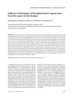
Similar to overall root diameter, root periderm thickness
increased non-linearly with distance from the terminal root tip
with a rapid increase in the first 50 to 70 mm and a much
slower increase in the subsequent 0.5 m (Fig. 2a). A multi-
layered periderm of 12 to 45 µm was present at 60 to 70 mm
distance from the terminal root tip in all species. At 500 mm
distance from the tip, periderm thickness had increased to
50 µm in beech and spruce, and to 70 µm in oak.
In beech and oak rootlets, the periderm consisted of only 2
to 3 cell layers close to the root tip but increased to 7–8 (beech)
or even 10 layers (oak) at distances > 200 mm (Fig. 2b).
Spruce roots, in contrast, had less than 4 peridermal cell layers
along the first 400 mm of a branch root. Thus, at distances
> 70 mm from the tip, oak fine roots possessed a significantly
thicker periderm with more cell layers than the two other spe-
cies. Spruce had a much smaller number of peridermal cell
layers than the two broad-leaved trees but the overall periderm
thickness was similar to that of beech because its peridermal
cork cells were comparably large.
3.3. Suberin and lignin contents of isolated root
peridermal cell wall samples
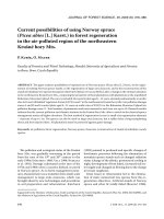
Suberin was detected at high concentrations (14–135 mg g
–1
DW or 1–14%) in isolated peridermal cell walls of oak, beech
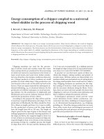
and spruce roots. When aliphatic suberin content is expressed
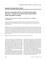
per root surface area, large differences were evident among the
species (Fig. 3). Oak roots had suberin contents that were three
times higher in the thinnest root diameter class (0–5 mm), and by
a factor of 10 higher in the largest diameter class (1.0–2.0 mm)
than those of spruce roots. Beech fine roots showed values
intermediate between spruce and oak for all diameter classes.
When the root diameter classes are compared, suberin content
showed a large and significant increase with diameter for oak,
a moderate increase for beech, and no change with diameter for
spruce (Fig. 3). Solvent extracts exhibited large quantities of
triterpenoids in concentrations of 1 to 9 g m
–2
, which varied
among species and root size classes in a pattern similar to that
found for suberin; long-chain aliphatic substances were, how-
ever, rare (data not shown).
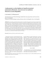
Figure 2. (a) Root peridermal thickness versus distance from the ter-
minal root tip. Square: oak, circle: beech, triangle: spruce (3 roots per
species). Oak: y = 89.72 × x/(0.025 + x), r = 0.62; beech: y =
55.56 × x/(0.038 + x), r = 0.93; spruce: y = 323.03 × x/(1.17 + x),
r = 0.93. (b) Number of peridermal cell layers versus distance from the
terminal root tip (3 roots per species). Oak: y = 12.83 × x/(0.021 + x),
r = 0.66; beech: y = 9.81 × x/(0.031 + x), r = 0.84; spruce: y =
2.39 × x/(0.010 + x), r = 0.60.
Figure 3. Amounts of aliphatic suberin in isolated peridermal cell
walls of oak, beech and spruce root segments expressed on a root sur-
face area basis. Three different diameter classes were distinguished
(mean ± SD, n = 3). Different capitals indicate significant differences
(P < 0.05) among the species for a root diameter class, different small
letters stand for significant differences among the diameter classes of
a species.
768 C. Leuschner et al.
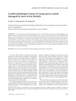
Lignin was detected in much smaller quantities in the peri-
dermal cell walls than suberin. In oak and beech roots, the
lignin content per surface area was roughly five times smaller
than that of suberin (Fig. 4). Relatively large amounts of lignin
were found in spruce fine roots where the thinnest rootlets
(< 0.5 mm) contained similar amounts of suberin and lignin
(about 1.5 mmol m
–2
root surface area for both components).
Relatively high lignin contents were also detected in the peri-
derm of thicker (1–2 mm) beech roots.
3.4. In situ water absorption rates of terminal branch
roots
Concurrent sap flow measurements on 5 oak, 4 beech and
4 spruce small-diameter roots in close vicinity to each other in
the organic Of and Oh horizons gave mean water absorption
rates of 201 (oak), 588 (beech) and 346 (spruce) g m
–2
d
–1
in
the period August 29–September 8, 1999 (averages over all
day and night hours, Tab. I). These numbers express the water
absorption per total surface area of all appending branch roots
(including root tips) distal to the gauge measuring point, and
are equivalent to 0.38 (beech), 0.13 (oak) and 0.22 (spruce)
mmol water m
–2
s
–1
. The daily mean absorption rates of
beech, oak and spruce roots were not significantly different
from each other; however, a trend with beech > spruce > oak
existed.
3.5. Root-soil water potential gradient and Lp
In an attempt to quantify root hydraulic conductivity Lp
for roots under in situ-conditions in the soil, measured root
water absorption rates were confronted with synchronous
measurements of root xylem (Ψ
root
) and soil water potentials
(Ψ
surface
) to characterise the principal driving force of water
uptake. On four days during summer 1999, the matric poten-
tial of the soil in close proximity to the studied roots varied
between –0.008 (moist soil) and –0.061 MPa (moderately dry
soil). The corresponding pressure chamber values of root
xylem pressure potential varied between –0.44 and –1.52 MPa
(Tab. II). Based on these measurements, the water potential
gradient between root surface and root xylem Ψ
surface
– Ψ
root
was estimated at 0.4 to 1.5 MPa with largest gradients appar-
ently existing in beech roots. Root hydraulic conductivity was
then obtained from Ψ
surface
– Ψ
root
and the corresponding flux
rate J
v
. The estimated Lp values of the three species ranged
Table I. Water absorption rates and related dry mass and surface area of 4 to 5 oak, beech and spruce branch roots as determined with
miniature sap flow gauges in the field. Given are mean values (± standard deviation) of measurements on 11 consecutive days in the period
August 29–September 8, 1999, in the mixed Unterlüss forest. The roots had a diameter of 3–4 mm at the gauge mounting point; the different
branch roots of a species belonged to the same tree individual. The standard deviation results from averaging over different days and roots.
Different letters indicate significantly different values among the three species (P < 0.05).
Beech Oak Spruce
Daily water uptake [g m
–2
d
–1
] 588 ± 387
a
200 ± 109
a
346 ± 117
a
Daily water uptake [mmol m
–2
s
–1
] 0.38 ± 0.25 0.13 ± 0.07 0.22 ± 0.08
Variation coefficient for uptake rates [%] 65.8 54.5 33.8
Total surface area [m
2
] 0.0852 ± 0.0434
a
0.0949 ± 0.0483
a
0.0871 ± 0.0412
a
Fraction d < 0.5 mm [%] 26.5 ± 3.8
a
26.6 ± 3.1
a
2.3 ± 0.38
b
Fraction d 0.5–1 mm [%] 41.0 ± 7.8
a
53.2 ± 4.5
b
32.7 ± 3.4
a
Fraction d 1–2 mm [%] 20.7 ± 5.5
a
14.8 ± 2.4
a
41.7 ± 2.0
b
Fraction d > 2 mm [%] 11.83 ± 6.5
ab
5.4 ± 4.7
a
22.2 ± 5.3
b
Total biomass [g dry weight] 6.26 ± 2.8
a
4.6 ± 1.9
a
9.8 ± 5.0
a
Fraction d < 1 mm [%] 46.4 ± 18.4
ab
65.4 ± 15.2
a
33.8 ± 11.5
b
Fraction d 1–2 mm [%] 19.4 ± 7.2
ab
16.6 ± 5.5
a
27.9 ± 2.8
b
Fraction d > 2 mm [%] 34.2 ± 17.27
a
18.0 ± 14.9
a
38.3 ± 12.1
a
Number of roots investigated 4 5 4
Figure 4. Amounts of lignin in isolated peridermal cell walls of oak,
beech and spruce root segments (data expressed on a root surface
area basis). Three different diameter classes were distinguished
(mean ± SD, n = 3). Different capitals indicate significant differences
(P < 0.05) among the species for a root diameter class, different small
letters stand for significant differences among the diameter classes of
a species.
Root water absorption 769
between 0.82 and 3.94 × 10
–8
m MPa
–1
s
–1
and revealed only
minor species differences. Oak tended to have smaller Lp values
than beech on all four days (difference significant on Septem-
ber 2), and spruce differed from beech on July 12.
4. DISCUSSION
This study profits from recent advances in two technolo-
gies which have a high relevance for the study of root hydrau-
lics, (i) the miniaturisation of sap flow gauges which allows
calculation of in situ-fluxes per root surface area, and (ii) the
chemical analysis of isolated root cell wall samples. By com-
bining these methods we were able, for the first time, to relate
in situ root water absorption rates to data on the chemical com-
position of the periderm, which is the principal apoplastic bar-
rier in mature tree fine roots. In contrast to earlier studies on
the chemistry of root cell walls (e.g. [52]), water absorption
and cell wall chemistry were both related to root surface area;
this enables a direct comparison.
It is remarkable that, even in direct vicinity of the root
apex, all fine roots had already reached the mature second stage
of root development with a multi-layered periderm. This con-
trasts with the results of [27] who found 100-110 and 60–
70 mm long sections without a closed periderm sheath in roots
of Pinus banksiana and Eucalyptus pilularis seedlings. The
nearly complete absence of primary white rootlets in the root
systems of our study may be understood in the light of the
remarkably great age of the studied terminal branch roots
(≥ 5 years for roots of 1 mm in diameter). It remains unclear
whether summer drought (as in July 1999), complete mycor-
rhizal infection of the root tips [4, 5], or other factors have inhib-
ited further growth of the root apex resulting in a comparably
high age of the rootlets close to the tip.
In the peridermal cell walls of spruce roots, aliphatic suberin
was detected at concentrations comparable to those in the endo-
dermis and hypodermis of corn roots (4–10 and ca. 21 mg g
–1
,
respectively; [50]). Beech and oak roots exhibited significantly
larger concentrations (40–60 and 90–135 mg g
–1
) than both
spruce and corn. In contrast to corn roots, aromatic suberin
occurred in the root periderm of the three tree species only in
traces (data not shown). Lignin was found in much smaller
amounts than suberin and showed less clear differences among
the three species. Because the studied roots contained no sec-
tions with white unsuberised rootlets lacking a periderm, we
conclude that water entering these roots must pass through peri-
dermal cell layers which contain at least 1.3 (spruce), 5.0
(beech) or 10.0 (oak) mmol suberin m
–2
.
Water absorption by the 4 to 5 roots of a species showed a
large spatial variability (coefficients of variation: 33.8 to
65.8%) despite the fact that the roots grew in a shared soil vol-
ume and the tree canopies were exposed to similar radiation
loads and atmospheric saturation deficits. According to the
much larger flux data set of [8, 10], large differences in water
uptake rates among neighbouring fine roots of a single tree are
a characteristic of the root systems of mature beech, oak and
spruce trees and do not reflect inaccuracies of the measuring
system. We hypothesise that large spatial variation in tree root
water uptake rates is a consequence primarily of small-scale
heterogeneity in soil structure and, thus, soil-root hydraulic
conductivities.
This novel technique for measuring root water absorption
does not allow a precise localisation of water uptake along the
root axis. The branch roots of this study (diameter: 3–4 mm)
had total surface areas of about 0.085 to 0.095 m
2
distal to the
gauge mounting point. On average, 80% (oak), 68% (beech)
and 35% (spruce) of the surface area of the potentially absorbing
Table II. Estimation of root hydraulic conductivity Lp of beech, oak and spruce branch roots under in situ-conditions in the undisturbed soil
based on sap flow measurements of root water absorption (J
v
), and synchronous water potential measurements in terminal branch roots
(pressure chamber values) and in the adjacent soil (tensiometer readings). See equation (1) in text. Different letters indicate significantly
different values among the three species on a measuring day (P < 0.05). In parentheses: standard deviation.
Ψ
surface
Ψ
root
A
r
J
v
Lp
MPa MPa m
2
m
3
s
–1
m s
–1
MPa
–1
× 10
–8
June 24
Beech –0.008 –0.65 0.09 (0.05) 1.31 (0.40) 2.66 (1.18)a
Oak –0.008 –0.44 0.28 (0.30) 1.50 (1.23) 1.70 (1.53)a
Spruce –0.008 –0.46 0.08 (0.06) n.d. n.d.
July 12
Beech –0.052 –1.52 0.09 (0.05) 3.77 (3.15) 3.22 (2.36)a
Oak –0.052 –0.76 0.28 (0.30) 1.82 (1.19) 1.57 (1.50)ab
Spruce –0.052 –0.89 0.08 (0.06) 0.84 (0.78) 1.06 (0.25)b
September 2
Beech –0.030 –0.81 0.09 (0.04) 1.72 (0.78) 2.87 (1.66)a
Oak –0.030 –0.64 0.09 (0.05) 0.58 (0.15) 1.27 (0.67)b
Spruce –0.030 –0.45 0.09 (0.04) 1.34 (0.48) 3.94 (1.08)a
September 9
Beech –0.061 –1.30 0.09 (0.04) 1.68 (0.64) 1.78 (0.96)a
Oak –0.061 –1.22 0.09 (0.05) 0.68 (0.13) 0.82 (0.54)a
Spruce –0.061 –0.66 0.09 (0.04) 0.86 (0.37) 1.87 (0.92)a
770 C. Leuschner et al.
branch roots referred to root sections with diameters < 1 mm,
the remaining surface being located on thicker root segments.
The specific role of fine and coarse roots in tree water uptake
is still a matter of dispute (e.g., [13, 32]), Recent research on
water uptake in different zones of onion roots has indicated
that apical root regions have a higher resistance to water
inflow than the more matured and stronger suberised zones
[3]. This agrees with a number of studies who reported a trans-
port of water and ions through peridermal woody roots [1, 6,
26, 46]. However, it is still an open question whether the dead
peridermal cork cells of tree roots are sufficiently permeable
to account for this flow, or whether passage occurs through
breaks in the periderm [27]. If the number of passage cells or
breaks were to determine radial water flow in tree roots, no
close relation between peridermal chemistry and water absorption
could be expected.
In this study, we observed a factor of about 2 between oak
and beech for suberin content in each diameter class, which
was close to the ratio of water uptake rate between the two spe-
cies. The larger suberin content of oak corresponded with a
thicker periderm and more periderm cell layers compared to
beech. This result might indicate that the degree of suberinisa-
tion of peridermal cell walls influences water absorption rates
in these species. A comparison of beech and spruce data, how-
ever, reveals that spruce with 2 to 6 times smaller suberin contents
had lower water absorption rates than beech with more
suberin. It appears that the relationship between periderm
chemistry or anatomy, and water absorption is only a weak
one in the three species.
We estimated root hydraulic conductivities (Lp) for absorb-
ing roots in the soil from measured water fluxes into the root
cylinder, and synchronous water potential measurements with
tensiometers and pressure chamber in soil and terminal branch
roots. The only other available conductivity data for beech, oak
and spruce roots were measured at decapitated sapling root sys-
tems in the laboratory with the root pressure probe technique
yielding data on root radial conductivity (Lpr) [34, 43, 44].
They are compared with our field-derived Lp data because we
expect total root hydraulic conductivity (Lp) and Lpr to be more
or less similar in the studied roots because root axial conduc-
tivity (Kh) typically is one or two orders smaller than Lpr if
expressed in the same unit [28]. The two approaches of conduc-
tivity measurement yielded roughly comparable results despite
largely different experimental setups (Tab. III). However, the
species comparison gave contrasting results with root pressure
probe data indicating a substantially higher Lpr in spruce than
in beech or oak roots which is not supported by our measure-
ments under in situ conditions. In the field, neither Lp nor water
absorption rates were higher in spruce than in beech roots.
Moreover, the pressure probe Lpr values are not fully consistent
with our data on periderm chemistry because they do not reflect
the high suberin content of the oak root periderm. If conduc-
tivity were a function of suberisation, Lpr values of oak should
have been much lower than those of beech which is not visible
from the pressure probe data.
One possible explanation of the discrepancy between root
pressure probe-derived hydraulic conductivities, anatomical
and chemical properties and in situ water absorption rates is
the fact that root systems of plants with highly different age
(saplings vs. mature trees) were investigated. The high degree
of suberisation found in this study is not a characteristic of tree
seedlings or saplings that were reared in a glasshouse. Moreo-
ver, it has to be kept in mind that root pressure probe measure-
ments are conducted under artifical conditions with water
being forced through the root by modification of xylem pres-
sure or applying osmotic gradients. This setup is highly different
from natural potential gradients that exist in the rhizosphere.
We suggest that the most likely explanation of a partial
mismatch among root hydraulics, chemical and anatomical
properties, and measured water absorption is the fact that addi-
tional factors, which may control water flow into the root, have
to be considered during the upscaling process from laboratory
to field. Root chemical and anatomical properties and even Lpr
may be less important in controlling in situ water absorption
than they are in a laboratory setup with excised root systems.
(i) The tips of oak, beech and spruce fine roots are nearly com-
pletely infected by ectomycorrhizal fungi. In a few studies,
ectomycorrhizas have been found to increase hydraulic conduct-
ance of tree roots (e.g. [29, 34]); whereas other authors reported
negative or neutral effects [31]. An only limited effect on water
uptake would be understandable because mycorrhizae affect
the outer part of the root rather than the stele and endodermis
which, for geometric and other reasons, may represent the bot-
tle neck. (ii) In dry soil, the hydraulic conductivity of the root-
soil contact zone has been found to be considerably lower than
that of the path between the root surface and the stele due to
incomplete root-soil contact in certain substrates [47]. A very
rapid decrease in hydraulic conductivity with increasing water
loss is to be expected in soil substrates with a high porosity,
such as the organic forest floor horizons of this study. Root
contraction in drought-stressed plants may contribute to a low
hydraulic conductivity in the perirhizal soil [14]. Therefore, it
is possible that a low conductivity in the root-soil interface has
masked variable Lp values of the three species under field condi-
tions in this study. (iii) Water absorption is also dependent on
the water potential in the root xylem. Comparative measure-
ments with the pressure bomb in terminal branch roots of co-
existing oak, beech and spruce roots showed considerable
Table III. Root hydraulic conductivity (Lp) of small-diameter roots
of beech, oak and spruce estimated under in situ-conditions compa-
red to laboratory measurements of root radial hydraulic conductivity
(Lpr, both in m s
–1
MPa
–1
× 10
–8
). Lp was measured with miniature
sap flow gauges in combination with root and soil water potential
measurements by pressure chamber and tensiometer techniques (this
study). Lpr was obtained from pressure relaxation measurements
with excised root systems of saplings in the laboratory. The field
data refer to intact terminal branch roots of mature trees that were
absorbing water under in situ conditions in the soil. Oak refers to
Q. petraea.
Laboratory-measured Lpr
1
Field-measured Lp
2
Beech 0.35–1.6 1.78–3.22
Oak 0.33–1.1 0.82–1.70
Spruce 4.9–7.8 1.06–3.94
1
After Rüdinger et al., 1994; Steudle and Meshcheryatov, 1996; and
Steudle and Heydt, 1997.
2
This study.
Root water absorption 771
differences among the species during periods of drought which
may be the result of differences in either leaf water status or
stem hydraulic conductivity [8]. Comparative measurements
of root water absorption in different soil types and during peri-
ods of low and high soil water deficits are needed to assess the
influence exerted by variable soil-root water potential gradi-
ents and soil-root hydraulic conductivities on root water
absorption. (iv) It has recently been suggested that a fine reg-
ulation of root water uptake is provided by water channels
(aquaporins) in the cell-to-cell passage of water flow in roots
[7, 45]. Opening and closing of the channels could alter Lpr
mainly when the water potential gradient has an osmotic
nature. However, a significant effect of water channels on conduc-
tivity could not be detected in corn roots [52]. Whether water
channels are a significant factor, that could explain species-
specific differences in water absorption among peridermal tree
roots, remains unclear.
There is the possibility that elevated suberin and lignin
contents in tree fine roots are more relevant for root drought
tolerance than they are for root water absorption. Several
authors have found an increase in suberisation of the endo- or
exodermis in herbaceous plant roots following drought or
salinity stress [11, 30]. Oak fine roots may be classified as
rather drought tolerant in the Unterlüss forest because sessile
oak fine root systems showed a more surface-directed distribution
pattern in the topsoil, and were less affected by drought-
induced fine root mortality during a dry summer, than co-
existing beech roots [16, 23]. This may correspond to the high
aliphatic suberin content of oak root cell walls. A low suberin
content in Norway spruce roots coincided with a high drought
sensitivity of this species in Central Europe [12]. Experimen-
tal testing in field studies with carefully controlled drought
intensities is needed to show whether oak, beech and spruce
indeed differ with respect to drought sensitivity of fine root
growth and mortality. Finally, it is necessary to increase the
spatial resolution of water uptake measurements because the
importance of different root diameter classes in root water
absorption is still unknown.
Acknowledgements: This work was supported by the Deutsche
Forschungsgemeinschaft (DFG) with grants to C.L. and L.S. (the
latter as part of the priority program “Apoplast”).
REFERENCES
[1] Addoms R.M., Entrance of water into suberized roots of trees,
Physiol. Plant 21 (1946) 109–111.
[2] Backes K., Leuschner Ch., Leaf water relations of competitive
Fagus sylvatica L. and Quercus petraea (Matt.) Liebl. trees during
four years differing in soil drought, Can. J. For. Res. 30 (2000) 335–
346.
[3] Barrowclough D.E., Peterson C.A., Steudle E., Radial hydraulic
conductivity along developing onion roots, J. Exp. Bot. 51 (2000)
547–557.
[4] Bledsoe C.S., Zasoki R.J., Effects of ammonium and nitrate on
growth and nitrogen uptake by mycorrhizal Douglas-fir seedlings,
Plant Soil 71 (1983) 445–454.
[5] Büttner V., Leuschner Ch., Spatial and temporal patterns of fine
root abundance in a mixed oak-beech forest, For. Ecol. Manage. 70
(1994) 11–21.
[6] Chung H H., Kramer P.J., Absorption of water and
32
P through
suberized and unsuberized roots of Loblolly pine, Can. J. For. Res.
5 (1975) 229–236.
[7] Clarkson, D.T., Carvajal M., Henzler T., Waterhouse R.N., Smyth
A.J., Cooke D.T., Steudle E., Root hydraulic conductance: diurnal
aquaporin expression and the effects of nutrient stress, J. Exp. Bot.
51 (2000) 61–70.
[8] Coners H., Wasseraufnahme und artspezifische hydraulische
Eigenschaften der Feinwurzeln von Buche, Eiche und Fichte:
In situ-Messungen an Altbäumen. Ph.D. thesis, Göttingen Univer-
sity, Germany, 2001.
[9] Coners H., Leuschner Ch., Water absorption by tree fine roots
measured in situ with miniature sap flow gauges, Funct. Ecol. 16
(2002) 696–703.
[10] Coners H., Leuschner Ch., Daily and seasonal variation of root
water uptake by three temperate tree species in its dependence on
plant, soil and atmospheric factors. Submitted.
[11] Cruz R.T., Jordan W.R., Drew M.C., Structural changes and
associated reduction of hydraulic conductance in roots of Sorghum
bicolor L. following exposure to water deficit, Physiol. Plant 99
(1992) 203–212.
[12] Ellenberg H., Vegetation Mitteleuropas mit den Alpen in ökologischer,
dynamischer und historischer Sicht. 5th ed. Stuttgart, Germany:
Ulmer Verlag, 1996.
[13] Escamilla J.A., Comerford N.B., Measuring nutrient depletion by
roots of mature trees in the field, Soil Sci. Soc. Am. J. 62 (1998)
797–804.
[14] Faiz M.A., Weatherley P.E., Root contraction in transpiring plants,
New Phytol. 92 (1982) 333–343.
[15] Green S.R., Clothier B.E., Root water uptake by kivifruit vines
following partial wetting of the root zone, Plant Soil 173 (1995)
317–328.
[16] Hertel D., Das Feinwurzelwerk von Rein- und Mischbeständen der
Rotbuche : Struktur, Dynamik und interspezifische Konkurrenz,
Dissertationes Botanicae 317 (1999) 1–190.
[17] Howard S.B., Ong C.K., Black C.R., Khan A.A.H., Using sap flow
gauges to quantify water uptake by tree roots from beneath the crop
rooting zone in agroforestry systems, Agrofor. Syst. 35 (1997) 15–29.
[18] Jackson R.B., Sperry J.S., Dawson T.E., Root water uptake and
transport: using physiological processes in global predictions,
Trends Plant Sci. 5 (2000) 482–488.
[19] Knievel D.P., Procedure for estimating ratio of living and dead root
dry matter in root core samples, Crop Sci. 13 (1973) 124–126.
[20] Kolattukudy P.E., Agrawal V.P., Structure and composition of
aliphatic constituents of potato tuber skin (suberin), Lipids 9 (1974)
682–691.
[21] Lapierre C., Pollet B., Monties B., Thiacidolysis of spruce lignin:
GC-MS analysis of the main dimers recovered after Raney nickel
desulphurication, Holzforschung 45 (1991) 61–68.
[22] Leuschner Ch., Changes in forest ecosystem function with
succession in the Lüneburger Heide, in: Tenhunen J., Hantschel R.,
Lenz R. (Eds.) Ecosystem Approaches to Landscape Management
in Central Europe, Ecol. Stud. 147 (2000) 517–568.
[23] Leuschner Ch., Hertel D., Coners H., Büttner V., Root competition
between beech and oak: a hypothesis, Oecologia 126 (2001) 276–284.
[24] Leuschner Ch., Rode M.W., The role of plant resources in forest
succession: changes in radiation, water and nutrient fluxes, and plant
productivity over a 300-yr-long chronosequence in NW Germany,
Perspectives in Plant Ecology, Evolution and Systematics 2 (1999)
103–147.
[25] Leuschner Ch., Backes K., Hertel D., Schipka F., Schmitt U.,
Terborg O., Runge M., Drought responses at leaf, stem and fine
root levels of competitive Fagus sylvatica L. and Quercus petraea
(Matt.) Liebl. trees in dry and wet years, For. Ecol. Manage. 149
(2001) 33–46.
[26] MacFall J.S., Johnson G.A., Kramer P.J., Comparative water uptake
by roots of different ages in seedlings of loblolly pine (Pinus taeda
L.), New Phytol. 119 (1991) 551–560.
772 C. Leuschner et al.
[27] McKenzie B., Peterson C.A., Root browning in Pinus banksiana
Lamb. and Eucalyptus pilularis Sm. 2. Anatomy and permeability
of the cork zone, Bot. Acta 108 (1995) 138–143.
[28] Moreshet S., Huang B., Huck M.G., Water permeability of roots,
in: Waisel Y., Eshel A., Kafkafi U. (Eds.) Plant Roots, The Hidden
Half, New York, Marcel Dekker, 1996, pp. 659–768.
[29] Muhsin T.M., Zwiazek J.J., Ectomycorrhizas increase apoplastic
water transport and root hydraulic conductivity in Ulmus
americana seedlings, New Phytol. 153 (2002) 153–158.
[30] North G.B., Nobel P.S., Radial hydraulic conductivity of concentric
root tissues of Agave deserti Engelm. under wet and drying
conditions, New Phytol. 130 (1996) 47–57.
[31] Oertli J.J., Transport of water in the rhizosphere and in roots, in:
Waisel Y., Eshel A., Kafkafi U. (Eds.) Plant Roots, The Hidden
Half, Marcel Dekker, New York, pp. 607–634.
[32] Peterson C.A., Cholewa E., Structural modification of the apoplast
and their potential impact on ion uptake, Z. Pflanzenernähr. Bodenkd.
161 (1998) 521–531.
[33] Raison R.J., Connell M.J., Khanna P.K., Methodology for studying
fluxes of soil mineral-N in situ, Soil Biol. Biochem. 19 (1987) 521–
530.
[34] Rüdinger M., Hallgren S.W., Steudle E., Schulze E D., Hydraulic
and osmotic properties of spruce roots, J. Exp. Bot. 45 (1994)
1413–1425.
[35] Sakuratani T., A heat balance method for measuring water flux in
the stem of intact plants, J. Agric. Meteorol. 37 (1981) 9–17.
[36] Senock R.S., Ham J.M., Heat balance sap flow gauge for small
diameter stems, Plant Cell Environ. 16 (1993) 593–601.
[37] Senock R.S., Leuschner Ch., Axial water flux dynamics in small
diameter roots of a fast growing tropical tree, Plant Soil 208 (1999)
57–71.
[38] Stavosky E., Peterson C.A., Effects of drought and subsequent
rehydration on the structure, vitality and permeability of Allium
cepa adventitous roots, Can. J. Bot. 71 (1993) 700–707.
[39] Schreiber L., Breiner H W., Riederer M., Düggelin M.,
Guggenheim R., The Casparian strip of Clivia miniata Reg. roots:
isolation, fine structure and chemical nature, Bot. Acta 107 (1994)
353–361
[40] Steudle E., Pressure probe techniques: basic principles and
application to studies of water and solute relations at the cell, tissue
and organ level, in: Smith J.A.C., Griffiths H. (Eds.), Water
deficits: plant responses from cell to community, Oxford, England:
Bios Scientific Publishers, 5–36, 1993.
[41] Steudle E., Water uptake by plant roots: an integration of views,
Plant Soil 226 (2000a) 45–56.
[42] Steudle E., Water uptake by roots: effects of water deficit, J. Exp.
Bot. 51 (2000b) 1531–1542.
[43] Steudle E., Heydt H., Water transport across tree roots, in: Rennenberg
H., Eschrich W., Ziegler H. (Eds.), Trees – Contributions to
Modern Tree Physiology, Leiden, The Netherlands: Backhuys
Publishers, 1997, pp. 239–255.
[44] Steudle E., Meshcheryakov A.B., Hydraulic and osmotic properties
of oak roots, J. Exp. Bot. 47 (1996) 387–401.
[45] Steudle E., Peterson C.A., How does water get through roots? J.
Exp. Bot. 49 (1998) 775–788.
[46] Van Rees K.C.J., Comerford N.B., The role of woody roots of slash
pine seedlings in water and potassium absorption, Can. J. For. Res.
20 (1990) 1183–1191.
[47] Veen B.W., Van Noordwijk M., De Willigen P., Boone F.R.,
Kooistra M.J., Root-soil contact of maize, as measured by a thin-
section technique, Plant Soil 139 (1992) 131–138.
[48] Von Wilpert K., Die Jahrringstruktur von Fichten in Abhängigkeit
vom Bodenwasserhaushalt auf Pseudogley und Parabraunerde,
Freiburger Bodenkundliche Abhandlungen 24 (1990) 1–184.
[49] Zeier J., Schreiber L., Chemical composition of hypodermal and
endodermal cell walls and xylem vessels isolated from Clivia
miniata: identification of the biopolymers lignin and suberin,
Physiol. Plant 113 (1997) 1223–1231.
[50] Zeier J., Schreiber L., Comparative investigation of primary and
tertiary endodermal cell walls isolated from the roots of five
monocotyledoneous species: chemical composition in relation to
fine structure, Planta 206 (1998) 349–361.
[51] Zeier J., Ruel K., Ryser U., Schreiber L., Chemical analysis and
immunolocalisation of lignin and suberin in endodermal and
hypodermal rhizodermal cell walls of developing maize (Zea mays
L.) primary roots, Planta 209 (1999) 1–12.
[52] Zimmermann M.H., Hartmann K., Schreiber L., Steudle E., Chemi-
cal composition of apoplastic transport barriers in relation to radial
hydraulic conductivity of corn roots (Zea mays L.), Planta 210
(2000) 302–311.
To access this journal online:
www.edpsciences.org