Báo cáo lâm nghiệp: "In vitro clonal propagation of Acacia mangium Willd. and its evaluation of genetic stability through RAPD marker" ppsx
Bạn đang xem bản rút gọn của tài liệu. Xem và tải ngay bản đầy đủ của tài liệu tại đây (534.07 KB, 6 trang )
381
Ann. For. Sci. 61 (2004) 381–386
© INRA, EDP Sciences, 2004
DOI: 10.1051/forest:2004031
Note
In vitro clonal propagation of Acacia mangium Willd. and its
evaluation of genetic stability through RAPD marker
Rashmi M. NANDA, Premananda DAS, Gyana Ranjan ROUT*
Plant Biotechnology Division, Plant Tissue Culture Laboratory, Regional Plant Resource Centre, Bhubaneswar- 751 015, Orissa, India
(Received 28 October 2002; accepted 5 June 2003)
Abstract – An in vitro propagation of a tropical leguminous tree, Acacia mangium, has been established. Induction of bud sprout was obtained
from mature nodal explants of 10-years-old tree of Acacia mangium on Murashige and Skoog (MS) [10] basal medium supplemented with
6-benzylaminopurine (BAP) (1.0 mg/L), gibberellic acid (GA
3
) 1.0 mg/L and indole-3-acetic acid (IAA) 0.05 mg/L. The rate of multiplication
was obtained on MS medium supplemented with 1.5 mg/L BAP, 0.05 mg/L IAA and 100 mg/L adenine sulfate (Ads). The multiplication rate
varied from 1 to 8 depending on the growth regulators used. Excised shoots were rooted on half-strength MS basal salts supplemented with
0.5 mg/L indole-3-butyric acid (IBA) or indole-3-acetic acid (IAA) and 20 g/L (w/v) sucrose after 13–14 days of culture. The micropropagated
plantlets have been acclimatized and successfully transferred to soil. The micropropagated plantlets appeared morphologically similar to the
mother plant. No variation was detected among the micropropagated plants by the use of Randomly Amplified Polymorphic DNA (RAPD)
markers.
in vitro / legume tree / multiplication / micro-propagation / RAPD marker
Résumé – Propagation de clones in vitro d’Acacia mangium Willd., et évaluation de leur stabilité génétique avec un marquage RADP.
On a réalisé la propagation in vitro d’un arbre, Acacia mangium, de la famille des légumineuses. L’induction de bourgeons débourrés a été
obtenue à partir d’explants de bois aoûté prélevés sur des arbres âgés de 10 ans, cultivés dans le milieu de base de Murashige et Skoog (MS)
auquel on a ajouté de la benzylaminopurine (BAP) (1,0 mg/L), de l’acide gibbérelique (GA
3
) (1,0 mg/L) et de l’acide indol-3 acétique (IAA)
(0,05 mg/L). Pour la phase multiplication on a utilisé le milieu MS avec addition de 1,5 mg/L de BAP, 0,05 mg/L d’IAA et 150 mg/L de sulfate
d’adénine (Ads). Le taux de multiplication a varié de 1 à 8 selon les régulateurs de croissance utilisés. L’enracinement des pousses a été obtenu
avec le milieu MS dilué de moitié, additionné de 0,5 mg/L d’acide indol butyrique (IBA) ou d’acide indol-3 acétique (IAA) et de 20 g/L de (w/v)
saccharose après 13 à 14 jours de culture. Les plantules issues de cette micropropagation ont été acclimatées et transférées avec succès sur le
terrain. Elles étaient apparemment similaires à la plante mère sur le plan morphologique. Aucune variabilité n’a pu être détectée entre plantules
par l’utilisation de marqueurs DNA (RAPD).
multiplication de légumineuses ligneuses / micropropagation / marqueurs RAPD
1. INTRODUCTION
Micropropagation of tree species offers a rapid means of
producing clonal planting stock for afforestation, woody bio-
mass production and conservation of elite germplasm. In gen-
eral, the woody plants are difficult to regenerate under in vitro
conditions but recently some success have been achieved in a
few leguminous tree species [5, 9, 11, 15, 16]. There were some
successful reports of development of plantlets from callus
raised through seedling shoot tip cultures and from cotyledon-
ary buds and nodal explant [11, 15]. Acacia mangium Willd.
(family Leguminosae) is a tropical multipurpose legume tree
native to Papua New Guinea and the Eastern provinces of Indo-
nesia, with extension to Northern Australia [6]. It grows in the
humid tropical countries of South East Asia, especially in acid
and degraded soils, to which it can restore fertility owing to its
natural nitrogen fixing ability. Besides, this tree is used as a
source of pulpwood production [6]. Conventional methods of
propagation of Acacia mangium, sexual as well as vegetative,
are beset with many problems that restrict their multiplication
on a large scale. Cloning of mature trees is generally preferred
over seedling explants because it is often not possible to deter-
mine whether these embryos or seedlings have the genetic
potential to develop the desired qualities later in their life cycle.
In vitro culture techniques have great potential for clonal prop-
agation of superior genotypes of this legume species for refor-
estation programmes as well as utilization in paper industries.
So far, there is no report on shoot multiplication from mature
* Corresponding author: ;
382 R.M. Nanda et al.
explant of Acacia mangium. This communication describes a
successful protocol on in vitro clonal propagation of Acacia
mangium from nodal explants of a mature tree and determines
the genetic stability of the micropropagated plants through
RAPD marker.
2. MATERIALS AND METHODS
2.1. Explant source
Semi-mature growing twigs (8–10 cm long) were collected from a
10-year-old tree of A. mangium and washed with 5% (v/v) detergent
solution ‘Teepol’ (Qualigen, India) for 10 min. Subsequently, the long
twigs were cut into smaller segments about 3–4 cm long and surface
sterilized with 0.1% (w/v) aqueous mercuric chloride solution for
20 min and washed 4–5 times in sterile distilled water. Each segment
was further cut into 1–2 cm having one node each.
2.2. Culture condition
The nodal explants were cultured on MS [10] medium supple-
mented with different concentrations of 6-benzylaminopurine (BAP:
0, 0.5, 1.0, 1.5 and 2.0 mg/L), Kinetin (Kn: 0, 0.5, 1.0, 1.5 and 2.0 mg/L),
adenine sulfate (Ads: 0, 50, 100 mg/L), indole-3-acetic acid (IAA: 0,
0.05, 0.10, 0.25 mg/L) or 1-naphthylacetic acid (NAA: 0, 0.05, 0.10,
0.25 mg/L) and gibberellic acid (GA
3
: 0, 0.5, 1.0, 1.5 mg/L) alone or
in combination. All media were adjusted to pH 5.7 using 0.1 N HCl or
0.1 N NaOH before autoclaving. The medium was gelled with 0.8%
(w/v) agar (Qualigen, India). Routinely, 20 mL of molten medium was
dispensed into a culture tube (25 × 150 mm) and plugged with non-
absorbant cotton wrapped in one layer of cheese cloth. The cultures
were steam sterilized at 1.06 kg·cm
–2
for 15 min. All cultures were
incubated in 16 h light / 8 h dark photoperiod under light intensity of
50 µEm
–2
s
–1
provided by cool white fluorescent light (Philips, India)
at 25 ± 2 °C with 55% relative humidity. Each treatment had 20 cul-
ture tubes and the experiment was repeated thrice. The effects of dif-
ferent treatments were quantified as the mean number of multiple
shoots/culture. The data were statistically analysed by the Duncan
Multiple range test [4].
2.3. Induction of rooting
In vitro raised shoots measuring about 2–3 cm growing on multi-
plication medium were excised and cultured on half-strength MS basal
salts supplemented with 0, 0.25, 0.5, 1.0 and 1.5 mg/L of IAA, IBA
and NAA alone or in combination for induction of rooting. In case of
rooting experiment, 10 replicates per treatment were taken and the
experiment was repeated thrice. The cultures were examined every
weeks and the morphological changes were recorded on the basis of
visual observations. The effects of different treatments were quanti-
fied as the mean percentage of rooting.
2.4. DNA extraction
DNA was extracted from fresh leaves derived from twenty in vitro
raised plants and from the field grown mother plant by the cetyltrime-
thyl ammonium bromide, EMERK (CTAB) method [2]. Approxi-
mately, 250 mg of fresh leaves were ground to powder in liquid nitro-
gen using a mortar and pestle. The ground powder was transferred to
a 25 mL tube with 10 mL of CTAB buffer: 2% (w/v) CTAB, 1.4 M
NaCl, 20 mM EDTA, 100 mM Tris-HCl pH 8.0, and 0.2% (v/v)
β-mercaptoethanol. The homogenate was incubated at 60 °C for 2 h,
extracted with an equal volume of chloroform: iso-amyl alcohol (24:1),
and centrifuged at 10 000 rpm for 20 min. DNA was precipitated from
the aqueous phase by mixing with an equal volume of isopropanol.
After centrifugation at 10 000 rpm for 10 min, the DNA pellet was
washed with 70% ethanol, air dried, and resuspended in 10 mM Tris
pH 8.0, 0.1 mM EDTA buffers. DNA quantity was estimated spectro-
photometrically by measuring the absorbance at 260 nm.
2.5. PCR amplification
Twenty arbitrary 10-base primers (Operon Technologies Inc.,
Alameda, California) were used for Polymerase Chain Reaction
(PCR). Amplification reactions were performed in 25 µL: 2.0 µL of
1.25 mM each of dNTP’s, 15 ng of the primer, 1× Taq polymerase
buffer, 0.5 U of Taq DNA polymerase (Genei, India) and 20 ng of
genomic DNA. DNA amplification was performed in a PTC 100 DNA
Thermal Cycler (M.J. Research, Inc., Watertown, MA, USA) pro-
grammed for 45 cycles: 1st cycle of 3.5 min at 94 °C, 1 min at 37 °C
and 2 min at 72 °C; then 44 cycles each of 1 min at 94 °C, 1 min at
37 °C, 2 min at 72 °C followed by one final extension cycle of 7 min
at 72 °C. Amplified products were electrophoresed in a 1.2% (w/v)
agarose (Sigma, USA) gels with 1× TAE buffer, stained with ethidium
bromide, and photographed under ultraviolet (UV) light. The size of
the amplification products were estimated for a 100-bp (100-bp to
3.0 kb) ladder (M B I. Fermentas Inc.). All the reactions were repeated
at least thrice.
2.6. Amplified DNA marker scoring
Amplified DNA markers were scored as present or absent both in
the regenerated and the mother plants. Electrophoretic DNA bands of
low visual intensity that could not be readily distinguished as present
or absent were considered ambiguous markers and were not scored.
2.7. Acclimatization
Rooted plantlets were removed from the culture tube. After thor-
ough washing of the roots in tap water, the plantlets were transferred
to sterile sandy soil at the ratio of 1:1 (garden soil: sand) into earthen
pots and kept under high humidity (70%) in a greenhouse with tem-
perature (30 ± 2 °C) for acclimatization.
3. RESULTS AND DISCUSSION
3.1. Multiplication rate
The present investigation was carried out to explore the mor-
phogenic potential of Acacia mangium by using different com-
binations of growth regulators. The nodal explants resumed
new bud growth by proliferating the axillary shoot within 2–
3 weeks of culture. Out of the various cytokinins tested, BAP
was the most effective for inducing bud break and growth in
axillary meristems. Kinetin alone or in combination did not
show any positive response on bud growth (data not shown).
No bud sprout was obtained in media without growth regulator.
The result also showed that the medium supplemented with
GA
3
alone or in combination with BAP favour positive
response on bud sprout (Tab. I). The maximum bud break was
observed in nodal explants cultured on MS medium supple-
mented with 1.0 mg/L BAP, 1.0 mg/L GA
3
and 0.05 mg/L IAA
(Fig. 1a). Within 4 weeks of culture, the axillary meristems
elongated up to 2–3 cm in height. Similar results were reported
in other legume trees [9, 16]. Regarding shoot multiplication,
Clonal propagation of Acacia mangium 383
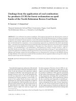
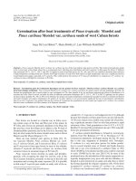
Figure 1. Micropropagation of Acacia mangium. (a) Development of bud sprout from nodal explants (arrow) of Acacia mangium on MS
medium supplemented with 1.0 mg/L BAP, 1.0 mg/L GA
3
and 0.05 mg/L IAA after 15 days of culture. (b) Induction of multiple shoots from
nodal explants on MS medium supplemented with 1.5 mg/L BAP, 100 mg/L Ads and 0.05 mg/L IAA after 4 weeks of culture. (c) Rooted plant-
let ready for ex vitro transfer. (d) RAPD profiles of micropropagated plants from nodal explants of Acacia mangium using primer OPC – 04
(5’-CCGCATCTAC-3’). Lane M shows RAPD bands from the field grown mother plant. Arrow indicates the size of the marker. Lanes 1–19 show
RAPD products from micropropagated plants.
Table I. Bud sprout responses of nodal explants of Acacia mangium on MS medium supplemented with different concentrations of BA, GA
3
,
IAA, NAA and 3% (w/v) sucrose under 16 h photoperiod at 25 ± 2 °C after one week of culture.
MS + growth regulator (mg/L) Percent of bud sprouting (%)
BAP GA
3
IAA NAA (mean ± S.E.)*
0
0.5
1.0
0
0
0
1.0
1.0
1.5
1.0
1.5
1.0
1.5
1.0
1.0
1.0
0
0
0
0.5
1.0
1.5
0.5
1.0
1.0
1.0
1.0
1.0
1.0
1.5
1.5
1.0
0
0
0
0
0
0
0
0
0
0.05
0.05
0
0
0.05
0
0.10
0
0
0
0
0
0
0
0
0
0
0
0.05
0.05
0
0.05
0
0
28.8 ± 1.2
42.6 ± 2.4
26.2 ± 1.4
36.8 ± 1.2
46.2 ± 2.2
58.6 ± 1.0
64.7 ± 2.3
68.2 ± 1.8
74.6 ± 2.4
72.4 ± 1.6
56.8 ± 1.2
58.4 ± 1.0
66.2 ± 2.3
54.3 ± 1.6
62.7 ± 1.8
b
d
a
c
e
h
j
l
n
m
g
h
k
f
i
+
+
+
* 20 replicates per treatment; repeated thrice. Means having same letters are not significantly different by Duncan’s multiple range test at the level of
5%. + Callusing at the basal end.
384 R.M. Nanda et al.
among the twenty combinations tried (BAP, Ads, IAA and
NAA), the combination of BAP (1.5 mg/L), Ads (100 mg/L)
and IAA (0.05 mg/L) proved to be the most effective treatment
for promoting shoot multiplication (6–8 shoots per nodal
explant, each shoot having 3–4 nodes) within 4 weeks of sub-
culture (Tab. II) (Fig. 1b). Analysis of variance for the effects
of cytokinins and auxins on production of multiple shoots and
average number of shoots/explant was presented in Table III.
The interaction of two cytokinins with low concentration of
auxin enhancing shoot multiplication has been reported by
many authors [3, 5, 12]. The inclusion of higher concentration
of auxin (either IAA or NAA) into the cytokinin rich medium
inhibited not only shoot multiplication but also produced some
compact callus at the base of the explants. Similar observations
were made in Pterocarpus santalinus [8] and in Acacia auricu-
liformis [9].
3.2. Rooting
Elongated shoots derived from nodal explants (grown on
MS medium supplemented with 1.0 mg/L BAP, 1.0 mg/L GA
3
and 0.05 mg/L IAA) were separated and cultured on half-
strength basal MS medium supplemented with IAA, IBA and
NAA alone or in combination for induction of rooting. Of the
three auxins tested, IBA and IAA induced rooting (Tab. IV).
The optimum concentration was 0.5 mg/L of IBA or IAA and
it resulted in 65 to 70% of root initiation within 14–18 days of
culture (Fig. 1c). At higher concentrations (1.0–1.5 mg/L) of
IBA or IAA, the percentage of rooting was reduced and callus
formation obtained at the basal cut end. Inhibition of rooting
was observed on medium containing different concentrations
of NAA. The combination of either IAA + NAA or IAA + IBA
or IBA + NAA did not show any positive response on rooting,
rather it formed callus at the cut end of the excised shoot and
also the shoot became yellowish. These observations have also
been reported for other woody species [14, 16, 17]. Monteuuis
and Bon [18] reported that rooting of the mature clone was
significantly increased by exposing the microshoots to 4–6 µM
IAA or IBA in darkness. In vitro grown rooted plants were
removed from the adhering gel, transplanted to earthen pots and
kept under high humidity for acclimatization. About 52% of the
plantlets survived under greenhouse condition after one month
of transfer.
Table II. Effect of cytokinins on shoot multiplication from nodal explants of Acacia mangium after 4 weeks of culture.
MS + growth regulators (mg/L) Av. no. of multiple
shoots/ explant
Av. length
of shoots (cm)
BAP Ads IAA NAA (mean ± S.E.)* (mean ± S.E.)*
0
1.0
1.5
2.0
1.5
1.5
1.5
1.5
1.5
2.0
1.5
1.5
1.5
1.5
2.0
2.0
1.5
1.5
2.0
2.0
0
50
50
50
50
50
50
50
100
100
100
100
100
100
100
100
100
100
100
100
0
0
0
0
0.05
0
0.1
0
0
0
0.05
0.10
0
0
0.05
0
0.25
0
0.25
0
0
0
0
0
0
0.05
0
0.1
0
0
0
0
0.05
0.10
0
0.05
0
0.25
0
0.25
0
0.24 ± 0.8 h
1.88 ± 0.7 g
2.14 ± 0.6 f
2.65 ± 0.6 e
1.62 ± 0.6 g
2.72 ± 0.7 e
1.32 ± 0.5 h
5.25 ± 0.6 c
5.54 ± 0.8 c
8.20 ± 0.9 a
6.15 ± 0.8 b
3.32 ± 0.6 d
2.14 ± 0.8 f
6.02 ± 0.8 b
2.76 ± 0.6 e
2.83 ± 0.5 e +
1.76 ± 0.7 g +
1.02 ± 0.6 h +
1.08 ± 0.7 h +
0
0.88 ± 0.4 f
1.12 ± 0.6 e
1.37 ± 0.7 d
1.24 ± 0.8 d
0.94 ± 0.8 e
1.16 ± 0.7 e
0.84 ± 0.6 f
2.03 ± 0.6 b
2.20 ± 0.8 b
3.22 ± 0.7 a
3.14 ± 0.6 a
1.65 ± 0.8 c
1.86 ± 0.5 c
2.12 ± 0.8 b
1.74 ± 0.9 c
1.12 ± 0.6 e
0.83 ± 0.5 f
1.06 ± 0.7 e
0.74 ± 0.6 f
* 20 replicates/treatment; repeated thrice. Shoots with less than 0.25 cm length were not taken into account. Means having same letters are not signifi-
cantly different by Duncan’s multiple range test at the level of 5%. + Callusing at the basal end.
Table III. Analysis of variance for the effects of BAP, Ads, NAA
and IAA on production of multiple shoots (A) and average number
of shoots per explant (B) from nodal explants of Acacia mangium
after 4 weeks of culture.
Source
of variation
df Mean squares
AB
Replication 19 4.56 0.635
Treatments 19 8 668.27* 1 207.82*
Error 361 2.02 0.234
* Significant at P < 0.05 as determined by the F-test.
Clonal propagation of Acacia mangium 385
3.3. Genetic stability
Out of the twenty different primers tested, three primers
(OPC-04, OPD-14 and OPC-19) produced good amplification
products that were monomorphic across all the micropropa-
gated plants. The size of the monomorphic fragments, produced
by primer OPC-04 ranged from 2.58 to 23.2 kb (Fig. 1d). Other
primers showed amplification with less number of monomor-
phic bands. There were no polymorphic DNA fragments
among the micropropagated plants as well as the mother plant.
The pattern of monomorphic bands in the in vitro raised plants
were reported earlier in other plants using different primers [1,
7]. Rout et al. [13] reported that there was no variation in plants
derived from meristem culture.
4. CONCLUSION
This study illustrates a successful micropropagation system
for Acacia mangium, a leguminous tree. The micropropagation
protocol is presented in Table V. The RAPD analysis indicates
that the plant raised directly from the axillary meristems were
genetically similar to the mother plant. Such a method will be
helpful for propagation of superior trees for reforestation pro-
grammes.
Acknowledgements: The authors wish to acknowledge to the Depart-
ment of Forests and Environment, Government of Orissa for providing
necessary facilities for this study.
REFERENCES
[1] Angel F., Barney V.E., Tohme J., Roca W.M., Stability of cassava
plants at the DNA level after retrieval from 10 years of in vitro stor-
age, Euphytica 90 (1996) 307–313.
[2] Bousquet J., Simon L., Lalonde M., DNA amplification from veg-
etative and sexual tissues of trees using polymerase chain reactions,
Can. J. For. Res. 20 (1990) 254–257.
Table IV. Effect of IAA, NAA and IBA on in vitro rooting of Acacia mangium.
½ MS + 2% sucrose (w/v) +
auxin concentration (mg/L)
Microshoot
rooted (days)
% of rooting
(mean ± S.E.)*
IAA
0
0.25
0.50
1.0
1.5
0
0
0
0
0
0
0
0
0.5
0.5
0
0
0.25
0.5
NAA
0
0
0
0
0
0.25
0.5
1.0
1.5
0
0
0
0
0
0
0.25
0.5
0.25
0.25
IBA
0
0
0
0
0
0
0
0
0
0.25
0.50
1.0
1.5
0.25
0.5
0.25
0.5
0
0
0
17–18
14
17+
19+
+
+
+
+
16–17
13
16+
18+
+
+
+
+
+
+
0
46.26 ± 3.2
70.78 ± 2.6
28.36 ± 1.8
11.32 ± 2.2
+
+
+
+
54.24 ± 2.4
65.23 ± 3.8
32.62 ± 1.3
24.18 ± 1.6
+
+
+
+
+
+
* 10 replicates/treatment; repeated thrice. + Callusing at the basal cut end.
Table V. Description of the micropropagation steps of Acacia mangium.
Step Time (weeks) Medium
(1) In vitro bud proliferation 2 1.0 mg/L BAP, 1.0 mg/L GA
3
and 0.05 mg/L IAA
+ 3% (w/v) sucrose
(2) Shoot multiplication 4 5 mg/L BAP, 100 mg/L Ads and 0.05 mg/L
+ 3% (w/v) sucrose
(3) In vitro rooting 2 ½ MS, 0.5 mg/L IBA + 2% (w/v) sucrose
(4) Plantlet establishment 4 Garden soil:sand (1:1), Greenhouse (30 °C)
386 R.M. Nanda et al.
[3] Dhar U., Upreti J., In vitro regeneration of a mature leguminous liana
(Bauhinia vahlii Wight and Arnott.), Plant Cell Rep. 18 (1999) 664–
669.
[4] Harter H.L., Critical values for Duncan’s Multiple range test, Bio-
metric 16 (1960) 671–685.
[5] Jaiwal P.K., Gulati A., In vitro high frequency plant regeneration of
a tree legume Tamarindus indica (L.). Plant Cell Rep. 10 (1991)
569–573.
[6] Kamis A., Taylor D. (Eds.), Acacia mangium: growing and utiliza-
tion, MPTS monograph series, No. 3, Winrock International and
FAO, Bangkok, Thailand, pp. 1–19.
[7] Kumar M.B., Barker R.E., Reed B.M., Morphological and molecular
analysis of genetic stability in micropropagated Fragaria × Anan-
assa cv. Pocahontas, In Vitro Cell. Dev. Biol. Plant 35 (1999) 254–
258.
[8] Lakshmi Sita G., Sreenatha K.S., Sujata S., Plantlet production from
shoot tip cultures of red sandalwood (Pterocarpus santalinus L.),
Curr. Sci. 62 (1992) 532–535.
[9] Mittal A., Agarwal R., Gupta S.C., In vitro development of plantlets
from axillary buds of Acacia auriculiformis – a leguminous tree,
Plant Cell Tissue Organ Cult. 19 (1989) 65–70.
[10] Murashige T., Skoog F., A revised media for rapid growth and bio-
assays with tobacco tissue cultures, Physiol. Plant. 15 (1962) 473–
497.
[11] Raghava Swamy B.V., Himabindu K., Lakshmi Sita G., In vitro
micropropagation of elite rosewood (Dalbergia latifolia Roxb.),
Plant Cell Rep. 11 (1992) 126–131.
[12] Rout G.R., Das P., Micropropagation of Madhuca longifolia
(Koenig) Mac Bride var. latifolia Roxb, Plant Cell Rep. 12 (1993)
513–516.
[13] Rout G.R., Reddy G.M., Das P., Studies on in vitro clonal propaga-
tion of Paulownia tomentosa STEUD. and evaluation of genetic
fidelity through RAPD marker, Silvae Genet. 50 (2001) 208–212.
[14] Skolmen R.G., Acacia (Acacia koa Gray.), in: Bajaj Y.P.S. (Ed.),
Biotechnology in Agriculture and Forestry, Trees-1, Springer-Verlag,
Berlin, 1986, pp. 375–384.
[15] Tomar U.K., Gupta S.C., In vitro plant regeneration of leguminous
trees (Albizia spp.). Plant Cell Rep. 7 (1988) 385–388.
[16] Upreti J., Dhar U., Micropropagation of Bauhinia vahlii Wight &
Arnott. – a leguminous liana, Plant Cell Rep. 16 (1996) 250–254.
[17] Yadav U., Lal M., Vijai S., Jaiswal V.S., Micropropagation of Morus
nigra L. from shoot tip and nodal explants of mature trees, Sci. Hor-
tic. 44 (1990) 61–67.
[18] Monteuuis O., Bon M.C., Influence of auxins and darkness on in
vitro rooting of micropropagated shoots from mature and juvenile
Acacia mangium, Plant Cell Tissue Organ Cult. 63 (2000) 173–177.
To access this journal online:
www.edpsciences.org