UNDERSTANDING THE COMPLEXITIES OF KIDNEY TRANSPLANTATION Part 10 pptx
Bạn đang xem bản rút gọn của tài liệu. Xem và tải ngay bản đầy đủ của tài liệu tại đây (12 MB, 52 trang )
Minimally Invasive Renal Transplantation
513
According to the authors, the case demonstrated that robotic assisted kidney transplantation
was feasible. However, at that time, technical and cost hindrances was suspected to retard
routine use of robots in future.
3.3 Further course/evolution of the ‘da Vinci surgical system’ in KTx
During recent years, the main application of the ‘da Vinci robotic system’ has been radical
prostatectomy. In most other fields of laparoscopy, refined suturing has not been necessary,
because ot the evolutionary development in stapling/clipsing devices, Ultracision and
LigaSure. This is the main reason why the ‘da Vinci system’ has not taken over in other
laparoscopic fields.
By close literature searches, the French group (nor any other group) does not seem to have
reported any further ‘da Vinci KTx’ cases during the last decade. For the sake of
completeness; the ‘da Vinci KTx’ case was mentioned in a review article about ‘Robotic renal
surgery’ by the same authors (Hoznek et. al., 2004).
In the ‘da Vinci KTx’ paper, the size of the incision used for kidney introduction, is not
indicated. The fact that a 6-9 cm incision is nevertheless required for decent implantation,
and 3 hours ‘da Vinci KTx’ operating time, may explain why this method for KTx was not
found worthy to pursue. In addition to the 6-9 cm implantation incision, the ‘da Vinci’
method is dependent on 2-3 laparoscopic ports (10-12 mm each), which are not necessary in
the MIKT setting.
In a recent publication (Khanna & Horgan, 2011) a laboratory training and evaluation
technique for robot assisted ex vivo KTx was demonstrated.
4. Minimally invasive KTx (MIKT); mostly without scopic aid – The Oslo
experience (2006)
In 2005, a MEDLINE search for recent publications (years 2000-2005) containing both
‘Kidney transplantation’ and ‘MIS’ yielded 227 hits. However, a careful look at these
references revealed that the great majority was about L-LDN, a few presented various MIS
procedures in transplanted patients, but none of them were concerned with the
transplantation procedure itself. The french da Vinci robot KTx report was not detected by
our searches, because ‘MIS’/’Laparoscopy’ had not been included as key words
The lack of MIKT publications in the literature was a bit surprising, for several reasons.
Firstly, because MIS procedures had been described for all kinds of abdominal surgery,
including sophisticated procedures, such as liver and pancreas resections. Secondly,
because the potential advantages of reducing incisions/tissue trauma are probably of
greater benefit in immunosuppressed patients, with significantly impaired wound healing.
Possible explanations might include the urge for safe handling of the kidney through
sufficient access, for total control during revascularization; and the present unfeasibility of
automating the vascular anastomoses.
4.1 Developing MIKT: Method/technique
During the first years of the 21th century a MIKT technique was developed in Oslo,
restricting to an appendectomy-like, approximately 8 cm long incision and with division
only of the conjoined tendon (Øyen et al., 2006).
A careful and meticulous “back table” preparation of the kidney prior to transplantation
was essential for MIKT, because of limited access to the parenchyma/hilus after
Understanding the Complexities of Kidney Transplantation
514
revascularization. All redundant fatty tissue outside the “hilus-plane” was removed, to get
undisturbed access for “complete” hemostatic control. All minor blood vessels, including
capsular vessels, were secured by ligation or diathermy. Furthermore, the lymphatic vessels,
mostly located alongside the artery, were ligated. The short right renal vein was extended
by reconstruction using part of the caval tube caudally.
In the recipient, a 7-9 cm transverse incision was placed 3-5 cm above the inguinal ligament,
with the medial end 2-3 cm from the midline. Only the ‘conjoined tendon’ and hardly any
muscular tissue was divided. The iliac vessels were dissected free extraperitoneally, in a
minimalistic fashion. A self-retracting system (Omnitract®) was introduced, giving medial,
vascular exposure while allowing space for the kidney lateral/cranial to the skin incision.
The meticulously prepared kidney was then placed in a small/fitting, lateral, retroperitoneal
pouch, which has been precooled by ice sludge. All three anastomoses were performed
with the kidney in this final “in situ” position. The renal vein was anastomosed to the
external iliac vein (‘end-to-side’). Therafter, the renal artery was anastomosed to the external
iliac artery (‘end-to-side’), or in most living donor cases (no aortic cuff) to the internal iliac
artery (‘end-to-end’). The MIKT access made it necessary to suture the back wall of the
vascular anastomoses from the inside. Clamping of the vessels was done in a simplified,
one-stage manner, using a Key-Lambert® clamp.
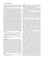
Fig. 3. Suturing the renal artery end-to-side to the external iliac artery (Clamp on renal vein).
Minimally Invasive Renal Transplantation
515
Fig. 4. MIKT scopic aid during the arterial end-to-side anastomosis.
Understanding the Complexities of Kidney Transplantation
516
In most cases the kidney was not moved from the neatly fitting retroperitoneal pouch after
revascularization. Reimplantation of the ureter was performed by extravesical technique
a.m. Lich-Gregoir, with minimal bladder dissection.
Scopic aid was only found necessary in a few cases under very deep, narrow circumstances.
The scope was then simply introduced through the same incision, alongside the
instruments, giving a “close up” of the anstomotic area.
A simplistic approach, with minimal dissection/tissue trauma was attempted at all stages.
Fig. 5. After revascularisation: The perfused renal artery and vein are seen, while the kidney
lies lateral to the skin incision.
Minimally Invasive Renal Transplantation
517
4.2 MIKT: Results
A series of patients, transplanted by strict MIKT technique was then compared with
matched controls subjected to conventional surgery. From December 2004 to July 2005, 21
kidney recipients were subjected to the new, minimally invasive technique. The MIKT
patients constituted a consecutive series of transplantations performed by a single surgeon.
A control group, subjected to conventional KTx (n=21) had been concurrently selected to
match the MIKT group regarding age, sex, donor source, and primary-/retransplant status.
No MIKT procedures were interrupted or converted to COKT. The results have been
summarized in Table 1.
RESULTS
[ mean (range)]
MIKT
n=21
Conventional Tx
n=21
Student
t-test
p-value
Skin incision length (cm) 8,1* (7 - 9) 20,5 (17-23) p<0,01
Operative time
(min)
118* (95-140) 187 (130-270) p<0,01
Analgesic requirementsPostop.
days 0 + 1+ 2
(Morphine Equiv.; mg)
35 (3-86) 56 (20-173)
n.s.
(p=0,053)
Hospitalization
(days in hospital postop.)
8,2* (6-13) 12,4 (7-29) p=0,02
Delayed graft function
10 % (2) 14 % (3)
Measured GFR
10-12 weeks post-Tx
(Cr-EDTA- Clearance; mean [range];
ml/min/1,73 m
2
)
57,4 (35 – 81) 51,2 (26 – 72)
n.s.
(p=0,053)
Peroperative incidents
No major No major
Surgical
complications/reinterventions
- Lymphocele: Reop.
- Wound dehiscence: Reop.
- Urinary obstruction: Reop.
- Perirenal hemorrhage: Reop.
- Bladder hemorrhage
- Total
2 (10 %)
0
0
1 (5 %)
0
3 (14 %)
3 (14 %)
1 (5 %)
1 (5 %)
1 (5 %)
2 (10 %)
8 (38 %)
Table 1. MIKT results. (extracted from Øyen et al., 2006)
Naturally, the MIKT skin incision was very much shorter. There were significant differences
in favour of MIKT regarding operative time and postoperative stay in hospital.
Furthermore, the analgesic requirements, expressed as morphine equivalents during
postoperative days 0+1+2 were less in the MIKT group, however at non-significant levels.
There were less complications and reinterventions in the MIKT recipients, totally 3 (14 %) -
versus 8 (38 %) in the open KTx group. Because of the high complication rate in the control
group, the total complication/reintervention rate of open KTx outside the study during the
inclusion period (n = 97) were investigated and found to be 30-40 % (data not shown).
Understanding the Complexities of Kidney Transplantation
518
Fig. 6. Exterior result after left-sided MIKT in a slim patient, through a 7,5 cm incision.
4.3 MIKT: Discussion
Compared with L-LDN employing a 6-9 cm skin incision for kidney harvesting, the MIKT
incision was only faintly larger (7-9 cm), and besides the L-LDN was dependent on 2-3
additional laparoscopic ports (5-12 mm each).
The first MIKT results were good, compared with the open, conventional KTx group and
indicated that the procedure might be executed fast (because of its simplicity) and safe. By
reducing incision, extent of dissection and thereby tissue trauma, the wound complications
would be suspected to be reduced accordingly. Potentially it may also reduce
hospitalization, and thereby the risk for nosocomial infections.
A major point about the MIKT approach (also when disregarding the results), was that
reduction of tissue trauma appeared particularly appropriate in these patients, with
significantly delayed wound healing and a high “background” complication rate. Due to the
immunosuppressive theraphy, the incidences of wound dehiscence and incisional hernia
were distinctly higher in Tx recipients, in particular after the introduction of
Sirolimus/Everolimus. For simple reasons, a significant reduction of the abdominal wall
incision would be anticipated to reduce these wound-related problems. Potentially, the
MIKT procedure might also counteract the huge lymphocele/lymph leakage problem, by
minimizing the dissection cavity and leaving less space available for fluid expansion.
Except from the single MIKT surgeon’s extensive Tx experience , the distinctly shorter MIKT
operating time might be explained by the simplified/minimalistic handling of the vessels,
the extravesical reimplantation technique, and fast closure of a small incision.
Our data did suggest the same beneficial effects on postoperative pain/analgesia and
recovery, that had been documented for a wide range of MIS procedures.
During recent years, Tx surgeons in Oslo have in part adopted the MIKT technique, by
significantly reducing the size of the incision, even though not conforming strictly to MIKT.
A significant reduction in overall KTx complication rates has been observed during 2008-
2011, which may be partly attributed to reduced incision size and thereby tissue trauma.
Minimally Invasive Renal Transplantation
519
5. Minimally invasive video-assisted KTx (MIVAKT) - The South Chorean
experience (2007)
In 2007 a minimally invasive, partly video-assisted KTx technique (MIVAKT) was described
by a South Chorean group (Seong-Pyo et al., 2007, Park et al., 2008) – obviously quite
independent of the previous ’da Vinci robot’ and MIKT reports.
5.1 MIVAKT: Method/Technique
The MIVAKT pocedure was carried out in 20 patients. Clinical variables were compared
with the conventional KTx method. A 7-8 cm skin incision was employed. By means of a
scopic balloon instrument a retroperitoneal space was created for the kidney. The vascular
anastomoses and ureteroneocystostomy were performed under both direct vision and
video-assisted aid.
5.2 MIVAKT: Results/Conclusion
The average length of the wound incision was 7-8 cm, placed below the belt line. The
average operating time were 186 min. Less analgesics was given compared with
conventional methods. There was one postoperative complication, a mild lymphocele. All
patients showed normalized serum creatinine levels within 4 days post-Tx and normal
findings on postoperative ultrasound and renal scintigraphy.
MIVAKT was shown to be technically feasible and might offer benefits in terms of better
cosmetic outcomes, less pain, and quicker recuperation, compared with conventional KTx.
Fig. 7. (A) The location and course of the external iliac vessels (thick arrow) and the contour
of the urinary bladder (thin arrow), marked preoperatively using ultrasound. (B) The 7–8 cm
oblique incision. (Seong-Pyo et al., 2007)
5.3 MIVAKT: Discussion
We consider the transverse (horizontal) MIKT incision to offer better access to the iliac
vessels, than the oblique MIVAKT incision. Furthermore, it is not at all necessary to use a
laparoscopic balloon dissector to create the retroperitoneal space. A kidney-fitting
retroperitoneal pouch is easily and safely made by hand/retractors through a minimal
incision.
Understanding the Complexities of Kidney Transplantation
520
The video-assisted MIVAKT approach is interesting. Though, in the MIVAKT series, it
seems like the vascular anastomoses for the most part were carried out under direct vision.
In the MIVAKT discussion it is stated that “The grafted kidney was hung over the skin
incision during the vascular anastomosis because the procedure is nearly impossible after
the placing of the grafted kidney in the retroperitoneal space.” This is not at all ‘impossible’;
but exactly what the MIKT technique is all about. Both the venous and arterial MIKT
anastomoses were performed with the kidney in its final retroperitoneal position, suturing
the back walls from the inside.
Fig. 8. (A) The circular retraction system and video-assisted TV monitoring. (B) The kidney
was placed just above the skin incision during the vascular anastomoses. The laparoscope
(thin white arrow) was found useful for visualisation and illumination. (Seong-Pyo et al.,
2007)
6. Laparoscopic KTx – A case report from Barcelona (2010)
In 2010 a spanish group presented a case report on KTx by means of regular laparoscopic
access, using 4 trocars and a Pfannenstiel incision (Rosales et al.).
6.1 Laparoscopic KTx: Method/Technique
With the recipient in the left lateral decubitus position, a hand-port was placed into a 7 cm
Pfannenstiel incision. One trocar was put through the hand-port, while three more trocars
were introduced in the right hemiabdomen.
Minimally Invasive Renal Transplantation
521
Fig. 9. Trocar positioning. Pfannenstiel incision. (Rosales et al., 2010)
Understanding the Complexities of Kidney Transplantation
522
Fig. 10. Laparoscopic venous and arterial end-to-side anastomoses (Rosales et al., 2010).
Minimally Invasive Renal Transplantation
523
By making a retroperitoneal, pelvic window ,the right external iliac vessels were dissected
free. The kidney was introduced through the hand-port, and end-to-side anastomoses were
performed by bulldog clamping through the hand-port and continous suture.
The ureterovesical anastomose was done by a modified Taguchi technique. Finally, the
kidney graft was placed extraperitoneally by continuous suture of the peritoneal window.
6.2 Laparoscopic KTx: Results
Surgical time was 240 min, with 300 cm
3
bleeding. Cold ischemia time was 182 min. The
postoperative course was uneventful and functionally satisfactory. Serum creatinine
decreased progressively, to 73 μmol/l on the day of discharge. Stay in hospital was 14 days.
6.3 Laparoscopic KTx: Discussion
A laparoscopic KTx operating time of 4 hours seems too much, when MIKT can be executed
in 2 hours, and with a total incision size that is probably smaller, when taking into account
the 3 additional laparoscopic ports. The transverse (7-9) cm MIKT incision in the iliac fossa
offers excellent direct access to the anastomotic area of the external iliac vessels. And
regarding safety towards vacular incidents, the laparoscopic approach must be considered
inferior.
Altogether, it seems unnecessary to perform the vascular anastomoses by laparoscopic
technique – when these can be performed openly by an incision that is nevertheless needed
for decent introduction/transplantation of the kidney.
7. Minimally invasive renal auto-transplantation (MI-Auto-KTx) (2010)
By combining ‘‘hand- assisted laparoscopic nephrectomy’’ and MIKT — using the same
incision (7–9 cm) for hand-assistance, kidney harvesting, and transplantation — we have
during 2009-2011 conducted ‘‘Minimally invasive renal auto-transplantation’’ (MI-Auto-
KTx) in 6 patients. The first two MI-Auto-KTx cases have allready been documented and
published (Øyen et al., 2010).
7.1 MI-Auto-KTx: Method
Laparoscopic hand-assisted nephrectomy: The handport incision (7-8 cm) was made
medially in the right iliac fossa; displaced laterally compared with the usual Pfannenstiel L-
LDN incision.
Extracorporeal ‘back bench’ preparartion: In the first case (female 38 years; renal artery
aneurysm) it was possible to maintain a single arterial stem, after resection of the 16 mm
aneurysm. In the second case (female 55 years; ureter lesion) three renal arteries had to be
reconstructed.
MIKT: We utilized the handport incision, targeted on the iliac vessels, without extension.
The meticulously prepared kidney was placed in a small/fitting, retroperitoneal pouch; and
anastomosed to the iliac vessels. Reimplantation of the ureter was performed by extravesical
technique.
7.2 MI-Auto-KTx: Results
Total operative times were 335 min and 434 min, respectively. In both cases the
postoperative course was uneventful, and the patients were transferred to the local hospital
Understanding the Complexities of Kidney Transplantation
524
Fig. 11. MI-Auto-KTx: Laparoscopic, right-sided, hand-assisted nephrectomy; by a 7-8 cm
medial “transplant incison”, using GelPort and 3 trocars. The right renal vein is stapled and
divided flush with the caval vein. (Øyen et al., 2010)
Minimally Invasive Renal Transplantation
525
at day 4/day 5. When examined 3 mts postoperatively, both auto-transplants were shown to
have excellent function by renal scintigraphy.
7.3 MI-Auto-KTx: Discussion
Our first two MI-Auto-KTx cases have demonstrated that a traditionally major surgical
procedure, with extensive incisions/tissue trauma, can be made minimally invasive, by a
similar incision as that used for L-LDN. Taking into regard the highly traumatic
conventional incisions, we expect the generally proven minimally invasive benefits to be
considerable.
8. Considerations about the future
The minimally invasive KTx procedures have so far not gained widespread acceptance and
still seem to be at a “pioneer stage”. However, considering the rapid evolution of MIS
during the last two decades, there is little reason to believe that KTx and Auto-KTx in future
will be excluded from this development.
Since a ≥ 6 cm incision will anyway be needed for decent introduction of the kidney (except
for the possibilty of introduction through natural orifices) , we think the MIKT procedure is
the most suited for further developments in this field.
9. References
Dubois F, Icard P, Berthelot G & Levard H (1991). Coelioscopic cholecystectomy:
preliminary report of 36 cases. Ann Surg, 211: 60-2.
Harrell AG, & Beniford BT. (2005). Minimally invasive abdominal surgery: lux et veritas
past, present, and future. Am J Surg, Vol 190, No 2, pp 239-43.
Hoznek A, Zaki SK, Samadi DB, Salomon L, Lobontiu A, Lang P & Abbou CC (2002).
Robotic assisted kidney transplantation: an initial experience J Urol, Vol 167, pp
1604–06.
Hoznek A, Hubert,J, Antiphon, Gettman MT, Hemal AK & Abbou CC (2004). Robotic renal
surgery. Urol Clin N Am, Vol 31, pp 731-76.
Khanna A & Horgan S (2011). A laboratory training and evaluation technique for robot
assisted ex vivo kidney transplantation. Int J Med Robot. Vol 7, No 1, pp 118-122.
McCullough CS, Soper NJ, Clayman RV, So SSK, Lendrisak MD, & Hanto DW (1991).
Laparoscopic drainage of a post-transplant lymphocele. Transplantation, Vol 51, No
3, pp 725-27.
Øyen O, Scholz T, Hartmann A & Pfeffer P (2006). Minimally invasive kidney
transplantation: The first experience Transpl Proc , Vol 38, pp 2798-2802.
Øyen O, Scholz T, Hartmann A & Pfeffer P (2006). Minimal invasive kidney transplantation
– The first experience. Transplantation, Vol 82, Suppl 2, pp 930-31.
Øyen O, Lien B, Line P-D, & Pfeffer P (2010). Minimally invasive renal auto-transplantation:
The first report (2010). J Surg Res, Vol 164, pp e181-84.
Park SC, Kim SD, Kim JI, & Moon IS (2008). Minimal skin incision in living kidney
transplantation. Transpl Proc, Vol 40, pp 2347-48.
Understanding the Complexities of Kidney Transplantation
526
Ratner LE, Ciseck LJ, & Moore RG.(1995) Laparoscopic live donor nephrectomy.
Transplantation;, Vol 60, No 9, pp 1047-49.
Rosales A, Salvador JT, Urdaneta G, Patino D, Montllo M, Esquena S, Caffaratti J, Ponce de
Leon J, Guirado L & Villavicencio H (2010). Laparoscopic kidney transplantation.
Eur Urol, Vol 5 7, pp 164-67.
Seong-Pyo M, Jeong-Whan C, Kuyong-Jong K, Gui-Ae J, Min-Woo C, Young-Joon A &
Seong-Whan K (2007). Minimally invasive video-assisted kidney transplantation
(MIVAKT) (2007). J Surg Res Vol 141, pp 204-10.
Wolf JSJ, Tchetgen MB & Merion RM (1998). Hand-assisted laparoscopic live donor
nephrectomy. Urology, Vol 52, No 5, pp 885-87.
25
Surgical Complications of
Renal Transplantation
Marcelo Ferreira Cassini, Murilo Ferreira de Andrade
and Silvio Tucci Junior
Ribeirão Preto Medical School,
São Paulo University
Brazil
1. Introduction
In the early era of kidney transplant, surgical complications were a major cause of graft loss.
Between 1960 and 1980, the estimated incidence was around 20%. With the improvement of
surgical techniques, the frequency of these complications has dropped significantly and this
subject until then common in the medical literature came to be seldom discussed (Botto V,
1993; Hernandez D, 2006). Currently, it is estimated that in large transplant centers the
incidence of surgical complications is less than 5%. In general, the results of renal
transplantation have improved primarily as a consequence of advances in medical and
immunosuppressive therapy and progress in surgical techniques. Posttransplant urologic
complications are unusual, with the range of 2.5% to 27% in most series, and can cause
significant morbidity and mortality (Zargar MA, 2005; Dalgic A, 2006) Results have
improved over the past decade as a direct application of less invasive endourologic
diagnostic and therapeutic techniques of the surgical complications (Streem SS, 1994).
However, the etiologies are the most common technical problems and association with
immunological complications. Surgical complications after renal transplantation can be
classified mainly as vascular (arterial and venous thrombosis, renal arterial stenosis,
lymphocele, hemorrhage) and urologic (ureteral obstruction, vesicoureteral reflux, urinary
fistula), although other types of complications are not uncommon, like graft’s rupture and
hematoma. These complications can occur early in the intra-operative, immediate
postoperative period or later, and imply in increase morbidity, hospitalization and costs
(Humar A, 2005).
Urologic complications are the most common surgical complication after renal
transplantation, causing significant morbidity and mortality. Recently, the incidence of
urologic complications after renal transplantation has decreased to 2.5% to 12.5% (Emiroglu
R, 2001).
Unfortunately, there is a still higher incidence of technical complications in
pediatric recipients, reaching approximately 20% with an associated 58% and 74% graft
survival rates for cadaveric and living-related transplantation (Salvatierra O Jr, 1997; US
Renal Data System, 1996).
Urologic complications represent an important cause of
morbidity, delaying normal graft functioning, and in some cases leading graft loss and/or
patient death (Beyga ZT, 1998; Colfry AJ Jr, 1974; Mundy AR, 1981; Hakim NS, 1994).
Understanding the Complexities of Kidney Transplantation
528
The most frequent urological complications after kidney transplantation involves the
ureterovesical anastomosis (fistula, stenosis and reflux), with a frequency ranging from 5%
to 10% in different séries.
2. Urologic complications
2.1 Urinary fistula
This is the leakage of urine from the collecting system. It can occur at the level of the
bladder, ureter or renal calices. The leakage of urine can be collected around the graft, move
to the retroperitoneum, scrotum or may manifest through the incision. His average
prevalence in many studies is around 5.7%. In general, most urinary leaks are the results of
ureteral problems, failure of ureterovesical anastomosis or ischemia and necrosis of the
distal ureteral stump.
Like the majority of surgeons now employ an extravesical ureteroneocystostomy technique
for implantation of the ureter, there are shorter ureter and decreased likelihood of ischemia,
and a limited cystostomy that rarely leads to leakage from the bladder (Gibbins WS, 1992;
Thrasher JB, 1990).
Clinical presentation:
In most cases, there is constant discharge of clear liquid (yellow citrus) through the drain, in
the immediate postoperative period, and sometimes the flow through the drain can even
surpass the diuresis the urinary catheter.
When later, after removal of the tubular drain, there may be bulging store kidney with
extension into the perineum and scrotum or decreased urine output with maintenance of
renal function. Unexplained graft dysfunction, pelvic fluid collection, fever, graft
tenderness, an lower extremity edema can also occur (Streen SB, 1994).
Early urinary leaks can be divided into two types: the first usually occurs within the first 1 to 4
days and is almost always related to technical problems with the implantation. In this case, the
ureter has usually pulled out of a tunnel caused by excessive tension at the anastomosis. This
complication appears to be more common with the extravesical ureteroneocystostomies
(Streen SB, 1994).
Some authors have recommended use of a ureteral stent to lessen the
likelihood of this complication (Gibbins WS, 1992).
The second type of early ureteral leak,
usually presents between 5 and 10 days, is associated with distal ureteral ischemia, which may
be a consequence of injury during the donor nephectomy, technical causes such as tunnel
hematoma or distal stripping of the blood supply (Rosenthal JT, 1994).
Diagnosis:
For being the most common surgical complication of kidney transplantation, urinary fistula is
easily diagnosed. In doubtful cases, where there is need to exclude the lymphocele as main
differential diagnosis, biochemical analysis of the liquid is characterized by having elevated
levels of creatinine, urea and potassium. In the lymphocele, creatinine should be similar of
blood. Urinary leak are often suspected because of increased drainage from the wound.
Radiographic tests of help include an abdominal ultrasound and nuclear renal scan. The
ultrasound is nonspecific for evaluating patients with suspected urinary fistula after kidney
transplantation. It will only reveal a fluid collection (anechoic image) around the graft. A renal
scan demonstrating extravasation (figures 1.1, 1.2) is the most sensitive method to differentiate
a urine leak from other fluid collections such lymphoceles or hematomas (Bretan PN Jr, 1989).
A cystogram should be performed if a bladder leak is suspected.
Surgical Complications of Renal Transplantation
529
Fig. 1.1. Renal scan with contrast early extravasation (urinary fistula).
Fig. 1.2. Late renal scan without contrast extravasation (no fistula).
In the evaluation of transplant patients, nuclear medicine can contribute in the earliest
complications that may arise in the period immediately following transplantation, as in the
late complications and complications of surgical nature. A landmark study, conducted an
initial assessment within the first 72 hours of surgery, is important so that we can better
assess possible changes in the course of evolution. Studies with DTPA or MAG3 are the ones
who will advise on the vascular phase and functional phase, and excretory phase, all
parameters of the utmost importance in the evaluation of the graft (Kahan BD, 1989; Luk
SH, 1999).
As surgical complications of kidney transplantation, the urinary fistulas are observed by
scintigraphy an accumulation of the radiotracer outside the kidney (Luk SH, 1999). In cases
of hematoma, other surgical complication, shows an area of low concentration of the tracer
near the kidney, which may cause displacement of large structures such as vessels, ureter,
Understanding the Complexities of Kidney Transplantation
530
bladder and collecting system obstruction. The diuretic renogram may help elucidate this
issue, because the transplanted kidney has the same performance as a native kidney
scintigraphy.
Management of the urinary fistula:
Disruption of urinary tract in a renal transplant patient or graft dysfunction requires rapid
diagnosis and treatment. Ureteral leakage needs careful and accurate diagnosis of the exact
cause and site. It is important to know if the problem has a physical cause such a leak or an
obstruction and is not associated with an acute rejection episode that required specific
treatment (Streen SB, 1994; Rosenthal JT, 1994).
Surgical treatment has to be performed in all patients except those presenting with minimal
extravasation at the ureteral reimplantation site and clinically stable. This group was initially
treated by urinary drainage. In cases of unfavorable outcome after clinical treatment, surgery
is indicated. Surgery is the initial aproach for big extravasation or when leaks arising from the
mid or upper ureter were suspected. We use the same incision of the transplant to access the
fistulae. The type of surgical reconstruction is based on the intraoperative evaluation of the
extent of the ureteral necrosis and local and systemic condition of the patient at the time of
surgery. Primary reconstruction with the ureter of the recipient or a new ureteral
reimplantation are performed preferentially when local and systemic condition allowed; if
local or systemic infection are present and the patient is clinically unstable an ureteral ligature
associated to a nephrostomy can be performed. Ureteral stenting alone is used exceptionally.
All patients received prophylactic or therapeutic antibiotic according to the antibiogram of the
collected fluid (Mazzucchi E, 2006).
The need for immediate open operative surgical intervention has been replaced, to a large
extent, by early endourologic intervention (Banowsky LHW, 1991).
The placement of a
percutaneous nephrostomy can divert a leak or relieve obstruction and allow more
definitive diagnosis. As described by Streem et al., endourologic management can select
patients for whom the likelihood of successful nonoperative management is good. In a few
cases, percutaneous access can offer long-term treatment with chronic stent management.
Percutaneous techniques like nephrostomy associated to antegrade ureteral stenting works
in 40% of a much selected group of patients presenting with small fistulae from the distal
ureter (Campbell SC, 1993).
Early open surgery is our preferred approach. Our policy is to perform primary urinary
tract reconstruction whenever local and systemic condition allows. Termino-lateral
anastomosis of the graft ureter or pelvis with the ureter of the recipient can be used as
technique for the correction of urinary leaks. Some groups use termino-terminal
anastomosis with the ureter of the recipient (Salomon L, 1999) with good results but can
results in ureterohydronephrosis of the native kidney after ureter ligation for reconstruction.
Ureteroneocystostomy “de novo” is used for reimplantation defects or for small distal
ureteral necrosis and can fail in many cases due to necrosis extension or incomplete ureteral
and bladder wall resection during surgery. Ureteral reimplantation remains an important
option for urinary fistulae management. Ureteral ligature and nephrostomy is performed
when there is gross infection of the fossa or when the patient presents in sepsis. There is also
described, in cases of infected urinary fistulas and to prevent distal ureteral ligature and
nephrostomy, the introduction of a Foley’s catheter throught the bladder wall. The
catheter’s balloon is inflated at the transplanted renal pelvis to occlude the pyeloureteral
junction and dry the region of the fistula (Suaid HJ, 2010).
Surgical Complications of Renal Transplantation
531
Recurrences are due to insufficient ureter resection, leaving an ischemic stump extension of
the process after the surgery or inadequate anastomosis. We recommend always leaving a
double J stent in these cases in order to reduce recurrences but stents do not work if the
necrosis extends. Recurrences were always managed surgically and an anastomosis with the
ureter of the recipient was the first choice. Some patients can need a third procedure due to
a new recurrence showing that the necrosis can extend after surgery and that extensive
resection of the ureter is frequently necessary.
Mortality directly related to the fistula or to its correction was high in the early
transplantation era (Dreikom K, 1992) and nowadays is reported to range from 0 to 8%
(Salomon L, 1999). These better results are due to an earlier and more aggressive approach,
reduction in the amount of corticosteroids in the immunosuppressive regimen and to better
antibiotics and clinical support. The increase in the experience with these cases can still
improve such results.
Routine ureteral stenting, to avoid urinary fistula, does not reduce significantly your
incidence and its use is recommended only in special cases (contracted bladder, difficult
anastomosis) (Campbell SC, 1993; Salomon L, 1999). In our center the modified Gregoir
technique has been the procedure of choice in the last 35 years and the incidence of ureteral
complications has been low.
2.2 Ureteral obstruction
Ureteral obstruction and ureteral leakage are the most common urinary complication after
renal transplantation (Azhar, Hassanain et al. 2010). The incidence related in literature
varies from 3 to 8% (Fontana, Bertocchi et al.; Smith, Windsperger et al.; Kaskarelis,
Koukoulaki et al. 2008). Obstruction may occur during the early postoperative course due to
blood clots, ureteral malrotation or kinking, tight submucosal tunnel, unsuspected donor
calculus (Poullain, Devevey et al. 2010) or perigraft fluid collection (Kahan and Ponticelli
2000; Campbell, Wein et al. 2007). Late ureteral obstructions generally after the first month
or even at years posttransplant are secondary to chronic ischemia which leads to chronic
fibrosis and strictures. Other cause includes compressive limphoceles or pelvic masses,
ureteral lithiasis and rarely obstruction by ureteral carcinoma (Huurman, Baranski et al.
2008)
or fungus ball (Vuruskan, Ersoy et al. 2005).
The clinical presentation includes pain over the surgical site, decreased urine volume
leading to oligoanuria and rise in blood pressure secondary to impaired renal function.
Diagnostic tests shows gradual rise in serum creatinine. The ultrasound demonstrates
pyelocaliectasis (fig. 2.1) or ureteropyelocaliectasis (fig. 2.2) in most of cases. Nuclear
scintigraphy is less sensitive because the obstructed kidney also displays impaired
radionuclide uptake, a sign often present in allograft rejection. When the diagnosis is
unclear the antegrade pyelogram must be performed, because is an accurate method to
define anatomically the site, degree of obstruction (Kahan and Ponticelli 2000).
The treatment must be instituted as early as possible to avoid loss of renal graft function.
Initially the nephrostomy by puncture must be done to ensure the patency of the kidney and
restore renal function to normal. The definitive treatment of the obstruction is oriented
according to the etiology. Stenosis ureteral at the site of bladder reimplantation is more
common and can be addressed by several endourology techniques such as ureteral
meatotomy or percutaneous ureteral dilation with balloon followed by angioplasty and
implant of stent at the ureters. Such techniques are at acceptable levels of success especially
Understanding the Complexities of Kidney Transplantation
532
Fig. 2.1. Ultrasound with moderate hidronefrosis.
Fig. 2.2. Ultrasound of transplant kidney with ureteroectasis secundary to distal ureteral
obstruction.
Surgical Complications of Renal Transplantation
533
when treat small lesions (Burgos, Bueno et al. 2009). However, open surgery with
reconstruction of the excretory pathway is still considered the gold standard. In distal
ureteral obstructions or when there is redundant ureter, we can review the
ureteroneocystostomy by extravesical Lich-Gregoir modified techniques (Campos Freire, de
Goes et al. 1974) or intravesical (Politano-Leadbetter, 1958).
When there are multiple, long stenosis of the ureter or even poor vascularization, it is
necessary to perform the anastomosis of the renal pelvis with the host ureter
(ureteropyelostomy) or the ureter with the host ureter (ureteroureterostomy). However, the
last technique has a higher rate of stenosis. When the native ureters cannot be used, the
“Boari flap” should be done joining the short ureteral stump or the renal donor pelvis,
allowing an adequate distance to the bladder. This allows tunneling the flap under the
ureter, decreasing reflux and bacterial contamination during episodes of infection at the
lower urinary tract. Extreme situations may require a pyelovesicostomy with anastomosis
the donor urinary pelvis directly to the bladder. In this circumstance there is direct
transmission of voiding pressure to the urinary collecting system as well as any urinary
infection, leading to chronic pyelonephritis and deteriorating renal graft (Kahan and
Ponticelli 2000).
3. Vascular complications
Although theoretically there is greater risk of surgical complications associated with living
donors and recipients of kidneys with multiple arteries, in actuality it has not been
considered more as a problem in laparoscopic (VLP) or open nephrectomy. This, indeed, is
standard procedure in many transplant centers, (Wilson CH, 2005; Hsu TH, 2003) showing
no significant adverse effects on the function and graft survival in VDL nephrectomies
without or with hand assistance which may lead to higher vascular extension. (Saidi R, 2009;
Hoda MR, 2010; Hoda MR, 2011) However, there is need for close attention to the anatomy
of the donor due to the possibility of having two or more arteries and veins, or early arterial
bifurcation (Benedetti E, 1995; Mazzucchi E, 2005; Harper JD, 2010). Furthermore,
knowledge of microsurgical techniques for careful arterial graft reconstruction with multiple
arteries and is essential for the reduction of vascular complications in these situations (Saidi
R, 2009; Beckmann JH, 2008).
4. Arterial renal thrombosis
The most worrisome of vascular complications, it occurs in about 1% of all kidney
transplants (Penny MJ, 1994; Bakir N, 1996) arterial thrombosis can reach values lower or
higher in different series (Salehipour M, 2009).
Usually results from technical difficulties in removing the organ or implant. In nephrectomy
and perfusion injury may occur in the endothelial layer, facilitating the process of
thrombosis. The anastomoses of small vessels or of very different sizes or twisting or
bending pressure are other predisposing factors for thrombosis, making demand for
assessing the floor space of the kidney as well as proper positioning of the graft at surgery.
With some frequency, there is a need to adjust the length of the renal artery to avoid kinking
of the same. A technical care is obliquely sectioning the end of the renal artery
(espatulating), which can reduce the risk of thrombosis and stenosis. Another factor to
consider is the quality of the receiver because the arterial embolization of atheromatous
Understanding the Complexities of Kidney Transplantation
534
plaques predispose to thrombosis. Lesions in the endothelial artery caused by vascular
clamp during anastomosis should also be considered (Gang S, 2009). Other situations of
greater risk for vascular complications are patients receiving three or four kidney
transplants, hyperacute rejection, and antiphospholipid antibodies (Gang S, 2009; Baños JLG
2005).
In children, either as donors or as recipients, renal transplantation deserves special
attention, or some authors recommend the exclusion of donors under the age of 3 years and
the best use of infusion solutions to reduce vascular complications and increase survival
rates graft (Irtan S, 2010).
Clinical presentation and diagnosis:
The hallmark of renal artery thrombosis is the absence of blood perfusion of the
parenchyma, which can still be identified intra-operatively. In the postoperative period the
most common clinical presentation is the sudden interruption of urinary flow, without pain
in the graft. Obstruction should be excluded from the catheter by blood clots. The renal
perfusion should be evaluated by DMSA renal scintigraphy, by ultrasound Doppler, and
even with arteriography, if needed (Nezami N, 2007).
The immediate surgical exploration may allow in a few cases, revascularization and
recovery of the graft, especially if the diagnosis of arterial thrombosis is done before closing
the incision. The loss of the graft is the most common consequence and nephrectomy should
be performed (fig. 3.3).
Fig. 3.3. Nephectomy: Arterial Renal Thrombosis.
Surgical Complications of Renal Transplantation
535
5. Renal artery stenosis
The prevalence of renal artery stenosis is around 2% to 10% (mean 3.7%) (Benoit G, 1990).
Clinical picture is suggested by onset severe hypertension post-renal transplant, dysfunction
or presence of acute renal failure with prolonged NTA. With a peak onset at six months,
renal artery stenosis can manifest itself as early as two days and as late as two years after
transplantation. Stenoses located in the line of anastomosis, especially in termino-terminal
anastomosis, the most frequent etiologic factor is technical failure. Other etiologic factors are
largely the same that lead to arterial thrombosis, but acting with less intensity.
Clinical picture and diagnosis:
The suspicion must always occur when a transplant patient started with a progressive
decline of renal function, heart murmur audible (or increasing its intensity) in the graft site
and hypertension refractory to medical treatment. The diagnosis may be suggested by non-
invasive techniques such as ultrasound associated with (color) Doppler (sensitivity 87 to
94%, specificity 86 to 100%). Doppler ultrasound is useful as screening and may show an
increased blood flow velocity > 6 kHz12 (Nezami N, 2007).
The arteriography still remains the gold standard for diagnosis of arterial stenosis renal
(Rengel M, 1998
)
The degree of stenosis is considered significant when more than 50% of the
arterial lumen. Recently, gadolinium-enhanced MRI has allowed a noninvasive and efficacy
comparable to that of renal arteriography convencional (Thornton MJ, 1999).
The test with
captopril, with plasma renin may be a method in the diagnosis of renal artery stenosis of
kidney transplantado (Glicklich D, 1990).
The therapy depends on the location and degree of stenosis. Conservative treatment can be
used in cases of mild stenosis in which blood pressure is controlled with medication and
serum creatinine level remained stable.
Invasive procedures are indicated when blood pressure is not controllable by medication,
there is progressive worsening of renal function or when noninvasive tests suggest the
progression of stenosis. In this situation, diagnostic arteriography is indicated in
combination with transluminal angioplasty and “stenting" (fig. 4.1, 4.2) (Leertouwer TC,
2000).
This technique allows restoring renal perfusion in most cases and its effectiveness is
confirmed immediately by a second angiography (Ghaffari S, 2009).
Intraluminal balloon dilatation with stenting is the preferred therapy for most patients,
especially recommended in cases of localized stenosis and distant > 1 cm of the anastomosis.
Surgery is reserved for lesions involving the anastomosis, or the surrounding area, and in
cases of early artery stenosis renal (Benoit G, 1990). Other surgical procedures are indicated
when the stenosis is severe and unsuitable for angioplasty or else, in this failure. Surgical
techniques include reviewing local resection of the stricture and reanastomosis, may or may
not be used autologous grafts (saphenous vein) or heterologous (Teflon) in the form of a patch
graft or bypass, with success rates ranging between 63 to 92% (Bruno S, 2004), (fig. 4.3).
6. Renal vein thrombosis
The renal vein thrombosis is uncommon but serious complication, with incidence ranging
between 0.9 and 4.5%, usually occurring in the first week after transplantation and with
great potential for graft loss (Giustacchini P, 2002). Because the transplanted kidney does
not have collateral circulation, venous stasis causes impairment of blood flow and
consequent loss of function.
Understanding the Complexities of Kidney Transplantation
536
Fig. 4.1. Arteriography (post-transplant) showing a renal stenosis artery.
Fig. 4.2. Result after “stent” angioplasty.
Surgical Complications of Renal Transplantation
537
Fig. 4.3. Bypass iliac-renal arteries.
As causative agents related are: angulation of the renal vein or anastomotic stricture,
dehydration, venous compression by lymphocele or hematoma, progression of ipsilateral
iliofemoral thrombophlebitis should also be considered. Late cases of renal vein thrombosis
have been associated with recurrence of membranous nephropathy, (Carrasco A
, 2008).
Clinical presentation and diagnosis:
The symptoms is nonspecific as the sudden onset of hematuria, oliguria or anuria,
accompanied by local pain and swelling of the graft. There may also increase the diameter of
the ipsilateral lower limb deep venous thrombosis associated. The evaluation of renal
Doppler ultrasound confirms the increase in renal volume and absence of venous flow. In
the arterial can be seen reverse diastolic flow. Although it has been reported that early
surgical exploration and thrombectomy allow the preservation of the graft in cases with
renal vein thrombosis, but usually the kidney is no longer viable at the time of surgical
exploration due to the spread intrarenal venous thrombus and prolonged hypertension. In
most cases the nephrectomy is performed (Fathi T, 2007).
A complication associated with renal vein thrombosis is the rupture of the graft, which may
cause hemorrhage and large hematoma perinephric (confirmed by ultrasonography),
together with signs of hypovolemia and circulatory shock. Physical examination usually
reveals bulging at the site. Nephrectomy is also standard procedure (figures 5.1, 5.2, 5.3).
However, in cases of rupture of the graft without thrombosis, should be attempted to suture
the parenchyma and preservation of the graft (Gang S, 2009).