Báo cáo khoa học: "Measuring the impact of Collybia fusipes system of oak trees" pps
Bạn đang xem bản rút gọn của tài liệu. Xem và tải ngay bản đầy đủ của tài liệu tại đây (1.58 MB, 9 trang )
Original
article
Measuring
the
impact
of
Collybia
fusipes
on
the
root
system
of
oak
trees
Benoit
Marçais*
Olivier
Caël,
Claude
Delatour
Unité des
ecosystèmes
forestiers,
laboratoire
de
pathologie
forestière,
Inra,
54280
Champenoux,
France
(Received
8
July
1998;
received
15
September
1998)
Abstract -
This
work
describes
the
aetiology
of
Collybia
fusipes
root
rot
and
the
impact
of
the
parasite
on
the
structure
of
mature
oak
root
systems.
The
collar
roots
were
examined
and
rated
for
C.
fusipes
infection
at
the
base of
26
Quercus
robur
and
20
Q.
rubra
trees.
Trees
were
then
felled
and
their
root
systems
were
up-rooted
with
a
mechanical
shovel.
Number
and
infection
status
of
the
roots
present
were
recorded
at
40,
60
and
80
cm
from
the
trunk
base.
C.
fusipes
drastically
reduced
the
number
of
living
roots.
At
80
cm
from
the
trunk
base,
on
cylinder
3,
Q.
robur
rated
as
lightly
and
heavily
damaged
had
only
52
and
25
%,
respectively,
the
fre-
quency
of
living
roots
of
undamaged
trees;
the
values
were
72
and
25
%,
respectively,
for
lightly
and
heavily
damaged
Q.
rubra
trees.
C.
fusipes
impacted
especially
the
vertical
roots
just
under
the
collar.
(©
Inra/Elsevier,
Paris.)
Quercus
/
Collybia fusipes
/
root
rot
/
incidence
Résumé -
Mesure
de
l’impact
de
Collybia fusipes
sur
le
système
racinaire
des
chênes.
Ce
travail
décrit
l’étiologie
du
pourridié
à
Collybia
fusipes
et
l’impact
du
parasite
sur
le
système
racinaire
des
chênes.
Le
départ
des
racines
maîtresses
a
été
examiné
et
noté
pour
l’infection
par
la
collybie
chez
26
Quercus
robur
et
20
Q.
rubra.
Les
arbres
ont
ensuite
été
abattus
et
leur
système
racinaire
extrait
avec
une
pelle
mécanique.
Le
nombre
de
racines
présentes
et
leur
état
sanitaire
ont
été
déterminé
à
40,
60
et
80
cm
du
collet.
La
collybie
diminuait
fortement
le
nombre
de
racines
vivantes
présentes.
Les
arbres
gravement
attaqués
à
l’examen
précédant
l’arra-
chage
n’avaient
plus,
à
80
cm
de
la
base
du
tronc,
que
25
%
du
nombre
de
racines
vivantes
des
arbres
non
attaqués.
Ceux
jugés
fai-
blement
attaqués
n’en
avaient
plus
que
52
à
72
%
selon
l’espèce.
La
destruction
par
le
parasite
touchait
plus
particulièrement
les
racines
verticales
situées
sous
le
tronc.
(©
Inra/Elsevier,
Paris.)
Quercus
/
Collybia fusipes
/
pourridié
/
impact
1.
INTRODUCTION
Oak
decline
has
been
a
chronic
problem
in
Europe
in
the
past
decades.
The
causes
of
this
decline
are
not
completely
clear.
Climatic
stress,
in
particular
droughts,
are
widely
accepted
to
be
important
factors
as
well
as
defoliation
by
insects
[4,
5].
Fungal
parasites
have
also
been
shown
to
be
involved.
One
of
them,
Collybia fusipes
(Bull.
ex
Fr.)
Quel.
is
a
basidiomycete
*
Correspondence
and
reprints
that
has
been
known
by
European
mycologists
for
a
long
time,
but
has
only
recently
been
reported
to
be
a
pathogen
of
mature
oak
roots
[1, 3].
It
was
often
found
associated
with
declining
oaks
in
France
[2].
Moreover,
it
was
shown
to
behave
as
a
primary
pathogen
on
Quercus
robur
L.
(pedunculate
oak)
and
Q.
rubra
L.
(red
oak)
seedlings
[8].
C.
fusipes
can
also
be
found
on
Castanea
sativa
Miller,
Carpinus
betulus
L.,
Corylus
avellana
L.
and
Fagus
sylvatica
L.
As
very
little
was
known
about
this
apparently
com-
mon
root
rot
fungus,
research
was
started
to
determine
the
impact
of
C.
fusipes
in
oak
forests
in
France.
Preliminary
results
showed
that
the
fungus
is
frequently
present,
most
of
the
time
not
in
connection
with
decline
[6].
In
two
of
the
three
surveyed
forests,
20-30
%
of
the
trees
with
C.
fusipes
fruit
bodies
had
poor
crown
condi-
tions,
while
in
the
third,
only
1
%
of
the
trees
with
fruit
bodies
had
poor
crowns.
Other
observations
suggest
that
the
relationship
between
crown
condition
and
root
infec-
tion
in
C.
fusipes
infected
trees
is
poor.
In
some
exam-
ined
red
oaks
where
most
of
the
main
lateral
roots
were
dead,
the
crown
did
not
show
any
pronounced
decline
in
the
following
7
years
(Delatour,
unpublished
results).
Also,
the
collar
roots
are
apparently
not
often
killed
in
pedunculate
oak
and
Q.
petrœa
(Matt.)
Liebl.
(sessile
oak).
They
can
have
bark
heavily
infected
by
C.
fusipes,
but
still
exhibit
little
evidence
of
cambial
death.
Therefore,
it
is
not
very
clear
whether
the
parasite
is
hav-
ing
a
significant
impact
on
the
tree
(e.g.
radial
growth,
decline
status).
To
clarify
this
question,
it
is
necessary
to
quantify
the
disease
in
the
roots.
Therefore,
we
wanted
to
know
if
we
could
predict
the
infection
status
of
the
entire
root
system
using
a
quick
rating
of
the
collar
roots.
For
that,
we
examined
the
main
collar
roots
of
a
sample
of
pedunculate
and
red
oak
trees
and
rated
them
for
infections,
then
up-rooted
them
and
studied
the
entire
root
system
in
more
detail.
2.
MATERIALS
AND
METHODS
2.1.
Study
plots
Trees
were
sampled
in
two
stands
from
central
and
north-eastern
France.
Quercus
rubra
trees
were
located
at
Les
Barres
(Loiret).
The
soil
consisted
of
a
60-90
cm
layer
of
podzolic
sand,
over
a
layer
of
soft
red
clay
in
which
a
fairly
large
number
of
roots
was
present.
In
win-
ter
the
water
table
is
close
to
the
surface.
There
was
no
major
physical
limit
to
vertical
root
growth
in
this
soil.
Tree
age
ranged
between
40
and
70
years.
The
peduncu-
late
oaks
were
located
at
Les
Aynans
(Haute-Saône),
in
a
pure
Q.
robur
stand.
The
soil
consisted
of
a
0.5-1
m
layer
of
sandy
loam
over
a
deep
layer
of
gravel.
Most
roots
over
1
cm
in
diameter
did
not
extend
into
the
grav-
el.
Tree
age
ranged
from
80
to
100
years.
Incidence
of
C.
fusipes
in
both
stands
was
known
to
be
high,
with
43
%
of
the
trees
with
fruit-bodies
at
the
trunk
base
at
Les
Barres
and
25
%
in
Les
Aynans
[6,
7].
2.2.
Sampling
of
the
trees
About
35
trees
with
diameter
of
20-33
cm
at
breast
height
were
chosen
in
each
stand.
On
most
trees,
C.
fusipes
infection
could
be
detected
quickly
by
scrap-
ing
the
collar
roots
with
a
knife
to
reveal
bark
necrosis.
Root
systems
were
studied
for
C.
fusipes
infection
in
the
following
way:
the
root
collar
was
partially
excavated
to
a
depth
of
20-30
cm
and
a
distance
of
80-100
cm
from
the
trunk
base.
The
infection
status
of
each
major
root
was
assessed
as:
0)
no
necrosis
detected;
1)
necrosis
pre-
sent,
but
covering
less
than
half
of
the
root
circumfer-
ence
(usually
superficial
for
Q.
robur,
with
penetration
of
C.
fusipes
in
the
bark
of
about
1-2
mm);
2)
necrosis
covering
one
side
of
the
root
entirely
(usually
2-5
mm
thick for
Q.
robur);
3)
C. fusipes
infection
over
the
entire
root
circumference
but
root
still
alive
(usually
more
than
4-5
mm
thick
for
Q.
robur);
4)
root
dead
with
decayed
wood.
Diameter
of
the
root
was
measured
at
about
10
cm
from
the
trunk
base.
The
root
infection
index
of
a
tree
was
computed
as:
Σ(root
diameter
x
root
rating)/Σ(root
diameter).
This
index
therefore
takes
values
from
0
to
4.
Trees
with
a
rating
of
0-0.5
will
be
referred
to
as
’not
damaged’,
having
no
or
very
limited
infection
by
C.
fusipes.
Those
with
a
rating
of
0.5-2
and
2-4
will
be
referred
to
as
lightly
and
heavily
damaged
trees,
respec-
tively.
A
sub-sample
of
20
red
oaks
and
26
pedunculate
oaks
was
selected
for
further
study.
It
consisted
of
nine
trees
undamaged
(five
Q.
robur
+
four
Q.
rubra),
21
lightly
damaged
(12 Q.
robur
+
nine
Q.
rubra)
and
16
heavily
damaged
(9
Q.
robur
+ 7 Q.
rubra).
Trunk
diameter
at
breast
height
was
recorded.
Tree
crowns
were
rated
as
damaged
if
large
dead
branches
were
present
in
the
upper
part
of
the
crown,
undamaged
otherwise.
This
rat-
ing
was
performed
in
March,
when
trees
had
no
leaves.
2.3.
Study
of
root
system
structure
and
of
infection
status
Trees
were
felled
to
leave
a
stump
40
cm
tall.
A
trench
1
m
deep
and
about 2
m
radius
was
dug
around
each
stump.
The
root
system
was
then
extracted
by
pulling
up
on
the
stump
with
a
mechanical
shovel
and
vigorously
shaking
it
to
remove
most
of
the
soil
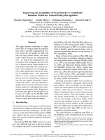
(figure
1a).
The
root
systems
were
washed
with
water
at
low
pressure
and
all
small
roots
(<
1
cm
in
diameter)
were
cut
and
discarded.
Root
system
structure
was
studied
using
a
method
adapted
from
Nielsen
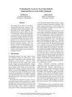
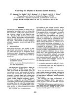
[10].
Briefly,
root
systems
were
placed upside
down
on
a
board
and
characterised
at
the
level
of
three
imaginary
surfaces
located
at
increasing
from
the
trunk
base,
cylinders
1,
2
and
3
(figure
2).
Cylinders
were
80,
120
and
160
cm
in
diame-
ter
and
extended
40,
60
and
80
cm
below
ground,
respec-
tively.
The
vertical
part
of
the
cylinder
was
referred
to
as
the
wall
and
the
horizontal
part
as
the
floor.
Cylinders
were
outlined
by
sticks
marked
at
the
level
of
the floor
and
placed
at
the
level
of
the
wall
(figure
1b,
c).
All
the
roots
passing
through
cylinder
3
were
cut
at
the
level
of
cylinder
3
floor
or
wall,
and
the
position,
diameter
and
infection
status
of
each
root
cross-section
were
recorded.
Position
of
the
root
sections
was
recorded
as:
i)
floor
or
wall
of
the
cylinder;
and
ii)
azimuth
(position
within
eight
compass
sectors).
The
largest
and
smallest
diame-
ters
of
each
root
section
were
measured
and
the
root
cross-section
was
estimated
as
the
geometric
mean
of
those
two
diameters.
Finally,
the
infection
status
of
the
section
was
recorded
as
healthy,
infected
or
dead.
This
procedure
was
repeated
for
cylinder
2
and
then
for
cylin-
der
1
(figure
2).
At
cylinder
1,
the
roots
were
cut
at
about
10-20
cm
from
the
place
where
they
join
the
stump.
Thirty-seven
root
pieces
with
lesion
margins
were
sampled
from
six
different
root
systems
(five
Q.
rubra
and
one
Q.
robur).
They
were
taken
to
the
laboratory,
washed
under
water;
surface
sterilised
for
1-2
min
in
sodium
hypochlorite
at
3.75
%
active
chlorine
and
rinsed
twice
in
sterile
water.
Chips
of
dead
bark
and
pieces
of
the
black
cord-like
fungal
structures
found
on
the
root
surface
were
placed
on
MAT
medium
(10
g.L
-1
of
malt
Difco,
100
mg.L
-1
penicillin,
100
mg.L
-1
streptomycin,
250
mg.L
-1
thiabendazole,
15
g.L
-1
agar).
2.4.
Data
analysis
The
frequency
(no.
per
m2)
and
total
cross-section
area
of
living
roots
was
computed
for
each
of
the
three
cylinders
and
for
wall
and
floor
of
the
cylinder.
The
root
frequency
for
a
cylinder
floor
was
considered
to
be
zero
if
the
absence
of
roots
could
not
be
explained
by
an
obvious
local
limit
to
root
extension.
When
it
could
be
explained
by
a
clear
local
limit
to
root
extension,
i.e.
all
roots
suddenly
changing
direction
or
branching
to
small
diameter
roots
at
a
lower
depth,
then
the
data
were
con-
sidered
missing.
This occurred
only
for
trees
from
Les
Aynans.
Root
frequencies
and
proportion
of
root
dead
were
log
transformed
and
analysed
by
linear
regression
analysis
using
SAS
Inc.
software
[11].
Differences
in
root
frequencies
between
trees
with
crown
damaged
or
undamaged
were
analysed
by
Student’s
t-test.
3.
RESULTS
On
standing
trees,
lesions
of
C.
fusipes
could
be
easily
detected
on
the
major
roots
as
patches
of
dead
bark
that
were
orange
in
colour
with
small
white
fans
of
mycelium
scattered
within
the
necrotic
inner
bark,
as
was
previous-
ly
mentioned
by
Guillaumin
et
al.
[3].
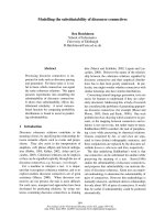
The
development
and
appearance
of
lesions
on
pedunculate
oaks
were
very
different
from
lesions
on
red
oaks.
Lesions could
be
very
extensive
on
pedunculate
oak
roots
before
the
cambium
was
attacked
(figure
3a).
Severely
attacked
large
roots
had
their
entire
surface
covered
with
thick
bark
lesions,
while
most
of
the
cambium
appeared
to
be
still
alive.
A
hypertrophy
response
of
the
bark
to
infection
could
be
observed
as
the
infected
bark
was
usually
thickened
up
to
3-4
cm,
most
of
it
being
necrotic.
The
cambium
was
first
reached
and
killed
at
several
scattered
locations,
then
areas
of
dead
cambium
enlarged
and
coalesced,
and
the
root
was
ultimately
killed.
By
contrast,
on
red
oak
C.
fusipes
induced
lesions
in
the
bark
were
always
asso-
ciated
with
a
similar
amount
of
cambial
death.
Also,
no
thickening
of
attacked
bark
tissues
was
observed
(see
figure
3b).
C.
fusipes
was
isolated
from
68
%
of
the
sampled
symptomatic
root
pieces.
Armillarla
was
isolated
from
two
root
pieces
of
one
of
the
Q.
rubra
trees
from
which
C.
fusipes
was
also
recovered.
It
was
determined
as
A.
mellea
(Vahl:
Fr.)
by
pairing
with
testor
monokaryons
of
known
Armillaria
species.
The
extension
of
A.
mellea
in
the
root
system
was
far
less
than
that
of
C.
fusipes,
and
it
was
a
located
on
small
root
at
the
periphery
of
the
root
system.
No
other
pathogenic
basidiomycete
was
iso-
lated.
At
the lesion
margin,
an
area
of
brown
necrotic
bark
1-10
cm
wide
was
usually
present
between
the
typ-
ical
orange
coloured
infected
bark
and
the
healthy
bark
tissues.
Isolation
success
of
C.
fusipes
from
the
brown
necrotic
tissue
was
poor
(six
successful
isolations
out
of
30
attempts).
Black
appressed
cord-like
structures
(about
0.5
mm
in
diameter)
with
globular
thickenings
(about
2-3
mm)
were
observed
on
the
surface
of
attacked
roots
of
pedunculate
oak
and
red
oak
(figure
3c).
This
ectotrophic
mycelium
was
present
over
all
the
necrotic
bark.
In
particular,
it
was
present
over
the
brown
necrotic
tissues,
closer
to
the
lesion
margin
than
the
orange
coloured
infected
bark.
C.
fusipes
was
difficult
to
isolate
from
the
very
thin
cords
(four
of
96
attempts).
However,
it
was
isolated
more
frequently
from
the
thickened
part
of
the
cord
structures
(14
of
34
attempts).
On
lightly
damaged
trees
of
both
species,
all
lesions
were
found
in
the
root
collar
area,
either
on
the
collar
itself,
or
on
a
large
horizontal
root
near
the
trunk
base.
No
C.
fusipes
lesions
were on
peripheral
roots
in
the
absence
of
root
collar
infection.
As
the
infection
increased,
lesions
quickly
reached
the
part
of
the
root
system
just
beneath
the
trunk
and
apparently
spread
from
there
to
the
entire
root
system.
On
seven
out
of
the
nine
lightly
damaged
red
oaks
investigated,
lesions
were
clus-
tered
on
one
part
of
the
root
system
(figure
1b).
Two
red
oak
trees
had
infections
located
in
two
distinct
parts
of
the
root
system
that
were
not
connected.
In
contrast,
no
unique
point
where
the
infection
might
have
started
could
be
distinguished
on
the
lightly
damaged
peduncu-
late
oaks
and
small
infections
were
usually
present
on
several
scattered
large
collar
roots.
Despite
a
similar
level
of
bark
infection
in
the
two
oak
species,
only
5
%
of
the
collar
roots
more
than
10
cm
in
diameter
were
killed
on
the
damaged
pedunculate
oaks,
while
32
%
were
killed
on
the
damaged
red
oaks.
In
con-
trast,
the
proportions
of
small
roots
(diameter
<
10
cm)
found
dead
and
colonised
by
C.
fusipes
on
the
damaged
pedunculate
and
red
oaks
were
similar
(28
and
29
%,
respectively).
The
total
proportion
of
dead
roots
was
much
higher
for
trees
of
both
species
with
high
root
infection
index
(figure
4),
whereas
the
frequency
of
liv-
ing
roots
decreased
(figures
1b,
c
and
5,
table
I).
At
80
cm
from
the
trunk
base,
on
cylinder
3,
Q.
robur
rated
as
lightly
and
heavily
damaged
had
only
52
and
25
%,
respectively,
the
frequency
of
living
roots
of
undamaged
trees;
the
values
were
72
and
25
%
for
lightly
and
heavi-
ly
damaged
Q.
rubra
trees,
respectively.
In
the
most
heavily
damaged
trees,
the
only
remaining
living
roots
were
recently
formed
adventitious
roots
while
all
the
original
root
system
was
killed
(figure
1d).
For
the
wall
of
cylinders
1-3
and
for
the
floor
of
cylinder
1,
there
were
no
significant
differences
between
the
two
oak
species
in
the
relationship
between
frequency
of
living
roots
and
infection
index,
and
the
data
were
pooled
for
the
regression
analysis.
The
decrease
in
living
root
fre-
quency
was
of
a
similar
order
of
magnitude
in
wall
of
cylinders
1,
2
and
3
(table
I).
Frequency
of
living
roots
decreased
very
quickly
for
low
root
infection
index
(fig-
ure
5 a-c).
Just
beneath
the
trunk,
on
the
floor
of
cylin-
der
1,
the
decrease
was
more
drastic
(figure
5d).
At
greater
depth,
on
the
floor
of
cylinder
2,
the
undamaged
red
oaks
had
a
higher
frequency
of
roots,
compared
to
undamaged
pedunculate
oaks.
Decrease
in
root
frequen-
cy
at
that
level
was
greater
for
Q.
rubra
attacked
by
C. fusipes
than
for
damaged
Q.
robur
(figure
5e).
On
the
floor
of
cylinder
3,
root
frequency
was
low
for
all
trees
and
even
some
undamaged
trees
had
no
roots
larger
than
1
cm
in
diameter
at
that
level.
No
relationship
between
infection
index
and
root
frequency
was
evident
at
that
depth
(figure
5f).
Trees
with
major
dead
branches
in
the
crown
had
much
fewer
living
roots
compared
to trees
with
undam-
aged
crowns
(table
II).
However,
the
relationship
between
root
infection
and
crown
damage
was
not
very
strong
because
some
trees
heavily
damaged
by
C.
fusipes
and
with
few
living
roots
had
crowns
with
no
major
damage,
i.e.
no
dead
branches
(table
II).
4.
DISCUSSION
For
both
oak
species,
the
root
infection
index
was
well
correlated
with
the
frequency
of
living
roots
left
on
the
tree,
and
thus
adequately
represented
the
state
of
the
entire
root
system.
The
main
reason
for
this
was
that
the
part
of
the
root
system
just
beneath
the
trunk
is
colonised
by
C.
fusipes
early
in
the
infection
process
and
so
the
root
infection
index,
measured
close
to
the
trunk,
reflects
well
what
occurs
deeper
in
the
soil.
Indeed,
if
C.
fusipes
causes
major
damages
in
all
the
root
system,
its
maxi-
mum
impact
occurred
on
the
floor
of
the
first
cylinder,
40
cm
below
soil
level
(figure
5d).
On
lightly
damaged
trees,
the
infection
was
always
limited
to
the
central
part
of
the
root
system,
and
thus
appeared
to
start
from
the
collar
area.
This
is
in
agree-
ment
with
previous
work
showing
that
in
infected
stands
each
tree
was
attacked
by
a
different
genet
of
C.
fusipes
and
thus
the
fungus
does
not
spread
from
tree
to
tree
by
root
contacts
[7].
C.
fusipes
lesions,
as
described
in
this
work,
corre-
spond
well
to
what
was
observed
on
inoculated
young
and
mature
oaks
([8];
Marçais,
unpublished
results).
In
particular,
both
the
ectotrophic
mycelium
(cord-like
structure)
and
the
brown
necrotic
area
at
the
lesion
mar-
gin
were
present
in
artificially
induced
infections.
C.
fusipes
spreads
at
the
bark
surface,
and
secondarily
toward
the
cambium.
Perhaps
the
ectotrophic
mycelium
is
involved
in
the
spread
of
the
fungus
at
the
bark
sur-
face,
as
for
Phellinus
noxius
G.H.
Cunn.,
P.
weirii
(Murr.)
Gilberson
and
Rigidoporus
lignosus
(Kl.)
Imaz
[9,
12].
However,
the
ectotrophic
mycelium
is
always
a
few
centimetres
back
from
the
lesion
margin.
Root
destruction
by
C.
fusipes
is
obvious
in
both
Q.
robur
and
Q.
rubra.
The
proportion
of
roots
dead
was
sometimes
very
high
in
the
heavily
damaged
trees
inves-
tigated,
and
the
total
living
root
biomass
was
drastically
reduced,
which
is
in
good
agreement
with
the
results
of
Guillaumin
et
al.
[3].
Although
pedunculate
oaks
showed
greater
capacity
than
the
red
oaks
to
keep
the
cambial
area
of
the
large
horizontal
collar
roots
alive,
their
small-
er
roots
were
killed
by
C.
fusipes
as
readily
as
those
of
Q.
rubra.
As
a
result,
the
root
system
of
heavily
dam-
aged
pedunculate
oaks
was
reduced
to
a
skeleton
of
large,
infected,
but
living
and
undecayed
large
roots.
This
might
explain
why,
despite
widespread
occurrence
of
C.
fusipes
in
oak
forests
in
France
[6],
problems
of
wind
thrown
infected
trees
have
never
been
reported
for
pedunculate
oaks.
In
contrast,
the
main
problem
induced
by
C.
fusipes
in
red
oak
stands
is
wind
thrown
trees
[2].
Despite
differences
in
disease
development
between
the
two
species,
the
relationship
between
the
root
infec-
tion
index
and
the
frequency
of
living
roots
was
the
same
pedunculate
and
red
oak
in
almost
all
parts
of
the
root
system.
The
only
exception
to
this
was
on
the
floor
of
cylinder
2
(the
horizontal
surface
60
cm
below
the
soil
surface),
where
the
impact
of
C.
fusipes
was
higher
for
red
oaks
than
for
pedunculate
oaks
(figure
5e).
This
can
probably
be
explained
by
the
presence
in
Les
Aynans
stand
of
a
gravel
layer
at
50-100
cm
beneath
the
soil
sur-
face
that
constituted
a
strong
physical
limit
to
rooting
for
the
pedunculate
oaks.
Since
even
the
undamaged
pedun-
culate
oaks
have
a
rather
low
root
frequency
60
cm
below
soil
level,
the
impact
of
the
infection
there
is
not
so
high.
There
was
a
relationship
between
crown
status
and
root
infection.
However,
there
were
a
number
of
excep-
tions,
i.e.
trees
with
very
few
living
roots
and
no
marked
symptoms
at
the
crown
level
(table
II).
Although
the
total
reduction
in
root
amount
is
important,
type
and
dis-
tribution
in
the
soil
of
the
remaining
roots
could
be
deci-
sive
for
the
future
of
the
infected
tree.
Our
results
demonstrate
that
the
pathogen
destroys
the
central
part
of
the
root
system,
which
is
mainly
composed
of
roots
pen-
etrating
deep
into
the
soil.
However,
other
roots
survive,
developed
from
the
large
lateral
roots,
which
are
able
to
pump
deep
soil
water.
The
weak
connection
between
decline
symptoms
and
root
reduction
suggests
that
the
remaining
roots
can
be
sufficient
for
heavily
infected
trees
to
live
for
a
long
time
in
the
absence
of
stressful
conditions
without
obvious
decline
symptoms.
Also,
adventitious
roots
often
develop
after
large
collar
roots
are
killed
and
could
mitigate
the
effect
of
root
loss.
However,
such
trees
are
probably
unable
to
overcome
abnormal
situations
such
as
water
shortage.
During
this
study,
we
rated
the
crown
status
in
winter
and
thus,
we
might
have
not
adequately
described
crown
decline.
Therefore,
one
cannot
make
definitive
conclu-
sions
from
our
study
on
this
point.
As
the
infection
index
we
tested
appears
to
measure
well
the
destruction
of
the
entire
tree
root
system
by
C.
fusipes,
we
now
have
a
tool
to
investigate
the
relationship
between
root
infection
and
crown
decline
in
infected
oaks
for
a
large
number
of
trees.
Acknowledgements:
We
would
like
to
thank
J.E.
Mộnard,
P.
Pộradon
and
F.
Cecconi
for
their
technical
assistance
and
E.
Hansen
for
reviewing
the
manuscript.
We
also
want
to
thank
D.
Piou
(ENGREF,
Arboretum
des
Barres)
and
the
Cemagref
for
their
help
at
the
Les
Barres
and
the
Office
National
des
Forờts
for
their
help
at
Les
Aynans.
REFERENCES
[1]
Delatour
C.,
Guillaumin
J.J.,
Un
pourridiộ
mộconnu :
le
Collybia fusipes
(Bull.
ex
Fr.)
Quel,
C.
R.
Acad.
Agric.
France
70 (1984)
123-126.
[2]
Dộpartement
de
la
santộ
des
forờts
(France),
La
santộ
des
forờts
(France)
en
1993,
Ministốre
de
lagriculture
et
de
la
pốche (DERF-DSF),
1994.
[3]
Guillaumin
J.J.,
Bernard
C.,
Delatour
C.,
Belgrand
M.,
Contribution
lộtude
du
dộpộrissement
du
chờne:
pathologie
racinaire
en
forờt
de
Tronỗais,
Ann.
Sci.
For.
42
(1985)
1-22.
[4]
Hartmann,
G.,
Blank,
R.,
Lewark,
S.,
Eichensterben
in
Norddeutschland
-Verbreitung,
Schadbilder,
mửgliche
Ursachen,
Forst
und
Holz
44
(1989)
475-487.
[5]
Landmann
G.,
Becker
M.,
Delatour
C.,
Dreyer
E.,
Dupouey
J.L.,
Oak
dieback
in
France:
historical
and
recent
records,
possible
causes,
current
investigations,
in:
Rundgesprọche
der
Kommission
fỹr
ệkologie,
Bd.
5
Zustand
und
Gefọhrdung
der
Laubwọlder,
1993,
pp.
97-114.
[6]
Marỗais
B.,
Caởl
C.,
Delatour
C.,
Investigation
on
the
distribution
and
impact
of
Collybia
fusipes
in
oak
forest,
in:
Delatour
C.,
Guillaumin
J.J.,
Lung-Escarmant
B.,
Marỗais
B.
(Eds.),
Proceedings
of
the
9th
International
Conference
on
Root
and
Butt
Rots
of
Forest
Trees,
Colloques
de
lInra
no.
89,
France,
1998,
pp.
215-222.
[7]
Marỗais
B.,
Martin
F.,
Delatour
C.,
Structure
of
Collybia
fusipes
population
in
two
infected
oak
stands,
Mycol.
Res.
102
(1998) 361-367.
[8]
Marỗais
B.,
Delatour
C.,
Inoculation
of
Oak
(Quercus
robur and
Q.
rubra)
with
Collybia fusipes,
Plant
Dis.
80
(1996)
1391-1394.
[9]
Nandris
D.,
Nicole
M.,
Geiger
J.P.,
Infections
artifi-
cielles
de
jeunes
plants
dHevea
brasiliensis
par
Rigidoporus
lignosus
(K1.)
Imaz
et
Phellinus
noxius
(Corner)
G.H.
Cunn,
Eur.
J.
For.
Path.
13
(1983)
65-73.
[10]
Nielsen
C.C.N.,
Detailed
instructions for
root
architec-
ture
assessments
with
ROOTARCH-method,
Arboretum,
Internal
Report
no.
7.
Royal
V.
and
Agric.
University
of
Kopenhagen,
Denmark,
1995.
[11]
SAS
Institute
Inc.,
SAS/STAT
(Users
Guide,
Version
6,
4th
ed.,
Vol.
1,
Cary,
NC,
USA,
1989.
[12]
Wallis
G.W.,
Reynolds
G.,
Inoculation
of
Douglas
fir
root
with
Poria
weirii,
Can.
J.
Bot.
40
(1962)
637-645.