Báo cáo y học: "Brain choline concentrations may not be altered in euthymic bipolar disorder patients chronically treated with either lithium or sodium valproate" pps
Bạn đang xem bản rút gọn của tài liệu. Xem và tải ngay bản đầy đủ của tài liệu tại đây (332.19 KB, 7 trang )
BioMed Central
Page 1 of 7
(page number not for citation purposes)
Annals of General Hospital
Psychiatry
Open Access
Primary research
Brain choline concentrations may not be altered in euthymic
bipolar disorder patients chronically treated with either lithium or
sodium valproate
Ren H Wu
1
, Tina O'Donnell
2
, Michele Ulrich
2
, Sheila J Asghar
2
,
Christopher C Hanstock
1
and Peter H Silverstone*
2
Address:
1
Department of Biomedical Engineering, University of Alberta, Edmonton, Alberta, Canada and
2
Department of Psychiatry, University
of Alberta, Edmonton, Alberta, Canada
Email: Ren H Wu - ; Tina O'Donnell - ; Michele Ulrich - ;
Sheila J Asghar - ; Christopher C Hanstock - ;
Peter H Silverstone* -
* Corresponding author
Bipolar disorderlithiumsodium valproatemagnetic resonance spectroscopycholine
Abstract
Background: It has been suggested that lithium increases choline concentrations, although
previous human studies examining this possibility using
1
H magnetic resonance spectroscopy (
1
H
MRS) have had mixed results: some found increases while most found no differences.
Methods: The present study utilized
1
H MRS, in a 3 T scanner to examine the effects of both
lithium and sodium valproate upon choline concentrations in treated euthymic bipolar patients
utilizing two different methodologies. In the first part of the study healthy controls (n = 18) were
compared with euthymic Bipolar Disorder patients (Type I and Type II) who were taking either
lithium (n = 14) or sodium valproate (n = 11), and temporal lobe choline/creatine (Cho/Cr) ratios
were determined. In the second part we examined a separate group of euthymic Bipolar Disorder
Type I patients taking sodium valproate (n = 9) and compared these to controls (n = 11). Here we
measured the absolute concentrations of choline in both temporal and frontal lobes.
Results: The results from the first part of the study showed that bipolar patients chronically
treated with both lithium and sodium valproate had significantly reduced temporal lobe Cho/Cr
ratios. In contrast, in the second part of the study, there were no effects of sodium valproate on
either absolute choline concentrations or on Cho/Cr ratios in either temporal or frontal lobes.
Conclusions: These findings suggest that measuring Cho/Cr ratios may not accurately reflect
brain choline concentrations. In addition, the results do not support previous suggestions that
either lithium or valproate increases choline concentrations in bipolar patients.
Published: 30 July 2004
Annals of General Hospital Psychiatry 2004, 3:13 doi:10.1186/1475-2832-3-13
Received: 30 September 2003
Accepted: 30 July 2004
This article is available from: />© 2004 Wu et al; licensee BioMed Central Ltd. This is an open-access article distributed under the terms of the Creative Commons Attribution License
( />), which permits unrestricted use, distribution, and reproduction in any medium, provided the original work is
properly cited.
Annals of General Hospital Psychiatry 2004, 3:13 />Page 2 of 7
(page number not for citation purposes)
Background
Bipolar disorder is a chronic severe mental illness affect-
ing approximately 1% of the adult population. The most
widely used mood stabilizer for this condition is lithium
[1], although the exact mechanism by which it is clinically
effective remains undetermined. One suggestion is that it
acts via effects on choline metabolism. This is based upon
findings that lithium can inhibit the membrane transport
of choline in both animals [2], and human post-mortem
brain tissue [3]. It also increases the accumulation of
erythrocyte choline in lithium-treated patients [4-7]. Also
of note is that choline concentrations increase signifi-
cantly in rats following electroconvulsive shock [8]. Based
upon this data choline has been used to treat mania in a
some small pilot studies [9], with one open label study
reporting that choline augmentation of lithium treatment
helped rapid-cyclers [10]. Patients treated with choline
also had increased basal ganglia concentrations of
choline, suggesting that externally administered choline
could alter brain concentrations [11,12].
The most appropriate method to measure brain choline
concentrations in vivo utilizes proton magnetic resonance
spectroscopy (
1
H-MRS). Previous studies of bipolar
patients utilizing this methodology have had mixed find-
ings. Overall, while some studies have suggested there
may be increased choline concentrations in specific situa-
tions [13-18], more have found no changes [19-27], and
one found a trend towards a decrease in concentrations
[28]. In both patients and volunteers lithium also doesn't
appear to alter choline/creatine peak ratios concentrations
[29,30]. Nonetheless, two reviews concluded that the evi-
dence to date suggests that lithium increases brain choline
concentrations [31,32], although as noted in these
reviews previous studies have varied considerably in terms
of patient populations, brain region studied, medications
administered, and MRS methodology. Many studies have
also examined differing patients (Type I and Type II) in
differing mood states (mixed, depressed, manic, and
euthymic). This may partially explain the varied results.
Sodium valproate is also widely used as a mood stabilizer,
both alone and in combination with lithium [33]. To date
there have been few studies which have examined the
effects of sodium valproate on choline concentrations or
activity. An in-vitro study suggested that valproate may
inhibit choline acetyltransferase activity [34]. In one study
9 patients taking either lithium or valproate were exam-
ined [35], and increased Cho/Cr ratios were seen in the
bipolar patients compared to controls. There were no dif-
ferences between the lithium and valproate treatment
groups, although the sample sizes were small. However,
another study in epilepsy patients treated with valproate
found no changes in choline concentrations [36]. None-
theless, given the lack of studies to date, the possibility
that valproate and lithium may both increase choline con-
centrations warrants further investigation.
Most of the previous studies have examined Cho/Cr
ratios. However, it should be noted that the "choline" res-
onance peak seen in
1
H-MRS spectra is composed prima-
rily of phosphocholine and glycerophosphocholine,
along with free choline, acetylcholine, and cytidine
diphosphate choline. Also, we have shown in animal
studies that both lithium and valproate can both decrease
creatine concentrations [37]. Therefore, when using Cho/
Cr ratios it is not possible to be certain that any changes in
this peak represent changes in brain choline concentra-
tions. We were therefore interested to determine if there
were any differences in results when using different meth-
odologies, and more specifically to determine if studies
using a ratio methodology may have different results from
studies utilizing metabolite concentrations.
Methods
In the first part of the study patients taking either lithium
or valproate were examined using the Cho/Cr ratio
method, and both Bipolar Type I and Bipolar Type II
patients were included who could also be taking other
medications. In the second part of this study only Bipolar
Type I patients on valproate monotherapy were included,
and quantification of choline concentrations was made.
Some of the data from the first part of this study has been
reported previously [38].
Subjects and Study Design
All subjects gave full informed consent, and both studies
were approved by the ethics committee at the University
of Alberta. Healthy controls were examined using a
detailed, but non-standardized, psychiatric interview.
They were excluded if there was any personal history, or
immediate family history, of psychiatric disorder. For
patients, diagnoses were made using DSM-IV criteria for
Bipolar Disorder Type I or Type II following detailed psy-
chiatric interview, with additional information being
available in almost all cases from long-term psychiatric
clinic records. They also had to be taking a dose of either
lithium or valproate which maintained their blood levels
within the ranges of 0.4–1.2 mmol/l for lithium and 200–
700 µmol/l for sodium valproate. Serum lithium and val-
proate levels were also measured on the day of MRS scan-
ning. Other medications taken by the patient were noted.
In the second part of the study the same criteria were used,
except that only patients meeting diagnostic criteria for
Bipolar Disorder Type I were included, and they had to be
on sodium valproate monotherapy. This was done to
examine Bipolar Type I patients in more detail, and to
remove a possible confounding variable. All patients had
to be euthymic for the previous 3 months, as determined
by interviews with the patient, and additional interviews
Annals of General Hospital Psychiatry 2004, 3:13 />Page 3 of 7
(page number not for citation purposes)
with their relatives and bipolar clinic records when avail-
able. MRS scans were carried out within 24 hours of this
interview.
Magnetic Resonance Spectroscopy Methodology
For both studies magnetic resonance experiments were
performed using a Magnex 3 T scanner with 80 cm bore
equipped with actively shielded gradient, and spectrome-
ter control was provided by an Surrey Medical Imaging
System (SMIS) console. The subjects head was immobi-
lized with a restraint system. Signal transmission and
reception were achieved using a quadrature birdcage reso-
nator for
1
H measurements.
Part 1 - Magnetic Resonance Spectroscopy
Initially, MRI data were acquired using gradient echo
imaging sequences to produce multiple slice images along
both coronal and transverse planes. This allowed registra-
tion of a 2 × 2 × 3 cm volume-of-interest (VOI) to be
selected in the temporal lobe.
1
H MR spectra were
acquired using the PRESS localization method [39,40],
with TE = 32 ms, TR = 3 s, and with 128 averages. Baseline
correction and deconvolution of the spectra was accom-
plished using the Peak Research (PERCH) spectrum anal-
ysis software package. The metabolite peaks of interest
[choline (Cho) and creatine (Cr)] in each spectrum were
fitted to a Gaussian line-shape for peak area estimation.
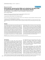
To determine changes in choline concentrations we exam-
ined the Cho/Cr ratio. Figure 1 shows an individual
1
H
MRS spectra in which all the major metabolite peaks can
be seen.
Study 2 - Magnetic Resonance Spectroscopy
To accurately quantify the brain concentration of creatine
we used a 125 ml glass sphere containing a solution of 4
mmol creatine as an external standard. The PRESS
sequence was used to acquire proton MRS data with TE1
= 25 msec, TE2 = 25 msec, TR = 3000 msec, and 128 scan
averages. The MRS data were acquired from three 2 × 2 ×
2 cm
3
voxels placed in the cortex of the left frontal lobe,
the cortex of the left temporal lobe, and in the external
standard solution. The average coordinates [41,42] of the
centers of the two brain voxels were determined: x = 0.5
mm (SD = 1.6), y = 63.5 mm (SD = 12.1), z = -25.5 mm
(SD = 4.2) in the frontal lobe, and x= 32.2 mm (SD = 6.3),
y = 20.5 mm (SD = 3.9), z = 10.7 mm (SD = 2.6) in the
temporal lobe. In order to measure T
1
and T
2
values of the
metabolites in the brain and external standard solution,
MRS data were collected with different TE values at a con-
stant TR and different TR values at a constant TE both for
the healthy volunteers and the patients and also from
external standard solution [42]. However, due to these
constraints, the fact that the two studies used different
populations at different times, and the size of the external
125 ml container (which limited voxel size to 2 × 2 × 2
cm
3
), it was not possible to exactly match the voxel size or
location between the two studies.
MRS Data Analysis
For quantitative measurement of brain metabolite con-
centrations we used previously described methodology
[42,43]. In this, [Met]
b
, in millimoles per kg of wet brain,
the CSF volume fraction, f
csf
, in the spectroscopic voxels
must be corrected. Thus, brain metabolite concentrations
were calculated as described in the following equation:
where V
voxel
is the volume of a 8 cm
3
spectroscopic voxel
[43], and N
b
represents the number of metabolite mole-
cules per unit voxel in brain.
Statistical Analysis for both MRS studies
Means ± SEM were used in the statistical analysis. Sex dif-
ferences were analyzed using chi-squared, and age differ-
ences with ANOVA with post-hoc Tukey tests. The MRS
data was analyzed using Student's unpaired t-test using a
A typical
1
H-MRS spectrum of the human brain at 3.0 T. A number of metabolites can be seenFigure 1
A typical
1
H-MRS spectrum of the human brain at 3.0 T. A
number of metabolites can be seen. 1: creatine (methylene) +
phosphocreatine, 2: glutamate + glutamine, 3: myo-inositol +
glycine, 4: taurine, 5: total choline compounds, 6: creatine
(methyl) + phosphocreatine, 7: N-acetylaspartate.
Met
N
1f V
b
b
csf voxel
[]
=
−
()
Annals of General Hospital Psychiatry 2004, 3:13 />Page 4 of 7
(page number not for citation purposes)
significance level of p < 0.05 comparing diagnostic groups
(patients vs controls) in each brain region (frontal and
temporal).
Results
Study 1
Subjects
A total of 18 healthy controls, 14 bipolar patients taking
lithium, and 11 bipolar patients taking valproate com-
pleted this study. Of the 14 bipolar patients taking lith-
ium, 7 were Type I and 7 were Type II. In the valproate
group, 7 were Type I and 4 were Type II. These groups were
studied both separately and together, but as there were no
statistically significant differences between the Type I and
Type II patients, the results for both types are presented
together. Of the 14 bipolar patients taking lithium 12
patients were taking other psychotropic medications:
these were benzodiazepines (7 patients), antidepressants
(5 patients), and antipsychotics (2 patients). Of the 11
patients taking sodium valproate 10 patients were taking
other psychotropic medications: these were benzodi-
azepines (5 patients), antidepressants (5 patients), and
antipsychotics (4 patients).
The mean age for the lithium group was 40.43 ± 2.96
years, for the valproate group 35.47 ± 2.27 years, and for
the control group was 31.35 ± 2.89 years. These differ-
ences were statistically significant (F = 3.68, df = 2, p =
<0.05), which was attributable to the lithium group being
significantly older than the control group (Tukey post hoc,
p < 0.05).
There were no gender differences within the groups: 10
females and 8 males in the control group (χ
2
= 0.167, df
1, p > 0.05), 5 females and 9 males in the lithium group
(χ
2
= 1.143, df 1, p > 0.05), and 6 females and 5 males in
the valproate group (χ
2
= 0.474, df 1, p > 0.05).
Mean serum lithium levels were 0.79 ± 0.06 mmol/l, and
the range was 0.46–1.08 mmol/l. The mean serum val-
proate levels were 508 ± 42 µmol/l, and the range was
210–912 µmol/l.
MRS Data
1
H MRS
We utilized the ratio of the choline peak to creatine peak
(Cho/Cr) as a primary correlate of Choline concentra-
tions. This result has been reported briefly in a previous
publication [38]. The mean Cho/Cr ratio with this meas-
ure was 1.46 ± 0.04 for controls, 1.18 ± 0.07 for lithium-
treated patients, and 1.12 ± 0.08 for valproate-treated
patients. These were statistically significant, with a reduc-
tion in ratios occurring in both the control vs. lithium
comparison (t = 3.628, df = 30, p = 0.001) and the control
vs. valproate comparison (t = 4.248, df = 27, p = 0.002).
Study 2
Subjects
A total of 11 healthy controls and 9 Bipolar Type I patients
taking valproate as monotherapy were entered into this
study. The mean age for the control group was 37.3 ± 2.2
years, and for the valproate patients 42.4 ± 3.0 years.
These differences were not statistically significant (F =
1.49, df = 1, p = 0.27).
There were no gender differences within the groups: 7
females and 2 males in the valproate group and 5 females
and 6 males in the control group (χ
2
= 0.474, df 1, p >
0.05). The mean serum valproate levels were 472 ± 36
µmol/l, and the range was 284–728 µmol/l.
In the frontal lobe the mean choline concentration for the
healthy controls was 2.21 ± 0.17 mmol/kg wet brain and
for the patients was 2.38 ± 0.12 mmol/kg wet brain. In the
temporal lobe the mean choline concentration for the
healthy controls was 2.35 ± 0.14 mmol/kg wet brain and
for the patients was 2.40 ± 0.19 mmol/kg wet brain. There
were no statistically significant differences between the
controls and patients in either the frontal (t = 0.78, df =
18, p = 0.44) or temporal (t = 0.203 df = 18, p = 0.84)
lobes (Table 1).
The Cho/Cr ratios in the frontal lobes were 0.27 ± 0.028
in controls and 0.28 ± 0.015 in patients. In the temporal
lobes the Cho/Cr ratios were 0.26 ± 0.021 in controls and
0.28 ± 0.016 in patients. There were no statistically signif-
icant differences between the controls and patients in
either the frontal (t = 0.367, df = 18, p = 0.72) or temporal
(t = 0.539, df = 18, p = 0.59) lobes (Table 1).
Discussion
The results from the present study vary considerably
between the two sections utilizing differing methodolo-
gies. This is despite the fact that both studies were carried
out by the same group on the same scanner with bipolar
patients coming from the same patient pool. This strongly
suggests that the methodology used to determine choline
concentrations can considerably alter the results. In the
first part of the study we found that both the lithium-
treated and valproate-treated patients had significantly
reduced Cho/Cr peak ratios compared to controls. This is
similar to the findings from one previous study which also
suggested that there may be a trend towards decreased
choline in grey matter [28]. This study was a frontal lobe
study that measured metabolite concentrations in a 1.5 T
scanner in bipolar type I patients hospitalized for manic
(n = 9) or mixed (n = 8) states. In this study most patients
were being treated with valproate and an atypical
antipsychotic.
Annals of General Hospital Psychiatry 2004, 3:13 />Page 5 of 7
(page number not for citation purposes)
These findings, however, differ from those in the second
part of the present study in which we found no differences
in choline concentrations between valproate-treated
patients and controls in either frontal or temporal lobes.
This second part of the study was much better controlled
in terms of the patients receiving valproate monotherapy,
only including bipolar Type I patients, and in using an
external choline solution to accurately quantify choline
concentrations. This finding of a lack of change is also in
keeping with most previous studies. Several studies which
have also previously measured metabolite concentrations
with 1.5 T scanners also found no changes. These include
a study of the hippocampus in 15 euthymic bipolar type
1 patients, of whom 10 were taking either lithium or val-
proate [19], a study of basal ganglia in 8 rapid cycling
patients on lithium [22], a study of the anterior cingulate
in 10 bipolar children [23], and a study in frontal lobes of
23 euthymic bipolar patients of whom 13 were on lithium
[25]. Several other studies have examined metabolite
ratios, mostly in patients on lithium, and those also found
no changes in choline concentrations [20,21,26,27]. In a
study using metabolite ratios in bipolar children who
were off medication for at least one week there was also
no change in choline concentrations [24]. In a double-
blind placebo-controlled human volunteer study before
and after one week of lithium administration we also
found no changes in cholinein 10 volunteers [30], which
is similar to a patient study which compared 7 patients on
lithium to 6 non-lithium treated controls and in which no
differences were seen [29].
In contrast, animal studies have suggested that lithium
may increase brain choline concentrations, and in lith-
ium-treated patients it also increases the accumulation of
choline within erythrocytes [4-7]. Nonetheless,
1
H-MRS
studies in patients examining this possibility is mixed. To
date 6 studies have suggested some support for this [13-
18], but in none of these studies were metabolite
concentrations measured, and most of the studies meas-
ured choline/creatine ratios [14-18], the other one meas-
uring metabolite intensity/tissue volume [13]. The first
study to examine brain choline in basal ganglia studied
only 4 patients, all of whom were on lithium [18].
Another study examined 19 euthymic inpatients and
found increased choline/creatine ratios in basal ganglia,
but only 10 of these patients were receiving lithium [17].
Table 1: Concentrations (mmol/kg wet brain) and ratios (Cho/Cre) in frontal and temporal lobes in healthy volunteers and in patients
chronically treated with valproate (Study #2)
Choline (Cho) Creatine (Cre) Cho/Cre
Frontal Temporal Frontal Temporal Frontal Temporal
Healthy Controls Age Sex
1 50 M 3.51 2.95 6.67 8.53 0.53 0.35
2 45 M 2.19 3.03 10.1 9.11 0.22 0.33
3 43 F 3.01 2.31 9.97 9.52 0.30 0.24
4 39 M 2.11 2.72 7.94 7.60 0.27 0.24
5 37 F 2.47 2.34 9.98 9.89 0.25 0.24
6 36 F 1.91 1.76 8.28 8.19 0.23 0.22
7 35 M 1.76 2.36 7.93 8.36 0.22 0.28
8 32 F 1.88 1.51 9.56 9.56 0.2 0.16
9 32 M 1.94 2.14 7.04 7.79 0.28 0.28
10 30 F 1.82 2.52 7.8 8.63 0.23 0.29
11 28 M 1.722.237.168.510.240.26
Mean 37.00 2.21 2.35 8.40 8.70 0.27 0.26
Valproate Treated Patients
1 58 F 2.72 2.1 9.16 10.13 0.30 0.21
2 50 M 2.61 3.42 8.17 10.53 0.32 0.33
3 49 F 2.03 1.79 8.56 7.48 0.24 0.24
4 48 F 2.44 1.88 9.93 8.19 0.25 0.23
5 36 M 2.60 2.53 7.84 7.51 0.33 0.34
6 35 F 2.07 2.77 9.26 10.39 0.22 0.27
7 35 F 2.78 1.89 8.35 9.79 0.33 0.19
8 34 F 1.76 2.93 7.26 8.01 0.24 0.37
9 34 F 2.43 2.27 7.75 7.23 0.31 0.31
Mean 42.11 2.38 2.40 8.48 8.81 0.28 0.28
Annals of General Hospital Psychiatry 2004, 3:13 />Page 6 of 7
(page number not for citation purposes)
The third study to report an increase in this ratio (in this
case in the left subcortical region) was in a mixed group of
patients receiving a wide range of medications [16]. Two
other studies have reported increased choline concentra-
tions, but only in limited circumstances. In one study in
11 bipolar children patients were examined before and
after lithium administration [14]. There were no signifi-
cant findings before or after lithium administration,
although there was a trend towards increased choline/cre-
atine ratios in the patients before lithium treatment. This
latter finding does not suggest that in patients lithium sig-
nificantly alters the choline/creatine ratio. The final study
examined 15 euthymic males who were on either lithium
or valproate [13]. This study found that thalamic choline
concentrations, determined by measuring metabolite
intensity/tissue volume ratios, were significantly
increased only if the right and left hemisphere were com-
pared separately, but not if they were compared together.
It is also conceivable that both lithium and valproate may
increase Choline concentrations, but that the differences
were not large enough for us to detect, or that without lith-
ium or valproate treatment patients would have lower
Choline concentrations. The cross-sectional nature of this
study does not allow this to be examined. It is also impor-
tant to recognize other limitations of the present study.
Firstly, these MRS studies are not pre- and post-treat-
ments, so may not accurately reflect changes that occur in
individual patients. Secondly, part of the study used a
ratio-method to assess choline concentrations, the limita-
tions of which are increasingly clear (particularly since
creatine concentrations may be altered by medication
[37]). Thirdly, the sizes of all groups are small and it there-
fore possible that a larger study may have been fully pow-
ered to identify differences between groups. Fourthly,
several patients in the first study (but not the second
study) were on other drugs which may have affected the
results of this study. Fifthly, we have not determined if age
affects the results, and in the first part the groups were not
all matched for age. In addition, the voxel locations were
not the same in both studies due to the reasons discussed
in the methodology section. Nonetheless, despite these
limitations we believe the results add significantly to the
literature in this under-researched area.
We conclude that, taking all current evidence together
including the findings from the present study, it is
unlikely that either lithium or valproate significantly alter
brain choline concentrations. However, given the large
differences in patients populations, medications received,
and MRS methodologies it is difficult to directly compare
all these studies. In addition, the methodology used to
measure choline concentrations can significantly alter the
results. Future MRS studies in bipolar patients should,
therefore, examine metabolite concentrations rather than
a ratio of choline compared to other metabolites.
Competing interests
None declared.
Acknowledgements
This work was supported in part by peer-reviewed grants from the Cana-
dian Institutes of Health Research (CIHR) and the Alberta Heritage Foun-
dation for Medical Research (AHFMR).
References
1. Vestergaard P, Licht RW: 50 Years with lithium treatment in
affective disorders: present problems and priorities. World J
Biol Psychiatry 2001, 2:18-26.
2. Lingsch C, Martin K: An irreversible effect of lithium adminis-
tration to patients. Br J Pharmacol 1976, 57:323-7.
3. Uney JB, Marchbanks RM, Reynolds GP, Perry RH: Lithium proph-
ylaxis inhibits choline transport in post-mortem brain. Lancet
1986, 2(Aug 23):458.
4. Jope RS, Jenden DJ, Ehrlich BE, Diamond JM: Choline accumulates
in erythrocytes during lithium therapy. N Engl J Med 1978,
299:833-834.
5. Brinkman SD, Pomara N, Barnett N, Block R, Domino EF, Gershon S:
Lithium-induced increases in red blood cell choline and
memory performance in Alzheimer-type dementia. Biol
Psychiatry 1984, 19:157-64.
6. Domino EF, Sharp RR, Lipper S, Ballast CL, Delidow B, Bronzo MR:
NMR chemistry analysis of red blood cell constituents in nor-
mal subjects and lithium-treated psychiatric patients. Biol
Psychiatry 1985, 20:1277-1283.
7. Stoll AL, Cohen BM, Hanin I: Erythrocyte choline concentra-
tions in psychiatric disorders. Biol Psychiatry 1991, 29:309-321.
8. Sartorius A, Neumann-Haefelin C, Bollmayr B, Hoehn M, Henn FA:
Choline rise in the rat hippocampus induced by electrocon-
vulsive shock treatment. Biol Psychiat 2003, 53:620-623.
9. Leiva DB: The neurochemistry of mania: a hypothesis of etiol-
ogy and rationale for treatment. Prog Neuropsychopharmacol Biol
Psychiatry 1990, 14:423-9.
10. Stoll AL, Sachs GS, Cohen BM, Lafer B, Christensen JD, Renshaw PF:
Choline in the treatment of rapid-cycling bipolar disorder:
clinical and neurochemical findings in lithium-treated
patients. Biol Psychiat 1996, 40:382-8.
11. Stoll AL, Renshaw PF, De Micheli E, Wurtman R, Pillay SS, Cohen BM:
Choline ingestion increases the resonance of choline-con-
taining compounds in human brain: an in vivo proton mag-
netic resonance study. Biol Psychiat 1995, 37:170-4.
12. Cohen BM, Renshaw PF, Stoll AL, Wurtman RJ, Yurgelun-Todd D,
Babb SM: Decreased brain choline uptake in older adults. An
in vivo proton magnetic resonance spectroscopy study. JAMA
1995, 274:902-7.
13. Deicken RF, Eliaz Y, Feiwell R, Schuff N: Increased thalamic N -
acetylaspartate in male patients with familial bipolar I
disorder. Psychiatry Res 2001, 106:35-45.
14. Davanzo P, Thomas MA, Yue K, Oshiro T, Belin T, Strober M,
McCracken J: Decreased anterior cingulated myo-inositol/cre-
atine spectroscopy resonance with lithium treatment in chil-
dren with bipolar disorder. Neuropsychopharmacology 2001,
24:359-369.
15. Moore CM, Breeze JL, Gruber SA, Babb SM, deB Frederick B, Villafu-
erte RA, Stoll AL, Hennen J, Yurgelun-Todd DA, Cohen BM, Renshaw
PF: Choline, myo-inositol and mood in bipolar disorder: a
proton magnetic resonance spectroscopic imaging study of
the anterior cingulate cortex. Bipolar Disord 2000, 2:207-216.
16. Hamakawa H, Kato T, Murashita J, Kato N: Quantitative proton
magnetic resonance spectroscopy of the basal ganglia in
patients with affective disorders. Eur Arch Psychiatry Clin Neurosci
1998, 248:53-58.
17. Kato T, Hamakawa H, Shioiri T, Murashita J, Takahashi Y, Takahashi
S, Inubushi T: Choline-containing compounds detected by pro-
ton magnetic resonance spectroscopy in the basal ganglia in
bipolar disorder. J Psychiatry Neurosci 1996, 21:248-254.
Publish with BioMed Central and every
scientist can read your work free of charge
"BioMed Central will be the most significant development for
disseminating the results of biomedical research in our lifetime."
Sir Paul Nurse, Cancer Research UK
Your research papers will be:
available free of charge to the entire biomedical community
peer reviewed and published immediately upon acceptance
cited in PubMed and archived on PubMed Central
yours — you keep the copyright
Submit your manuscript here:
/>BioMedcentral
Annals of General Hospital Psychiatry 2004, 3:13 />Page 7 of 7
(page number not for citation purposes)
18. Sharma R, Venkatasubramanian PN, Barany M, Davis JM: Proton
magnetic resonance spectroscopy of the brain in schizo-
phrenic and affective patients. Schizophr Res 1992, 8:43-49.
19. Deicken RF, Pegues MP, Anzalone S, Feiwell R, Soher B: Lower con-
centration of hippocampal N-acetylaspartate in familial
bipolar I disorder. Am J Psychiat 2003, 160:873-882.
20. Bertolino A, Frye M, Callicott JH, Mattay VS, Rakow R, Shelton-
Repella J, Post R, Weinberger DR: Neuronal pathology in the hip-
pocampal area of patients with bipolar disorder: a study with
proton magnetic resonance spectroscopic imaging. Biol
Psychiat 2003, 53:906-913.
21. Chang K, Adleman N, Dienes K, Barnea-Goraly N, Reiss A, Ketter T:
Decreased N-acetylaspartate in children with familial bipo-
lar disorder. Biol Psychiat 2003, 53:1059-1065.
22. Lyoo IK, Demopulos CM, Hirashima F, Ahn KW, Renshaw PF: Oral
choline decreases brain purine levels in lithium-treated sub-
jects with rapid-cycling bipolar disorder: a double-blind trial
using proton and lithium magnetic resonance spectroscopy.
Bipolar Disord 2003, 5:300-306.
23. Davanzo P, Yue K, Thomas MA, Belin T, Mintz J, Venkatraman TN,
Santoro E, Barnett S, McCracken J: Proton magnetic spectros-
copy of bipolar disorder versus intermittent explosive disor-
der in children and adolescents. Am J Psychiatry 2003, 160:.
24. Castillo M, Kwock L, Courvoisie H, Hooper SR: Proton MR spec-
troscopy in children with bipolar affective disorder: prelimi-
nary observations. Am J Neuroradiol 2000, 21:832-838.
25. Hamakawa H, Kato T, Shioiri T, Inubushi T, Kato N: Quantitative
proton magnetic resonance spectroscopy of the bilateral
frontal lobes in patients with bipolar disorder. Psychological Med
1999, 29:639-644.
26. Ohara K, Isoda H, Suzuki Y, Takehara Y, Ochiai M, Takeda H, Igarashi
Y, Ohara K: Proton magnetic resonance spectroscopy of the
lenticular nuclei in bipolar I affective disorder. Psych Res Neu-
roimag Sect 1998, 84:55-60.
27. Bruhn H, Stoppe G, Staedt J, Merboldt KD, Hänicke W, Frahm J:
Quantitative proton MRS in vivo shows cerebral myo -inosi-
tol and cholines to be unchanged in manic-depressive
patients treated with lithium [abstract]. Proc Soc Mag Res Med
1993:1543.
28. Cecil KM, DelBello MP, Morey R, Strakowski SM: Frontal lobe dif-
ferences in bipolar disorder as determined by proton MR
spectroscopy. Bipolar Dis 2002, 4:357-365.
29. Stoll AL, Renshaw PF, Sachs GS, Guimaraes AR, Miller C, Cohen BM,
Lafer B, Gonzalez RG: The human brain resonance of choline-
containing compounds is similar in patients receiving lithium
treatment and controls: an in vivo proton magnetic reso-
nance spectroscopy study. Biol Psychiatry 1992, 32:944-949.
30. Silverstone PH, Hanstock CC, Rotzinger S: Lithium does not alter
the choline/creatine ratio in the temporal lobe of human vol-
unteers as measured by proton magnetic resonance
spectroscopy. J Psychiatry Neurosci 1999, 24:222-226.
31. Stoll AL, Renshaw PF, Yurgelun-Todd DA, Cohen BM: Neuroimag-
ing in bipolar disorder: what have we learned? Biol Psychiatry
2000, 48:505-517.
32. Strakowski SM, DelBello MP, Adler C, Cecil KM, Saz KW: Neuroim-
aging in bipolar disorder. Bipolar Disord 2000, 2:148-164.
33. Pies R: Combining lithium and anticonvulsants in bipolar dis-
order: a review. Ann Clin Psychiatry 2002, 14:223-232.
34. Sher PK, Neale EA, Graubard BI, Habig WH, Fitzgerald SC, Nelson
PG: Differential neurochemical effects of chronic exposure of
cerebral cortical cell culture to valproic acid, diazepam, or
ethosuximide. Pediatr Neurol 1985, 1:232-7.
35. Moore CM, Breeze JL, Gruber SA, Babb SM, deB Frederick B, Villafu-
erte RA, Stoll AL, Hennen J, Yurgelun-Todd DA, Cohen BM, Renshaw
PF: Choline, myo-inositol and mood in bipolar disorder: a
proton magnetic resonance spectroscopic imaging study of
the anterior cingulate cortex. Bipolar Disord 2000, 2:207-216.
36. Simister RJ, McLean MA, Barker GJ, Duncan JS: Proton MRS
reveals frontal lobe metabolite abnormalities in idiopathic
generalized epilepsy. Neurology 2003, 61:897-902.
37. O'Donnell T, Rotzinger S, Nakashima TT, Hanstock CC, Ulrich M, Sil-
verstone PH: Chronic lithium and sodium valproate both
decrease the concentration of myo-inositol and increase the
concentration of inositol monophosphates in rat brain. Brain
Research 2000, 880:84-91.
38. Silverstone PH, Asghar SJ, O'Donnell T, Ulrich M, Hanstock CC:
Lithium protects against dextro-amphetamine induced
brain choline concentration changes in bipolar disorder
patients. World J Biol Psychiat 2004, 5:35-41.
39. Gordon RE, Ordidge RJ: Volume selection for high resolution
NMR studies [abstract]. Proc Soc Magn Reson Med 1984:272.
40. Bottomley PA: Spatial localization in NMR spectroscopy in
vivo. Ann NY Acad Sci 1987, 508:333-348.
41. Talairach J, Tournoux P: Co-planar stereotaxic atlas of the
human brain. New York: Thieme Medical 1988:51-110.
42. Huang W, Alexander GE, Daly EM, Shetty HU, Krasuski JS, Rapoport
SI, Schapiro MB: High brain myo -inositol levels in the prede-
mentia phase of Alzheimer's disease in adults with Down's
syndrome: a
1
H MRS study. Am J Psychiatry 1999, 156:1879-1886.
43. Vermathen P, Capizzano AA, Maudsley AA: Administration and
(1)H MRS detection of histidine in human brain: application
to in vivo pH measurement. Magn Reson Med 2000, 43:665-675.