Báo cáo y học: "Platelet-Activating Factor Antagonists Decrease Follicular Dendritic-Cell Stimulation of Human B Lymphocytes" pptx
Bạn đang xem bản rút gọn của tài liệu. Xem và tải ngay bản đầy đủ của tài liệu tại đây (560.12 KB, 9 trang )
49
Antigen presentation is a crucial part of any
immune response. Antigen-presenting cells coor-
dinate the interaction between antigens and effec-
tor cells such as Tlymphocytes and B lymphocytes.
Follicular dendritic cells (FDCs) are specific anti-
gen-presenting cells that interact with B lympho-
cytes. These cells, found in lymph node germinal
centres (GCs), trap antigens in immune complexes
and present them to surface immunoglobulin
receptors on B lymphocytes. This leads to the
interaction of B lymphocytes with antigens and is
a crucial step in the generation of long-lasting
antibody responses and memory B lymphocytes.
1
However, FDCs provide additional signals via
adhesion receptors and through a network of chan-
nels that rescue B lymphocytes from apoptosis,
allowing them to proliferate and ultimately secrete
immunoglobulin. These points of attachment
include adhesion molecules such as VLA-4, the
complement receptor CR2, and other molecules
potentially.
2
There are also multiple tight con-
junction links between the B lymphocytes and
Original Article
Platelet-Activating Factor Antagonists
Decrease Follicular Dendritic-Cell Stimulation
of Human B Lymphocytes
Isaac Halickman, MD; Yolande Bastien, MS; Qianli Zhuang, MD, PhD;
Monty B. Mazer; Baruch Toledano, MD; Bruce D. Mazer, MD
Abstract
Both B-lymphoblastoid cell lines and tonsillar B lymphocytes express receptors for platelet-activating fac-
tor (PAF). In lymph node germinal centres, B lymphocytes interact with follicular dendritic cells (FDCs),
which present antigen-containing immune complexes to B lymphocytes. FDCs have phenotypic features
that are similar to those of stromal cells and monocytes and may therefore be a source of lipid media-
tors. In this study, we evaluated the effects of the PAF antagonist WEB 2170 on the activation of tonsil-
lar B lymphocytes by FDCs. FDCs were isolated from tonsils by Bovine Serum Albumin (BSA) gradient
centrifugation. After being cultured for 6 to 10 days, they were incubated with freshly isolated B cells in
the presence or absence of the specific PAF receptor antagonist WEB 2170. B-lymphocyte proliferation
was assessed by [
3
H]-thymidine incorporation, and immunoglobulin (Ig) G and IgM secretion was
assessed by enzyme-linked immunosorbent assay (ELISA). WEB 2170 (10
Ϫ6
to 10
Ϫ8
M) inhibited [
3
H]-
thymidine incorporation by up to 35% ± 3%. Moreover, the secretion of IgG and IgM was inhibited by up
to 50% by WEB 2170 concentrations ranging from 10
Ϫ6
to 10
Ϫ8
M. There was no evidence of toxicity by
trypan blue staining, and the addition of WEB 2170 to B cells in the absence of FDCs did not inhibit the
spontaneous production of IgG or IgM. The effect of the PAF antagonist is primarily on B lymphocytes,
as reverse transcription polymerase chain reaction detected little PAF receptor messenger ribonucleic
acid (mRNA) from FDCs. These data suggest that endogenous production of PAF may be important in
the interaction of B lymphocytes with FDCs.
I. Halickman, Y. Bastien, Q. Zhuang, M. B. Mazer,
B. Toledano, B. D. Mazer—Meakins-Christie Laboratories
and the McGill University/Montreal Children’s Hospital
Research Institute, Montreal, Quebec
Correspondence to: Dr. Bruce Mazer, 3626 St. Urbain,
Montreal, QC Canada, H2X 2P2 email:
50 Allergy, Asthma, and Clinical Immunology / Volume 1, Number 2, Spring 2005
the FDCs, and it is presumed that molecules such
as soluble mediators or lipids pass through these
tight junctions and enhance the communication
between B lymphocytes and the FDCs.
3
The lineage of FDCs is unclear. They may
arise from bone marrow stem cells similar to those
that interact with B lymphocytes in their early
development. However, a second possible lineage
is monocyte or macrophage lineage, similar to
the lineage of dendritic cells that interact with T
lymphocytes.
4
This confusion persists because
FDCs have both features of stromal cells and fea-
tures of monocytes such as CD14 and adhesion
molecules such as VLA-4.
5,6
We have determined that platelet-activating
factor (PAF), a potent lipid mediator, can abrogate
apoptosis and elevate immunoglobulin levels in B-
lymphoblastoid cell lines.
7,8
More recently, we
demonstrated that GC-like B lymphocytes iso-
lated from tonsils had a high level of PAF recep-
tor (PAFR) messenger ribonucleic acid (mRNA)
expression when compared to more mature man-
tle-zone B lymphocytes and that PAF induced
tonsillar B lymphocytes to produce the cytokine
interleukin-4 (IL-4).
9
Finally, following antigen
receptor ligation, PAFR was irreversibly down-reg-
ulated on immortalized B lymphocytes, suggest-
ing that the optimal time for a B lymphocyte to
respond to PAF is upon entering the GC.
10
The
source for PAF in the lymph node that might stim-
ulate GC B lymphocytes is unknown. Because
both cells of monocyte or stromal cell origin have
been shown to produce lipid mediators,
11–14
it is
possible that mediators such as PAF may assist
FDCs in attracting or activating B lymphocytes.
In these studies, we determined that a pharmaco-
logic antagonist of PAF, WEB 2170, could alter
the ability of FDCs to stimulate proliferation and
immunoglobulin secretion in B lymphocytes.
Methods
Media and Reagents
RPMI-1640 was purchased from Life Technolo-
gies (Burlington, ON) and was supplemented with
10% fetal bovine serum (Hyclone, Logan, UT) and
with penicillin (50 U/mL), streptomycin
(50 g/mL), L-glutamine (10 g/mL), and sodium
pyruvate (1 g/mL) (all purchased from Life
Technologies). PAF (1-alkyl-2-acetyl-sn-3-glycero-
phosphocholine, C-16) was purchased from BIO-
MOLInternational (Plymouth Meeting, PA). The
specific PAFR antagonist, WEB 2170, was cour-
tesy of Boehringer-Ingelheim (Ingelheim-am-
Rhein, Germany).
Fractionation of B Lymphocytes
and FDCs from Tonsils
Human FDCs were isolated from tonsils excised
surgically for routine indications. After mincing,
the mononuclear cell fraction was isolated by
Ficoll-Paque density centrifugation (Pharmacia,
Toronto, ON). Tonsillar mononuclear cells were
then separated into T- and B-lymphocyte frac-
tions by rosetting once with neuraminidase-treated
sheep red blood cells, followed by a second Ficoll-
Paque gradient. Monocytes were removed by
adherence depletion. The B-lymphocyte fraction
was subsequently applied to a 1.5% albumin gra-
dient and centrifuged for 5 minutes at 400 rpm at
4°C, with no brake. The pellet contained primar-
ily FDCs and some associated B lymphocytes.
FDCs were plated in six well plates in complete
medium; after 3 days, the nonadherent B cells
were removed, and the remaining FDCs were kept
in culture. They were fed by the addition of com-
plete medium twice weekly prior to use.
Cell Culture
FDCs were maintained in six well plates until
use, trypsinized, counted, and resuspended in
complete medium at 4 ϫ 10
5
per millilitre. B lym-
phocytes were isolated from tonsils as described
above and were resuspended in complete medium
at 0.5 ϫ 10
6
per millilitre. To obtain low-density
GC-like cells, mixed B lymphocytes were applied
to Percoll (Amersham Biotec, Piscattawa, NJ)
gradients, and the fraction obtained between
30% to 50% Percoll was collected. B lymphocytes
and FDCs were cultured together in complete
medium, with or without the addition of the PAF
antagonist WEB 2170 in 96 well plates.
Platelet-Activating Factor Antagonists Decrease Stimulation of B Lymphocytes — Halickman et al 51
[3H]-Thymidine Incorporation
B lymphocytes and FDCs were cultured together
for 90 to 114 hours at 37°C, in 5% carbon diox-
ide. [
3
H]-Thymidine was then added (1 CU per
well), and the cells were incubated an additional
6 hours. The cells were harvested by water lysis
(PHD Cell Harvester, Cambridge Technology,
Cambridge MA), and [
3
H]-thymidine incorpora-
tion was measured by a liquid scintillation beta-
counter (Wallac, Gaithersburg, MD).
Measurement of Immunoglobulins M and G
Cell culture supernatants were harvested after
7 days, and immunoglobulin (Ig) G and IgM
were measured by enzyme-linked immunosor-
bent assay (ELISA), as described.
7
Briefly, super-
natants from cell culture or standard sera (Bind-
ing Site, Birmingham, UK) were applied at
appropriate dilutions on Nunclon round-bottom
ELISA plates (Nunc A/S, Roskilde, Denmark)
precoated with goat antihuman IgM or IgG
(Biosource, Camarillo, CA) and blocked with
1% Bovine serum albumin (BSA) in a
tris(hydroxymethyl)aminomethane (TRIS)–based
buffer (pH, 7.2), then incubated overnight at 4°C.
After extensive washing with water, the appropriate
dilution of the alkaline phosphatase conjugated
goat antihuman second antibody (Biosource) was
added, and the plates were incubated for 1 hour at
37°C and then equilibrated to room temperature.
After extensive washing, the plates were developed
with alkaline phosphatase substrate 105 (Sigma
Corp., St. Louis, MO) and read at wavelength
405 (Anthos 2001 ELISAReader, Anthos Labtec
Instruments, Salzburg, Germany).
Detection of PAFR by
Polymerase Chain Reaction
Total cellular ribonucleic acid (RNA) was extracted
from 15 ϫ 10
6
cells with TRIzol (Life Technolo-
gies) with the modifications for reverse tran-
scriptase polymerase chain reaction (RT-PCR).
RNA was dissolved in DEPC H
2
O and stored at
Ϫ80°C until use. RT-PCR was performed for
30 cycles in a Hybaid Omnigene thermal cycler
(Hybaid Ltd., Middlesex, UK) using
the PAFR-specific primers 5´-CGGACAT-
GCTCTTCTTGATCA-3´ (sense) and 5´-GTC-
TAAGACACAGTTGGTGCTA-3´ (antisense),
as described.
11
Polymerase chain reaction (PCR)
products were applied to a 2% agarose gel, stained
with ethidium bromide, and visualized under ultra-
violet (UV) light.
Results
Isolation and Identification of FDCs
FDCs were purified from tonsils and maintained
in tissue culture for at least 6 days prior to use. This
ensured that most FDC-associated B lymphocytes
from the original tonsil were removed. The FDCs
exhibited the typical morphologic appearance of
spindle-shaped adherent cells in culture and stained
negatively for CD20 and CD3 and positively for
CD14 and VLA-4 by flow cytometry (data not
shown). In addition, viability testing indicated
that by day 6, all B cells had died in culture, with-
out added B-cell mitogens, but the FDCs remained
viable and multiplied steadily.
Effect of PAF Antagonists
on Cell Proliferation
FDCs maintain B-cell growth in culture, even
without the addition of mitogens.
15
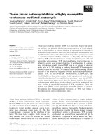
We cultured
purified FDCs with freshly isolated B lympho-
cytes and assessed cellular proliferation by [
3
H]-
thymidine incorporation following 120 hours
of culture. In the absence of FDCs, B lympho-
cytes alone did not incorporate [
3
H]-thymidine
significantly whereas FDCs alone did show some
baseline [
3
H]-thymidine incorporation (Figure
1, upper panel). Treatment with mitomycin C
(15 g/mL for 2 hours) did not significantly
decrease background thymidine incorporation
by FDCs. The combination of mixed tonsillar B
lymphocytes and FDCs caused morphologically
larger B-lymphocyte clusters (data not shown)
52 Allergy, Asthma, and Clinical Immunology / Volume 1, Number 2, Spring 2005
and significantly greater uptake of [
3
H]-
thymidine than either of the two cell types alone
(see Figure 1, upper panel). Addition of the PAF
antagonist WEB 2170 (10
Ϫ6
to 10
Ϫ8
M) to mixed
B lymphocytes consistently diminished cell pro-
liferation (see Figure 1, upper panel). Significant
inhibition was also achieved when we separated
tonsillar B lymphocytes into characterized frac-
tions by Percoll density centrifugation. The low-
density Percoll fraction contains cells that have
the highest PAFR expression, based on mRNA
and functional studies.
9
Culturing of the low-
density fraction and FDCs also led to significant
increases in [
3
H]-thymidine incorporation, which
was inhibited 35% ± 3% by WEB 2170
(see Figure 1, lower panel). However, the other
fractions resulting from Percoll density separa-
tion (medium and high density) were not
significantly affected by the addition of
WEB 2170 (data not shown). No toxicity resulted
from WEB 2170 administration, as was demon-
strated by direct observation of the cultures and
by trypan blue staining. Inhibition was not found
with WEB 2170 doses ≤ 10
Ϫ10
M (data not
shown).
Effect of PAF Antagonists on Production
of Immunoglobulin
Maintenance of B lymphocytes in culture by FDCs
leads to an increase in Ig production.
16
To assess
if PAF antagonists would decrease the ability of
FDCs to sustain Ig production, we added WEB
2170 to B lymphocytes, FDCs, or the combination
of B lymphocytes and FDCs at the initiation of cul-
ture, and supernatants were harvested after 7 days
of incubation. In keeping with the purity of our
FDCs, no IgG or IgM was detected from these cells
in culture alone (data not shown). B lymphocytes
alone made detectable amounts of Ig, but the com-
bination of FDCs and B lymphocytes produced
three to five times more IgM (91 ± 68 ng/mL vs
308 ± 175 ng/mL) and IgG (220 ± 72 ng/mL vs
875 ± 448 ng/mL). The addition of WEB 2170 at
doses of 10
Ϫ8
to 10
Ϫ6
M led to a significant inhi-
bition of Ig production, from 44 to 75% of the base-
line IgM or IgG production (Figure 2).
Detection of PAFR mRNA
from FDCs, by PCR
We have demonstrated that both B-lymphocyte cell
lines and freshly isolated tonsillar B lymphocytes
express high levels of PAFR mRNA
9
and that stim-
ulation of the PAFR leads to demonstrable intra-
cellular signalling and increased Ig production.
8,9
In
addition, PAF rescued B-lymphocyte cell lines from
apoptosis
7,17
and induced expression of IL-4 and
IL-13 mRNA in freshly isolated B lymphocytes.
9
Radiolabelled WEB 2170 was taken up on PAFR
expressed by B lymphocytes and B-lymphocyte
cell lines.
10
However, it is unknown whether FDCs
express PAFR and would thus be directly affected
by the PAF antagonist. RNAwas extracted from cul-
tured B lymphocytes and cultured FDCs that were
morphologically free of contaminating B lympho-
cytes, as detailed above. The results of the PCR
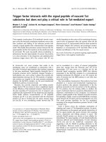
analysis are shown in Figure 3. Although PAFR
mRNAwas detectable from FDCs (see Figure 3, lane
3), only a small amount was present as compared
with that detected from B lymphocytes (see Figure
3, lane 4). This suggests that the predominant action
of WEB 2170 is on B-lymphocyte PAFR.
Discussion
The interaction between FDCs and B lympho-
cytes is a crucial one for the generation of high-
affinity antibody secreting memory B lympho-
cytes.
18–21
FDCs provide crucial elements for
B-lymphocyte survival in the GC of lymph nodes,
such as antigen and accessory molecules. As anti-
gen-presenting cells, FDCs trap IgG-coated anti-
gens, primarily in immune complexes, and present
them on their surfaces for long durations.
1
B lym-
phocytes that have a high affinity for the antigen
are selected, and these are allowed to further
develop in a process known as affinity maturation.
The surfaces of FDCs display other accessory
molecules, including complement receptors and
adhesion molecules such as VLA-4, that provide
stimulatory signals for B-cell proliferation and Ig
production. In addition, FDCs can rescue B lym-
phocytes from apoptosis, independently of adhe-
sion molecules or CD40 ligation.
22,23
We have shown that B lymphocytes from ton-
sils express receptors for the potent ether lipid
PAF. The highest level of PAFR mRNA expres-
sion was found in the low- and medium-density
layers from Percoll-fractionated subsets of B lym-
phocytes, and the addition of PAF to the low-den-
sity fraction engendered the highest increases in
intracellular calcium and in the production of Ig.
9
These two populations represent GC B lympho-
cytes as well as rapidly proliferating centroblasts
and centrocytes. Because FDCs are potentially of
hematopoietic-cell origin, related to monocytes or
macrophages, we hypothesized that they might be
sources of lipid mediators such as PAF.
24
Indeed,
early work on FDCs indicated a possible role for
prostaglandins in their function.
12
In these pre-
liminary studies, we tested whether the PAF antag-
onist WEB 2170 could inhibit FDC-mediated
stimulation of B lymphocytes.
FDCs support B-lymphocyte growth and the
synthesis of Ig without additional mitogens.
19
This
allowed direct observation of the role of the PAF
antagonist. The high-affinity water-soluble PAF
antagonist WEB 2170
25,26
clearly had an effect,
decreasing cell proliferation (as measured by [
3
H]-
thymidine incorporation) by 35% and the produc-
tion of IgM and IgG by the isolated tonsillar cells
by 45 to 75%. This was seen over doses ranging
from 10
Ϫ6
to 10
Ϫ8
M; doses exceeding this were
ineffective. High exogenous doses of a pharma-
cologic PAF antagonist are probably needed to
saturate the PAFR sites on B lymphocytes and to
overcome the large local production known to
occur where lipid mediators are synthesized. This
dose is comparable to the effective dose for inhibit-
ing PAF-mediated elevation of free cystolic calcium
(Ca
2+
) concentrations ([Ca
2+
]i) or in vitro Ig pro-
duction.
27,28
Although PAF antagonists had a con-
sistent effect in FDC B-lymphocyte cultures, it is
not surprising that they did not completely inhibit
[
3
H]-thymidine incorporation or Ig production.
First, the source of Ig secretion from cultured ton-
Platelet-Activating Factor Antagonists Decrease Stimulation of B Lymphocytes — Halickman et al 53
Figure 1 WEB 2170 diminished
the proliferation of B lymphocytes.
Tonsillar B lymphocytes were cul-
tured with isolated follicular den-
dritic cells for 120 hours with and
without WEB 2170 (10
Ϫ8
M). After
114 to 116 hours, [
3
H]-thymidine
(1.0 CU per well) was added, and
the cells were harvested 4 to
6 hours later. The histograms rep-
resent cell proliferation as
expressed by incorporation of [
3
H]-
thymidine. Upper panel shows
assessment of mixed tonsillar B
lymphocytes( n = 4); lower panel
shows assessment of low-density
tonsillar B lymphocytes The gray
and black bars represent two sep-
arate experiments performed in
triplicate (n = 2).
sillar cells includes a small number of plasma cells
that secrete immunoglobulins, as well as other
mature B cells that may die in culture and release
a basal level of IgG and IgM. Second, the inter-
action of FDCs and B lymphocytes is mediated by
numerous adhesion molecules, chemokines, and
other mediators
18,29
; these studies indicate that PAF
may be an important contributor to this process.
The effect of the PAF antagonist WEB 2170
is most likely directly on B lymphocytes and not
on the FDCs themselves. Whereas monocytes and
other dendritic cells such as those derived from
CD34
+
peripheral blood cells have been shown to
express high levels of PAFR mRNA,
30
FDCs
expressed little PAFR mRNA. Expression of PAFR
mRNA was low as compared to B lymphocytes
(see Figure 3), even when the mRNAwas ampli-
fied to plateau by RT-PCR.
9,31
The RT-PCR used
in this study has been validated
10
against a semi-
quantitatve RT-PCR and accurately reflects
changes in the level of PAF mRNA.
9
Although no
contaminating B lymphocytes were visible in our
cultures, it is possible that the trace amounts of
PAFR that were detected were due to cells other
than the FDCs themselves. However, to date, no
other populations (apart from FDCs) isolated from
tonsils propagate under the culture conditions
described.
In light of our recent observations regarding
PAFR expression on GC B lymphocytes,
9
the
action of the PAF antagonist in these studies sug-
gests that bioactive lipid mediators may play a role
in chemoattraction or GC cell organization, includ-
ing the interaction of FDCs and B lymphocytes.
The presence of the PAF antagonist did not dimin-
ish aggregation between B lymphocytes and FDCs
(data not shown). Further investigation into the link
54 Allergy, Asthma, and Clinical Immunology / Volume 1, Number 2, Spring 2005
Figure 2 WEB 2170 diminished the production of
immunoglobulin (Ig) in cultures of mixed follicular den-
dritic cells (FDCs) and B lymphocytes. Mixed tonsil-
lar B lymphocytes and FDCs were cultured together
with or without the indicated concentrations of WEB
2170 in 24 well plates for 7 days. On day 7, supernatants
were harvested and used for measuring total IgG or IgM.
The histograms indicate the percentage decrease in
IgG (A) and IgM (B) from cultures untreated by WEB
2170 (values are indicated in the text) (n = 5). B = B
lymphocytes; F = FDCs; W = WEB 2170. *p < .05.
Figure 3 Results of reverse transcription polymerase
chain reaction for the detection of platelet-activating fac-
tor receptor (PAFR) in follicular dendritic cells (FDCs)
and B lymphocytes. Total RNA was extracted from
purified FDCs or B lymphocytes, and PAFR messen-
ger RNA(mRNA) was detected with specific primers
(see “Methods”). The figure shows ethidium bro-
mide–stained gel that is representative of two identi-
cal experiments. Lane 1 shows DNA ladder; lane 2,
negative control (B lymphocytes without reverse tran-
scriptase); lane 3, FDCs; lane 4, human tonsillar B
lymphocytes; lane 5, LA350 B-lymphocyte cell line.
Both sources of B lymphocytes have a much stronger
mRNA signal than the FDCs have. Equal lane loading
was verified by using -actin as a housekeeping gene
(not shown).
between FDCs and B lymphocytes may answer
some heretofore unanswered questions. For exam-
ple, although FDCs rescue B lymphocytes from
apoptosis, the molecule that mediates this is still
unknown. This effect is independent of CD40 and
VLA-4 ligation,
22
and FDCs do not produce
cytokines, such as IL-4, that rescue B lymphocytes
from apoptosis.
32,33
A novel factor secreted by
FDCs, 8D6, is not involved in rescue from apop-
tosis and is not solely responsible for maintaining
the growth of B-lymphocyte lines cultured with
FDCs.
2
We have demonstrated that PAF rescues
the GC-like B-cell line Ramos from programmed
cell death induced by B-cell receptor (BCR) lig-
ation
7,17
; the role of PAF antagonists in the rescue
of apoptosis is being explored in our laboratory.
We have undertaken to determine the source
of PAF in these cultures. FDCs have features of
cells of myeloid-cell origin
34
although some inves-
tigators have suggested an endothelial or fibrob-
last origin for FDCs.
35,5
FDCs also have gap junc-
tions that are areas of molecular transport for
intercellular communication.
36
B lymphocytes are
also a source of bioactive ether lipids, including
palmitoyl-2-acetoyl-sn-glycero-3-phosphocholine
(PAGPC).
28
The presence of this PAF analogue,
which is a potent mitogen for Ig synthesis by B
lymphocytes, has to date made it difficult to purify
PAF in mixed FDC/B-lymphocyte cultures. The
rapidity of GC formation
19
predicts that rapidly pro-
duced or preformed chemoattractants, including
chemokines or lipid mediators, are needed for
direction of cellular traffic. Recent studies have
established that FDCs produce monocyte chemoat-
tractant protein-1
37
and B cell chemoattractant -1
(BCA).
38
Thus, a number of candidate molecules
are available for assisting the assembly of GCs.
Definition of the ability of FDCs to produce
chemoattractant lipid mediators such as PAF or
PAGPC is important for a full comprehension of
interactions that lead to GC development in lym-
phoid tissue.
References
1. Tew JG, Kosco MH, Burton GF, Szakal AK.
Follicular dendritic cells as accessory cells.
Immunol Rev 1990;117:185–211.
2. Li L, Zhang X, Kovacic S, et al. Identification
of a human follicular dendritic cell molecule that
stimulates germinal center B cell growth. J Exp
Med 2000;191:1077–84.
3. Krenacs T, Rosendaal M. Immunohistological
detection of gap junctions in human lymphoid
tissue: connexin43 in follicular dendritic and
lymphoendothelial cells. J Histochem Cytochem
1995;43:1125–37.
4. Kapasi ZF, Qin D, Kerr WG, et al. Follicular den-
dritic cell (FDC) precursors in primary lymphoid
tissues. J Immunol 1998;160:1078–84.
5. Imal Y, Yamakawa M. Morphology, function and
pathology of follicular dendritic cells. Pathol Int
1996;46:807–33.
6. Haley ST, Tew JG, Szakal AK. The monoclonal
antibody FDC-M1 recognizes possible follicu-
lar dendritic cell precursors in the blood and bone
marrow. Adv Exp Med Biol 1995;378:289–91.
7. Toledano BJ, Bastien Y, Noya F, et al. Platelet-
activating factor abrogates apoptosis induced by
cross-linking of the surface IgM receptor in a
human B lymphoblastoid cell line. J Immunol
1997;158:3705–15.
8. Mazer B, Clay KL, Renz H, Gelfand EW. Platelet-
activating factor enhances Ig production in B
lymphoblastoid cell lines. J Immunol
1990;145:2602–7.
9. Bastien Y, Toledano BJ, Mehio N, et al. Detection
of functional platelet-activating factor receptors
on human tonsillar B lymphocytes. J Immunol
1999;162:5498–505.
10. Zhuang Q, Bastien Y, Mazer BD. Activation via
multiple signaling pathways induces down-reg-
ulation of platelet-activating factor receptors on
human B lymphocytes. J Immunol 2000;165:
2423–31.
11. Butch AW, Kelly KA, Willbanks MS, Yu X.
Human follicular dendritic cells inhibit super-
antigen-induced T-cell proliferation by distinct
mechanisms. Blood 1999;94:216–24.
12. Heinen E, Cormann N, Braun M, et al. Isolation
of follicular dendritic cells from human tonsils
and adenoids. VI. Analysis of prostaglandin
secretion. Ann Inst Pasteur Immunol 1986;
137D:369–82.
13. Schwaeble W, Schafer MK, Petry F, et al.
Follicular dendritic cells, interdigitating cells,
and cells of the monocyte-macrophage lineage
are the C1q-producing sources in the spleen.
Identification of specific cell types by in situ
Platelet-Activating Factor Antagonists Decrease Stimulation of B Lymphocytes — Halickman et al 55
hybridization and immunohistochemical analy-
sis. J Immunol 1995;155:4971–8.
14. Hourton D, Stengel D, Chapman MJ, Ninio E.
Oxidized low density lipoproteins downregulate
LPS-induced platelet-activating factor receptor
expression in human monocyte-derived
macrophages: implications for LPS-induced
nuclear factor-kappaB binding activity. Eur J
Biochem 2001;268:4489–96.
15. Grouard G, de Bouteiller O, Banchereau J, Liu
YJ. Human follicular dendritic cells enhance
cytokine-dependent growth and differentiation
of CD40-activated B cells. J Immunol
1995;155:3345–52.
16. Clark EA, Grabstein KH, Gown AM, et al.
Activation of B lymphocyte maturation by a
human follicular dendritic cell line, FDC-1. J
Immunol 1995;155:545–55.
17. Toledano B, Bastein Y, Noya F, Mazer B.
Characterization of B lymphocytes rescued from
apoptosis by platelet-activating factor. Cell
Immunol 1999;191:60–8.
18. Tew JG, Wu J, Fakher M, et al. Follicular den-
dritic cells: beyond the necessity of T-cell help.
Trends Immunol 2001;22:361–7.
19. Qin D, Wu J, Vora KA, et al. Fc gamma recep-
tor IIB on follicular dendritic cells regulates the
B cell recall response. J Immunol 2000;164:
6268–75.
20. Wu J, Qin D, Burton GF, et al. Follicular den-
dritic cell-derived antigen and accessory activity
in initiation of memory IgG responses in vitro.
J Immunol 1996;157:3404–11.
21. Szakal AK, Tew JG. Follicular dendritic cells:
B-cell proliferation and maturation. Cancer Res
1992;52:5554s–6s.
22. Koopman G, Keehnen RM, Lindhout E, et al.
Germinal center B cells rescued from apoptosis
by CD40 ligation or attachment to follicular den-
dritic cells, but not by engagement of surface
immunoglobulin or adhesion receptors, become
resistant to CD95-induced apoptosis. Eur J
Immunol 1997;27:1–7.
23. Lindhout E, Lakeman A, de Groot C. Follicular
dendritic cells inhibit apoptosis in human B lym-
phocytes by a rapid and irreversible blockade of
preexisting endonuclease. J Exp Med 1995;
181:1985–95.
24. Bussolati B, Rollino C, Mariano F, et al. IL-10
stimulates production of platelet-activating factor
by monocytes of patients with active systemic
lupus erythematosus (SLE). Clin Exp Immunol
2000;122:471–6.
25. Fray MJ, Cooper K, Parry MJ, et al. Novel antag-
onists of platelet-activating factor. 1. Synthesis
and structure-activity relationships of benzodi-
azepine and benzazepine derivatives of
2-methyl-1-phenylimidazo[4,5-c]pyridine. J Med
Chem 1995;38:3514–23.
26. Casals-Stenzel J. Thieno-triazolo-1,4-diazepines
as antagonists of platelet-activating factor: pre-
sent status. Lipids 1991;26:1157–61.
27. Mazer B, Domenico J, Sawami H, Gelfand EW.
Platelet-activating factor induces an increase in
intracellular calcium and expression of regula-
tory genes in human B lymphoblastoid cells. J
Immunol 1991;146:1914–20.
28. Mazer BD, Toledano BJ, Saririan M, Bastien Y.
Dose dependent agonist and antagonist effects
of the platelet-activating factor analogue 1-palmi-
toyl-2-acetoyl-sn-glycero-3-phosphocholine on
B lymphocytes. J Allergy Clin Immunol 1998;
102:231–7.
29. Cyster JG, Ansel KM, Reif K, et al. Follicular
stromal cells and lymphocyte homing to folli-
cles. Immunol Rev 2000;176:181–93.
30. Sozzani S, Longoni D, Bonecchi R, et al. Human
monocyte-derived and CD34+ cell-derived den-
dritic cells express functional receptors for platelet
activating factor. FEBS Lett 1997;418:98–100.
31. Bastien Y, Mazer BD. Detection of the human
platelet-activating factor receptor mRNA in
human B lymphocytes by polymerase chain reac-
tion on reverse transcripts (RT-PCR) [published
erratum appears in Biochem Biophys Res
Commun 1994;204:436]. Biochem Biophys Res
Commun 1994;202:1373–9.
32. Skibinski G, Skibinska A, Deckers M, James K.
Tonsil stromal-cell lines expressing FDC-like
properties: isolation, characterization, and inter-
action with B lymphocytes. Dev Immunol
1998;6:273–84.
33. Toellner KM, Scheel-Toellner D, Sprenger R, et
al. The human germinal centre cells, follicular
dendritic cells and germinal centre T cells pro-
duce B cell-stimulating cytokines. Cytokine
1995;7:344–54.
34. Szakal AK, Kapasi ZF, Haley ST, Tew JG. A
theory of follicular dendritic cell origin. Curr Top
Microbiol Immunol 1995;201:1–13.
35. Muretto P. Immunohistochemical study of ton-
sils from newborn infants with emphasis on
56 Allergy, Asthma, and Clinical Immunology / Volume 1, Number 2, Spring 2005
Platelet-Activating Factor Antagonists Decrease Stimulation of B Lymphocytes — Halickman et al 57
follicular dendritic reticulum cells. Eur J
Histochem 1998;42:189–95.
36. Krenacs T, van Dartel M, Lindhout E, Rosendaal
M. Direct cell/cell communication in the lym-
phoid germinal centre: connexin43 gap junctions
functionally couple follicular dendritic cells to
each other and to B lymphocytes. Eur J Immunol
1997;27:1489–97.
37. Husson H, Carideo EG, Cardoso AA, et al. MCP-
1 modulates chemotaxis by follicular lymphoma
cells. Br J Haematol 2001;115:554–62.
38. Shi K, Hayashida K, Kaneko M, et al. Lymphoid
chemokine B cell-attracting chemokine-1
(CXCL13) is expressed in germinal center of
ectopic lymphoid follicles within the synovium
of chronic arthritis patients. J Immunol
2001;166:650–5.