Báo cáo y học: "A case of IgG4-related tubulointerstitial nephritis concurrent with Henoch-Schönlein purpura nephritis" pot
Bạn đang xem bản rút gọn của tài liệu. Xem và tải ngay bản đầy đủ của tài liệu tại đây (1.89 MB, 5 trang )
CAS E REP O R T Open Access
A case of IgG4-related tubulointerstitial nephritis
concurrent with Henoch-Schönlein purpura
nephritis
Rukako Tamai
1
, Yoshiyuki Hasegawa
1
, Satoshi Hisano
2
, Katsuhisa Miyake
3
, Hitoshi Nakashima
3*
and Takao Saito
3
Abstract
We describe a 72-year-old man, who had been suffered from Henoch-Schönlein purpura (HSP) several times,
presented with hematoproteinuria with granular cast, and general lymphadenopathy. The immunological
examination of the serum showed polyclonal hypergammagloburinemia with high value of IgG4. The renal biopsy
revealed interstitial inflammato ry cell infiltration, in cluding infiltration of lymphocytes and plasma cells, and
segmental glomerulonephritis. Direct immunofluorescence microscopy revealed apparent positive staining with
anti-human IgA, and anti-human IgG in glomeruli, anti-human IgG4 antibody staining showed many positive
plasma cells in the interstitium. The patient was diagnosed with HSP nephritis that was complicated by IgG4-
related nephropathy. As a result of the treatment with 30mg prednisolone, the swelling of the LNs decreased, but
the patient continued to have persistent hematoproteinuria.
Introduction
A novel clinicopathological entity of IgG4-related auto-
immune disease characterized by extensive IgG4-positive
plasma cell infiltration of organs t ogether with CD4- or
CD8-positive T lymphocytes is proposed [1]. Renal
involvement in th is entity was also suggested, and three
patterns of renal involvement have been described:
1) extraparenchymal involvement such as hydronephro-
sis associated with retroperitoneal lesions; 2) diffuse
tubulointerstitial nephritis (T IN); and 3) renal l esions
composed of focal lymphoplasmacytic infiltration of the
renal interstitium [2]. In this report we describe a rare
case diagnosed with HSP nephritis that was complicated
by IgG4-related nephropathy.
Case report
A 72-year-old man presented with cervical, axillary, left
subclavian, and inguinal lymph nodes (LNs) swelling.
The LNs gradually increased in size for 1 month. During
this period, the patient often had a low-grade fever and
arthralgia. He also experienced a marked weight loss of
7 kg in 3 months. In June 2009, he developed an erythe-
matous rash predominantly on his lower legs and was
admitted to the hospital. In 2005, he ha d developed
similar erythematous rashes in t he lower extremities
several times. In 2006, the patient was diagnosed with
Henoch-Schönlein purpura (HSP) on the basis of histo-
logical examination of skin biopsy samples, which
showed leukocytoclastic vasculitis. Immunohistochem-
ical study with anti-IgA antibody was not performed.
A treatment with prednisolone (PSL; 25 mg) had been
effective (Figure 1). He had no history of allergic dis-
eases such as bronchial asthma, atopic dermatitis, and
allergic rhinitis. In 2002, he underwent gastrectomy for
gastric cancer.
On admission, he was febrile, and the rash was palp-
able and purpuric in nature. A physical examination
showed no abnormalities in t he lungs, heart, abdomen,
and central nervous system. Laboratory findings showed
an increased erythrocyte sedimenta tion rate (73 mm/h)
and the value of C-reactive protein was 0.22 mg/dL.
The hemoglobi n concentration was 11.0 g/dL, the white
blood cell count was 8,900/mm
3
(neutrophils 66.8%,
lymphocytes 21.5%, monocytes 4.1%, eosinophils 7.0%,
and b asophils 0.6%), and the platelet count was 45.1 ×
10
4
/mm
3
. Hematuria and proteinuria with granular cast
were detected. The results of the serum chemistry
* Correspondence:
3
Division of Nephrology and Rheumatology, Department of Internal
Medicine, Faculty of Medicine, Fukuoka University, Nanakuma7-45-1, Johnan-
ku, Fukuoka city, 814-0180, Japan
Full list of author information is available at the end of the article
Tamai et al. Allergy, Asthma & Clinical Immunology 2011, 7:5
/>ALLERGY, ASTHMA & CLINICAL
IMMUNOLOGY
© 2011 Tamai et al; licensee BioMed Central Ltd. This is an Open Access article distributed under the terms of the Creative Commons
Attribution License ( which permits unrestricted use, distribution, and reproduction in
any medium, provided the original work is properly cited.
analyses are as follows: serum creatinine, 0.96 mg/dL
(normal, 0.4-1.2 mg/dL); blood urea nitrogen, 16.7 mg/
dL; total serum protein 8.6 mg/dL (normal, 6.5-8.2 g/
dL); and serum albu min 3.6 g/dL (normal, 3.7-5.2 g/dL).
Serum transaminase, amylase, and lactate dehydrogenase
(LDH) levels were within normal limits. The immunolo-
gical tests were positive for antinuclear antibody at a
titer of 80 dil, and the immunofluorescence patterns
were speckled and homogeneous. Anti-double-stranded
DNA antibody, rheumatoid factor, anti-Sjögren’ ssyn-
drome A (anti-SS-A), anti-SS-B antibodies, anti-Sm anti-
body, anti-Jo-1 antibody, and anti-RNP antibody were all
absent. The serum level of immunoglobulin G (IgG) was
abnormally high, but IgA and IgM were within normal
limits (4,359 mg/dL, 242 mg/dL, and 64 mg/dL, respec-
tively). The serum IgE level was elevated (537 U/mL).
Molecules of the subclass IgG4 accounted for 25%
(1,100 mg/dL) of the IgG molecules. Serum protein elec-
trophoresis revealed poly clonal hypergammaglobuline-
mia. Serum levels of C3, C4, and total complement
hemolytic activity (CH50) were 55 mg/dL (normal, 86-
160 mg/dL), 3 mg/dL (normal, 17-45 mg/dL), and less
than 12.0 U/mL (normal, 25-48 U/mL), respectively.
Myeloperoxidase antineutrophil-cytoplasmic antibody
(MPO-ANCA) was d etected at a titer 22 EU (normal,
<10EU), but proteinase-3 antineutrophil cytoplasmic
antibody was not detected. Serologic specimens also
tested negative for cytomegalovirus, herpes simplex
virus, Epstein-Barr virus, mycoplasma, hepatitis C virus
(HCV) antibody, and hepatitis B virus surface (HBs)
antigen. The tuberculin skin test was negative for the
purified protein der ivativ e. Although se veral small L Ns
swelling in the para-aortic and bilateral renal artery
branching area were detected in an abdominal CT scan,
any abnormal finding was not confirmed in FDG-PET.
Chest CT showed no finding such as interstitial pneu-
monia. Systemic lymphadenopathy, polyclonal hyper-
gammaglobulinemia associated with IgE and IgG4
elevation, hypocomplementemia, and renal dysfunction
reminded us of development of IgG4 related disease,
and echo-guided percutaneous kidney biopsy was per-
formed on the 7th hospital day. Four out of 28 glomer-
uli showed glob al sclerosis, and 2 glomeruli collapsed
with periglomerular fibrosis. The other glomeruli
showed mild or no mesangial proliferative change. The
biopsy revealed interstitial inflammatory cell infiltra-
tion, including infiltration of lymphocytes and plasma
cells, and concurrent segmental glomerulonephritis
2004 200
5
200
6
200
7
20092008 2010
IgG (mg/dl) 5440
Purplish-red spot
4359
2.0
4.0
6.0
8.0
10.0
12.0
TP
Alb
1425
PSL
25mg
30mg
(g/dl)
Year
-
U
rine
Protein
Blood
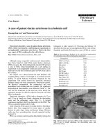
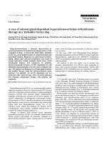
Figure 1 Clinical course of the patient. Purplish-red spot in the lower extremities as a picture had been developed 3 times in 6 years
(downward bald arrow). Hematoproteinuria has been detected since 2006. TP; serum total protein, Alb; serum albumin, PSL; prednisolone.
Tamai et al. Allergy, Asthma & Clinical Immunology 2011, 7:5
/>Page 2 of 5
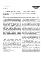
(Figure 2A, B and 2C). Direct immunofluorescence
microscopy revealed a pparent positive staining wi th
anti-human IgA (Figure 2D) and anti-human IgG anti-
bodies in the mesangium, Complement 3 deposition
was also recognized. Anti-human IgG4 antibody stain-
ing revealed many positive plasma cells in the intersti-
tium (Figure 2E). The ratio of IgG4-positive plasma
cells to IgG-positive plasma cells was more than 50%
(data not shown). Electron micrograph revealed
numerous electron-dense deposits in the mesangium.
Subepithelial electron-dense deposit in the capillary
wall was not detected (Figure 2F).
The patient was diagnosed with HSP nephritis that
was complicated by IgG4-related TIN. The patient was
treated with PSL (30 mg/day) for 14 days, followed by
tapering of PSL. As a result of the treatment, the swel-
ling of the LNs decrea sed, but t he patient continued to
have persistent hematoproteinuria.
Discussion
HSP has been re cognized as a distinct clinical condition.
The syndrome is also referred to as anaphylactoid pur-
pura and allergic purpura because of circumstantial
evidence implicating hypersensitivity to bacteria or
viruses as a possible cause. Histopathological examina-
tions revealed that the cutaneous le sions result from
leukocytoclastic vasculitis. Immunofluorescence studies
have revealed immunoglobulin (Ig) and complement
component deposits in the cutaneous blood vessels and
kidney, but serum complement levels are usually nor-
mal. IgA is the most abundant and sometimes the only
Ig found in the skin and kidney lesions. The morpholo-
gic and immunopathologic features are similar in HSP
nephritis and IgA nephropathy (IgAN), which i s charac-
terized by various degrees of focal or d iffuse mesangial
proliferation, diffuse deposition of IgA in the mesan-
gium, and electron-dense deposits in the mesangium [3].
It has became well known that the elevat ion of serum
IgG4 concentration and abundant IgG4-positive plasma
cell infiltration in the pancreas are characteristic find-
ings in auto immune pancreatitis (AIP) [4], and IgG4-
related TIN is also considered to belong to the same
disease spectrum. Accordingly, the concept of IgG4-
related systemic disease have not been established
[1,5-11], the patients with this diseases share many
common features; (1) elevated serum IgG4 level,
A
B
C
E
F
D
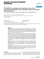
Figure 2 Representative images of the renal biopsy samples. A. Mild mesangial proliferation is observed. (PAS ×200) B.Obvious
inflammatory cell infiltration including lymphocytes, plasma cells and eosinophils are found in the interstitium. (PAS ×200) C. Massive fibrosis
and inflammatory cell infiltration are observed in the interstitium. (Masson Trichrome staining ×200) D. Immunofluorescence photomicrographs
showing IgA (×400). E. Immunostaining reveals abundant IgG4-positive plasma cells in the interstitium. F. Electron micrograph shows numerous
electron-dense deposits in the mesangium. ×3,000.
Tamai et al. Allergy, Asthma & Clinical Immunology 2011, 7:5
/>Page 3 of 5
(2) abundant IgG4-positive plasma cell infiltration in the
affected organs, and (3) marked improvement with cor-
ticosteroid therapy [5,7,9,10,12-16]. Our patient exhib-
ited these 3 features. Further, immunohistological
studies revealed IgG, IgA, and C3 deposition in the glo-
meruli resembling IgAN. Although IgA nephropathy
associated with MPO-ANCA positive glomerulonephritis
has also been reported previously [17,18], renal biopsy
of this case did not show any finding of necrotizing or
crescentic glomerulonephritis. This patient had symp-
toms of HSP systemic ally. Therefore we made a diagno-
sis of concomitant HSP nep hritis and IgG4-related TIN.
Recently, it has been reported that several IgG4-related
TIN complicated with glomerular disease [9,19]. How-
ever, this may be the first case of IgG4-related TIN with
HSP nephritis.
Allergy research has elucidated the relationship among
IgG antibodies, allergens, and the IgG4 subclass in
patients undergoing allergen-specific immunotherapy
[20],[21],andithasbeenshownthatextendedand
high-dose exposure to occupational or injected allerge ns
can induce an increase in IgG and IgG4 antibodies and
a d ecrease in IgE antibodies [22-2 4]. IgG4 is produced
in response to repeated exposure to environmental anti-
gens [25], [26]. Our patient had been experiencing
relapsing HSP for 4 years, and this episode might indi-
cate that he may have been repeatedly exposed to the
allergen (Figure 1). Although the nature of the allergen
that triggers HSP i s unknown, the facts that amounts
of IgG were e xtremely high at the point of purpura
development indicate protection with the production
of IgG4 might be induced by repetitive allergen expo-
sures, and this hard prote ction may related with the
development of IgG4 related TIN. Nevertheless, HSP
developed 3 times, and therefore HSP nephritis might
be complicated.
We described a rare case of HSP co mplicated by con-
current IgG4-related TIN. A biopsy of the collected spe-
cimens revealed IgG, IgA, and C3 deposition in the
glomeruli and IgG4-producing plasma cell infiltration in
the interstitium. We speculate that HSP resulting from
repeated allergen exposure might induce the develop-
ment of IgG4-related TIN and also HSP nephritis.
Consent
Written informed consent was obtained from the patient
for publication of this case report and accompanying
images. A copy of the written consent is available for
review by the Editor-in-Chief of this journal.
Acknowledgements
This work was suppor ted in part by grants from the Ministry of Education,
Science, Technology, Sports and Culture of Japan (HN, KM, and TS) and also
supported in part by a grant for the Progressive Renal Diseases Research
Projects from the Ministry of Health, Labor and Welfare, Japan (TS).
Author details
1
Department of Internal Medicine, Saiseikai Futsukaichi Hospital, Yumachi 3-
13-1, Chikushino city, 818-8516, Japan.
2
Department of Pathology, Faculty of
Medicine, Fukuoka Universi ty, Nanakuma7-45-1, Johnan-ku, Fukuoka city,
814-0180, Japan.
3
Division of Nephrology and Rheumatology, Department of
Internal Medicine, Faculty of Medicine, Fukuoka University, Nanakuma7-45-1,
Johnan-ku, Fukuoka city, 814-0180, Japan.
Authors’ contributions
RT and YH provided clinical care, HN conceived the report, and SH
performed all the immunochemistry. KM and TS participated in the design
of this report. All authors have read and approved the final manuscript.
Competing Interests
The authors declare that they have no competing interests.
Received: 14 January 2011 Accepted: 31 March 2011
Published: 31 March 2011
References
1. Kamisawa T, Funata N, Hayashi Y, Eishi Y, Koike M, Tsuruta K, Okamoto A,
Egawa N, Nakajima H: A new clinicopathological entity of IgG4-related
autoimmune disease. J Gastroenterol 2003, 38:982-4.
2. Saeki T, Nishi S, Ito T, Yamazaki H, Miyamura S, Emura I, Imai N, Ueno M,
Saito A, Gejyo F: Renal lesions in IgG4-related systemic disease. Intern
Med 2007, 46:1365-71.
3. Appel GBRJ, D’Agati V: Secondary glomerular disease. Brenner and Rector’s
The Kidney , 8 2006, 1094-7.
4. Hamano H, Kawa S, Horiuchi A, Unno H, Furuya N, Akamatsu T,
Fukushima M, Nikaido T, Nakayama K, Usuda N, Kiyosawa K: High serum
IgG4 concentrations in patients with sclerosing pancreatitis. N Engl J Med
2001, 344:732-8.
5. Kamisawa T: IgG4-related sclerosing disease. Intern Med 2006, 45:125-6.
6. Hamano H, Kawa S: Are there any other organs in which autoimmune
pancreatitis-associated lesions remain to be identified? Intern Med 2006,
45:883-4.
7. Kitagawa S, Zen Y, Harada K, Sasaki M, Sato Y, Minato H, Watanabe K,
Kurumaya H, Katayanagi K, Masuda S, Niwa H, Tsuneyama K, Saito K,
Haratake J, Takagawa K, Nakanuma Y: Abundant IgG4-positive plasma cell
infiltration characterizes chronic sclerosing sialadenitis (Kuttner’s tumor).
Am J Surg Pathol 2005, 29:783-91.
8. Yamamoto M, Harada S, Ohara M, Suzuki C, Naishiro Y, Yamamoto H,
Takahashi H, Imai K: Clinical and pathological differences between
Mikulicz’s disease and Sjögren’s syndrome. Rheumatology (Oxford) 2005,
44:227-34.
9. Saeki T, Imai N, Ito T, Yamazaki H, Nishi S: Membranous nephropathy
associated with IgG4-related systemic disease and without autoimmune
pancreatitis. Clin Nephrol 2009, 71:173-8.
10. Watson SJ, Jenkins DA, Bellamy CO: Nephropathy in IgG4-related systemic
disease. Am J Surg Pathol 2006, 30:1472-7.
11. Deshpande V, Chicano S, Finkelberg D, Selig MK, Mino-Kenudson M,
Brugge WR, Colvin RB, Lauwers GY: Autoimmune pancreatitis: A systemic
immune complex mediated disease. Am J Surg Pathol 2006, 30:1537-45.
12. Chari ST, Smyrk TC, Levy MJ, Topazian MD, Takahashi N, Zhang L, Clain JE,
Pearson RK, Petersen BT, Vege SS, Farnell MB: Diagnosis of autoimmune
pancreatitis: The Mayo clinic experience. Clin Gastroenterol Hepatol 2006,
4:1010-6, quiz 934.
13. Saeki T, Saito A, Hiura T, Yamazaki H, Emura I, Ueno M, Miyamura S, Gejyo F:
Lymphoplasmacytic infiltration of multiple organs with
immunoreactivity for IgG4: IgG4-related systemic disease. Intern
Med
2006, 45:163-7.
14. Yamamoto M, Takahashi H, Ohara M, Suzuki C, Naishiro Y, Yamamoto H,
Shinomura Y, Imai K: A new conceptualization for Mikulicz’s disease as an
IgG4-related plasmacytic disease. Mod Rheumatol 2006, 16:335-40.
15. Muraki T, Hamano H, Ochi Y, Komatsu K, Komiyama Y, Arakura N,
Yoshizawa K, Ota M, Kawa S, Kiyosawa K: Autoimmune pancreatitis and
complement activation system. Pancreas 2006, 32:16-21.
Tamai et al. Allergy, Asthma & Clinical Immunology 2011, 7:5
/>Page 4 of 5
16. Okazaki K, Chiba T: Autoimmune related pancreatitis. Gut 2002, 51:1-4.
17. Haas M, Jafri J, Bartosh SM, Karp SL, Adler SG, Meehan SM: ANCA-
associated crescentic glomerulonephritis with mesangial IgA deposits.
Am J Kidney Dis 2000, 36:709-18.
18. Allmaras E, Nowack R, Andrassy K, Waldherr R, van der Woude F, Ritz E:
Rapidly progressive IgA nephropathy with anti-myeloperoxidase
antibodies benefits from immunosuppression. Clin Nephrol 1997,
48:269-73.
19. Morimoto J, Hasegawa Y, Fukushima H, Uesugi N, Hisano S, Saito T,
Kaneoka H: Membranoproliferative glomerulonephritis-like glomerular
disease and concurrent tubulointerstitial nephritis complicating IgG4-
related autoimmune pancreatitis. Intern Med 2009, 48:157-62.
20. Devey ME, Wilson DV, Wheeler AW: The IgG subclasses of antibodies to
grass pollen allergens produced in hay fever patients during
hyposensitization. Clin Allergy 1976, 6:227-36.
21. van der Giessen M, Homan WL, van Kernbeek G, Aalberse RC, Dieges PH:
Subclass typing of IgG antibodies formed by grass pollen-allergic
patients during immunotherapy. Int Arch Allergy Appl Immunol 1976,
50:625-40.
22. Aalberse RC, van der Gaag R, van Leeuwen J: Serologic aspects of IgG4
antibodies. I. Prolonged immunization results in an IgG4-restricted
response. J Immunol 1983, 130:722-6.
23. Rowntree S, Platts-Mills TA, Cogswell JJ, Mitchell EB: A subclass IgG4-
specific antigen-binding radioimmunoassay (RIA): Comparison between
IgG and IgG4 antibodies to food and inhaled antigens in adult atopic
dermatitis after desensitization treatment and during development of
antibody responses in children. J Allergy Clin Immunol 1987, 80:622-30.
24. Platts-Mills T, Vaughan J, Squillace S, Woodfolk J, Sporik R: Sensitisation,
asthma, and a modified Th2 response in children exposed to cat
allergen: A population-based cross-sectional study. Lancet 2001,
357:752-6.
25. Heiner DC: Significance of immunoglobulin G subclasses. Am J Med 1984,
76:1-6.
26. van der Zee JS, van Swieten P, Aalberse RC: Inhibition of complement
activation by IgG4 antibodies. Clin Exp Immunol 1986, 64:415-22.
doi:10.1186/1710-1492-7-5
Cite this article as: Tamai et al.: A case of IgG4-related tubulointerstitial
nephritis concurrent with Henoch-Schönlein purpura nephritis. Allergy,
Asthma & Clinical Immunology 2011 7:5.
Submit your next manuscript to BioMed Central
and take full advantage of:
• Convenient online submission
• Thorough peer review
• No space constraints or color figure charges
• Immediate publication on acceptance
• Inclusion in PubMed, CAS, Scopus and Google Scholar
• Research which is freely available for redistribution
Submit your manuscript at
www.biomedcentral.com/submit
Tamai et al. Allergy, Asthma & Clinical Immunology 2011, 7:5
/>Page 5 of 5