Báo cáo y học: "Adult mesenchymal stem cells and cell-based tissue engineering" doc
Bạn đang xem bản rút gọn của tài liệu. Xem và tải ngay bản đầy đủ của tài liệu tại đây (1.19 MB, 14 trang )
32
APC = adenomatous polyposis coli; bHLH = basic helix–loop–helix; BMP = bone morphogenetic protein; Cbfa1 = core binding factor alpha 1;
ECM = extracellular matrix; ERK = extracellular signal-regulated kinase; ES = embryonic stem; GDF = growth/differentiation factor; IBMX =
3-isobutyl-1-methylxanthine; IL = interleukin; JNK = c-Jun N-terminal kinase; LRP = low-density lipoprotein receptor-related peptide; MAPK =
mitogen-activated protein kinase; MSC = mesenchymal stem cell; PLA = poly-L-lactide; PLGA = poly-L-lactide-co-glycolide; PPAR-γ = peroxisome
proliferator-activated receptor-γ; SMAD = vertebrate homologue of Drosophila Mothers Against Decapentaplegic (MAD); TGF-β = transforming
growth factor beta; WISP = Wnt-1-inducible protein.
Arthritis Research and Therapy Vol 5 No 1 Tuan et al.
Introduction
Despite the pluripotency of embryonic stem (ES) cells,
legal and moral controversies concerning their use for
therapeutic and clinical application have prompted active
examination of the reservoirs of progenitor cells harbored
within the adult organism. In principle, such unspecialized
cells are considered to be quiescent, but capable of self-
renewing; their asymmetric division produces one identical
daughter stem cell and a second progenitor cell that
becomes committed to a lineage-specific differentiation
program [1]. These cells remain in their ‘undifferentiated’
state from suppression by some intrinsic or extrinsic
factor, until stimulated. Such adult stem cells have been
discovered and characterized in a multitude of tissues,
suggesting the potential for therapeutic application in their
host tissue [2–4]. As these cells are capable of differentia-
tion along specific lineages and of being recruited to
tissues in need, the promise for autologous clinical implan-
tation or genetically engineered stem cells for protein or
drug delivery without the risk of immunorejection looms on
the horizon. However, the success of future clinical appli-
cations depends critically upon a thorough understanding
of the biology of these cells, and new findings are continu-
ously being reported. For example, there is recent evi-
dence suggesting that the pluripotent stem cell, once
thought to be restricted to the fates of a lineage hierarchy,
is capable of transdifferentiation. Some recent examples
include the observations that the hematopoietic stem cells
of bone marrow have been shown to become hepatic oval
cells [5–7]; that muscle satellite cells exhibit hematopoi-
etic potential [8]; that neural stem cells have been shown
to produce lineage-committed hematopoietic progenitors
[9]; and that mesenchymal stem cells from bone marrow
have traveled to skeletal muscle [10], differentiated into
neuronal tissue [11,12], supplied mesangial cells during
repair processes [13], and given rise to cardiomyocytes
in vitro [14,15]. These observations strongly imply a criti-
cal influence of microenvironmental cues on cell fate.
Sources of mesenchymal stem cells
This review focuses on the adult mesenchymal stem cell
(MSC), which has the potential to differentiate into
The identification of multipotential mesenchymal stem cells (MSCs) derived from adult human tissues,
including bone marrow stroma and a number of connective tissues, has provided exciting prospects for
cell-based tissue engineering and regeneration. This review focuses on the biology of MSCs, including
their differentiation potentials in vitro and in vivo, and the application of MSCs in tissue engineering.
Our current understanding of MSCs lags behind that of other stem cell types, such as hematopoietic
stem cells. Future research should aim to define the cellular and molecular fingerprints of MSCs and
elucidate their endogenous role(s) in normal and abnormal tissue functions.
Keywords: cell differentiation, cell signaling, mesenchymal stem cells, stem cells, tissue engineering
Review
Adult mesenchymal stem cells and cell-based tissue engineering
Rocky S Tuan, Genevieve Boland and Richard Tuli
Cartilage Biology and Orthopaedics Branch, National Institute of Arthritis, and Musculoskeletal and Skin Diseases, National Institutes of Health,
Bethesda, Maryland, USA
Corresponding author: Rocky S Tuan (e-mail: )
Received: 7 October 2002 Accepted: 1 November 2002 Published: 11 December 2002
Arthritis Res Ther 2003, 5:32-45 (DOI 10.1186/ar614)
© 2003 BioMed Central Ltd (Print ISSN 1478-6354; Online ISSN 1478-6362)
Abstract
33
Available online />chondrocytes, osteoblasts, adipocytes, fibroblasts,
marrow stroma, and other tissues of mesenchymal origin.
Interestingly, these MSCs reside in a diverse host of
tissues throughout the adult organism and possess the
ability to ‘regenerate’ cell types specific for these tissues
(Table 1). Examples of these tissues include adipose
tissue [16], periosteum [17,18], synovial membrane [19],
muscle [20], dermis [21], pericytes [22–24], blood [25],
bone marrow [26], and most recently trabecular bone
[27,28]. Currently, bone marrow aspirate is considered to
be the most accessible and enriched source of MSCs,
although trabecular bone may also be considered an alter-
native source, in view of recent efficient isolation of multi-
potential cells from this tissue [29]. Given the wide
distribution of the sources of MSCs, the bone marrow
stroma may be considered to be the source of a common
pool of multipotent cells that gain access to various
tissues via the circulation, subsequently adopting charac-
teristics that meet the requirements of maintenance and
repair of a specific tissue type. In fact, the presence of
MSCs in tissues other than the marrow stroma strongly
suggests the existence of cell populations with more
limited capacity for differentiation; specifically, monopo-
tent or bipotent cells may have differentiation potentials
developmentally adapted to (and perhaps restricted to)
the tissues in which they reside.
Bone marrow contains three main cell types: endothelial
cells, hematopoietic stem cells, and stromal cells. In a
ground-breaking study, Friedenstein et al. [30] isolated
cells, clonogenic fibroblast precursor cells (CFU-F), from
whole bone marrow and showed that they were capable
of forming bone- and cartilage-like colonies. Long-term
bone marrow cultures also revealed the presence of
adherent stromal cells that supported and maintained the
hematopoietic component as a feeder layer [31]. After the
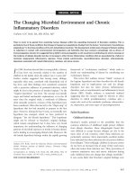
Table 1
Sources of multipotential adult mesenchymal stem cells
Source tissue Multilineage differentiation potential Representative references
Bone marrow Adipocyte [26,143]
Astrocyte, neuron [12,137]
Cardiomyocyte [14,15, 131–133]
Chondrocyte [26,46,76,79]
Hepatocyte [6,7,144]
Mesangial cell [13]
Muscle [10,35]
Neuron [11,12]
Osteoblast [26,33,43,145–147]
Stromal cell [148]
Various embryonic tissue lineages [139]
Muscle Adipocyte, myotubes, osteocyte [138]
Endothelial cell, neuron [20,149]
Chondrocyte [112]
Osteocyte [20]
Trabecular bone Adipocyte, chondrocyte, osteoblast [27-29]
Dermis Adipocyte, chondrocyte, muscle, osteoblast [21]
Adipose tissue Chondrocyte, muscle, osteoblast [16]
Stromal cell [150]
Periosteum Chondrocyte, osteoblast [17,18]
Pericyte Chondrocyte [22]
Osteoblast [23,24]
Blood Adipocyte, fibroblast, osteoblast, osteoclast [25]
Synovial membrane Adipocyte, chondrocyte, muscle, osteoblast [18]
34
Arthritis Research and Therapy Vol 5 No 1 Tuan et al.
endothelial cells, monocytes, and lymphocytes were
removed using negative immunoselection, these long-term
cultures revealed stromal cells that coexpressed pheno-
typic characteristics of the osteoblastic and adipocytic lin-
eages, thereby indicating their progenitor status [32].
Many subsequent studies have substantiated the multipo-
tent mesenchymal progenitor nature of cells isolated
according to Friedenstein’s method [e.g. 33–35]. These
studies have prompted interest not only in the differentia-
tion potential of MSCs, but also in the mechanisms gov-
erning their lineage-specific differentiation, particularly to
bone and cartilage. For example, Pittenger et al. [26]
showed that cells isolated from human marrow aspirates
were capable of remaining in a stable undifferentiated
state when cultured long-term in vitro, and that colonies
derived from single isolated cells could be induced to dif-
ferentiate along osteogenic, adipogenic, and chondro-
genic lineages when provided the appropriate cues.
Concurrent with such discoveries, varying methods of iso-
lation or preparation of more homogeneous, potentially
clonally derived MSC populations have emerged.
Kuznetsov et al. [36] found the stromal-cell population to
be capable of forming colonies in response to the follow-
ing growth factors: platelet-derived growth factor, trans-
forming growth factor beta (TGF-β), basic fibroblast
growth factor, and epidermal growth factor when cultured
in serum-containing medium. More recently, Digirolamo
et al. [37] have shown that cells with the highest colony-
forming efficiency exhibited the greatest replicative poten-
tial, and also readily differentiated into osteoblasts and
adipocytes. As such, techniques for the isolation and
in vitro culture expansion of bone-marrow-derived MSCs
range from aspiration and density-gradient centrifugation
to simple, direct plating methods to size sieving [38,39].
Although preliminary studies suggest that cells isolated
using different methodologies are, in fact, the same and
appear to retain similar potentials for differentiation, there
is as yet no clear-cut definition of the human MSC, in view
of the multitude of methods and procedures used in their
isolation and characterization.
Characteristics of mesenchymal stem cells
MSCs are described as multipotent because of their ability,
even as clonally isolated cells, to exhibit the potential for
differentiation into a variety of different cells/tissue lineages
(Fig. 1). However, in most studies, it remains to be deter-
mined whether true stem cells are present, or whether the
population is instead a diverse mixture of lineage-specific
progenitors. Inconsistency in published reports of the
growth characteristics and differentiation potential of
MSCs underscores the need for a functional definition of
these cells. At present, there is lack of a unifying definition
as well as information on specific markers that define the
cell types characterized as MSCs, with the sole definition
being their ability to differentiate along specific mesenchy-
mal lineages when induced to do so, to remain in a quies-
cent undifferentiated state until provided the signal to
divide asymmetrically, and finally, to undergo many more
replicative cycles than normal, fully differentiated cells.
Some groups have used the term ‘marrow stromal cell’
interchangeably with ‘mesenchymal stem cell’ [40]. While
these two types of cell are likely to have a common ances-
tor, the stromal characteristic can be thought of as a com-
mitted lineage with limited potential for differentiation.
Studies examining and comparing the morphology, pheno-
type, and in vitro function of MSCs and marrow-derived
stromal cells have shown the MSCs to be more homoge-
neous and fibroblastoid, while the marrow-derived stromal
cells were less homogeneous, with both fibroblastic and
hematopoietic characteristics present to varying degrees.
Although both cell types were able to support
hematopoiesis, the undifferentiated MSCs were not as
efficient, and while the cells displayed similar mRNA and
cytokine profiles, their individual responses to IL-1 treat-
ment differed [41]. Therefore, we propose that the stromal
compartment of the bone marrow itself contains MSCs
and that the stromal cell is actually an early differentiated
progeny of the MSC.
Despite improvements in long-term culture expansion,
MSCs display finite life spans, uncharacteristic of immortal-
ized ‘stem’ cells. Although MSCs are present throughout
life, their total number is inversely correlated to the age of
the patient and depends upon the site of extraction and the
systemic disease state [42]. Bruder et al. [43] character-
ized the long-term growth kinetics and osteogenic differen-
tiation potential of MSCs aspirated from bone marrow of
the iliac crest; the cells averaged 38 ± 4 population dou-
blings following extensive subcultivation and cryopreserva-
tion before they reached senescence. Retroviral
transduction of human MSCs with the human telomerase
gene has successfully extended the life span to more than
260 population doublings, while allowing the cells to
remain stably undifferentiated with full multilineage differen-
tiation potential [44,45]. For the purpose of further eluci-
dating the mechanisms regulating the lineage-specific
differentiation pathways of MSCs, immortalized clonal sub-
lines have been established using the human papilloma
virus E6/E7 genes with and without transduced telomerase
reverse transcriptase [28,46,47]. Okamoto et al. have
shown the immortalized parental population to be com-
posed of a heterogenous combination of uni-, bi-, and tri-
potential progenitor cells [47]. These findings again point
to the intrinsic heterogeneity as well as the need for thor-
ough characterization of the MSC population.
At present, the characterization of human MSCs lags sig-
nificantly behind that of bone marrow hematopoietic cells.
MSCs isolated directly from bone marrow are positive for
CD34, the hallmark antigen for positive immunoselection
of the hematopoietic stem cell, but lose this antigen upon
35
in vitro culture. While these results suggest a common
precursor for these two cell populations, they can be dis-
tinguished based upon the CD50 surface antigen, which
is common only to the hematopoietic stem cell [48]. Isola-
tion and enrichment of the MSC population has been
greatly facilitated by the Stro-1 monoclonal antibody [49].
Stro-1 immunoselection of cells derived from human bone
marrow revealed all fibroblast-colony-forming units to be
positive for Stro-1 but to lack the CD34 antigen, indicating
CD34 to be a nonspecific marker of human MSCs [50].
The Stro-1-positive population of bone-marrow-derived
cells has been shown to be capable of differentiating into
multiple mesenchymal lineages, including hematopoiesis-
supportive stromal cells with a vascular-smooth-muscle-
like phenotype, adipocytes, osteoblasts, and chondro-
cytes [51]. In addition, the cell-surface antigen activated
leukocyte-cell adhesion molecule, which reacts with the
monoclonal antibody SB-10, has been shown to be
expressed in undifferentiated cells but is lost during mes-
enchymal differentiation. This surface antigen has been
suggested to act as a cell adhesion molecule involved in
osteogenesis during bone morphogenesis [52]. The pres-
ence of specific, distinct antigens that are identified by the
monoclonal antibodies SH2, SH3, and SH4 on the cell
surface of marrow-derived MSCs and that are absent from
osteocytes and osteoblasts suggests that these recog-
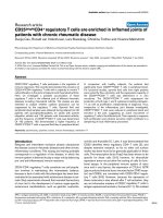
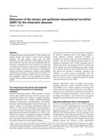
Available online />Figure 1
Lineage potential of adult human MSCs. MSCs are characterized by their multilineage differentiation potentials, including bone, cartilage, adipose
tissue, muscle, tendon, and stroma. This figure depicts some of the in vitro culture conditions (boxed) that promote the respective differentation
process into a specific lineage. Signaling pathways and/or components or events shown to be involved in lineage-specific differentiation are in
italics. See text for details. Dotted arrowheads denote potential ‘reverse’ differentiation events. bFGF, basic fibroblast growth factor; bHLH, basic
helix–loop–helix; BMP, bone morphogenetic protein; Cbfa1, core binding factor alpha 1; ECM, extracellular matrix; FGF, fibroblast growth factor;
GDF, growth/differentiation factor; IBMX, 3-isobutyl-1-methylxanthine; LRP, low-density lipoprotein receptor-related peptide; MAPK, mitogen-
activated protein kinase; PDGF, platelet-derived growth factor; SMAD, vertebrate homologue of Drosophila Mothers Against Decapentaplegic
(MAD); TGF-β, transforming growth factor beta; WISP, Wnt-1-inducible protein.
36
nized epitopes are developmentally regulated [53]. More
recently, the antigen binding the SH2 antibody was identi-
fied as endoglin (CD105), the receptor for TGF-β3, which
potentially plays a role in mediating the chondrogenic dif-
ferentiation of MSCs as well as their interactions with
hematopoietic cells [54]. The SH3 and SH4 antibodies
have been shown to react with CD73 (ecto-5′-nucleoti-
dase), which plays a role in the activation of B lympho-
cytes in lymphoid tissue but whose role has yet to be
elucidated in human MSCs [55]. As progress in phenotyp-
ing the MSC and its progeny continues, the use of selec-
tive markers has resulted in the enhanced propagation
and enrichment of the MSC population, while maintaining
them in an undifferentiated state without diminishing the
differentiation potential. Walsh et al. [56] found that
fibroblast growth factor-2 increases the proliferative
potential of human-bone-marrow-derived MSCs ex vivo.
This increase in colony size and overall cell number in
response to treatment with fibroblast growth factor-2 was
accompanied by an increase in the expression of Stro-1
and in the abundance of alkaline phosphatase-positive
cells, suggesting that osteoblast progenitor cells are pref-
erentially targeted by the growth factor [56]. Other
studies, however, have shown the differential potential of
human MSCs to be unaffected by fibroblast growth
factor-2 treatment, notwithstanding the proliferative effects
[57]. The phenotypic characterization of MSCs from
human bone marrow has been further realized through the
identification of the cytokine expression profile of undiffer-
entiated cells. Constitutive expression of cytokines, such
as granulocyte-colony stimulating factor, stem cell factor,
leukemia inhibitory factor, macrophage-colony stimulating
factor, and IL-6 and IL-11 is consistent with the ability of
MSCs to support hematopoiesis and provide factors that
regulate the marrow milieu itself [58].
Applications of mesenchymal stem cells in
tissue engineering and regenerative medicine
Bone
The challenges of engineering a tissue with numerous cell
types, each expressing individual differentiation patterns,
are significant for bone. The regeneration of bone is a key
issue at the forefront of current tissue engineering applica-
tions, owing to the ease of use and accessibility of osteo-
progenitor cells. The molecular mechanisms of human
MSC regulation and the importance of specific growth
factors during the different stages of osteogenic differenti-
ation are subjects of intensive investigation.
Molecular regulation of osteogenic differentiation
The induction of MSC osteogenesis is a highly pro-
grammed process, best illustrated in vitro. Treatment with
the synthetic glucocorticoid dexamethasone stimulates
MSC proliferation and supports osteogenic lineage differ-
entiation [59,60]. Organic phosphates, such as β-glyc-
erophosphate, also support osteogenesis by playing a role
in the mineralization and modulation of osteoblast activi-
ties [61,62]. Free phosphates can induce the mRNA and
protein expression of osteogenic markers such as osteo-
pontin, and these phosphates have known effects on the
production and nuclear export of a key osteogenesis regu-
latory gene, Cbfa1 (core binding factor alpha 1) [63–65].
Other supplements, such as ascorbic acid phosphate and
1,25-dihydroxyvitamin D
3
, are commonly used for
osteogenic induction, with the latter involved in increasing
alkaline phosphatase activity in osteogenic cultures and
promoting the production of osteocalcin [66]. In addition
to established supplements, members of the bone mor-
phogenetic protein (BMP) family of growth factors are also
routinely used for osteoinduction. BMP-2 alone appears to
increase bone nodule formation and the calcium content
of osteogenic cultures in vitro, while concomitant applica-
tion of BMP-2 and basic fibroblast growth factor increases
MSC osteogenesis both in vivo and in vitro [67].
A number of signaling pathways have been shown to par-
ticipate in MSC osteogenesis. The secreted signaling pro-
teins known as Wnts have been implicated in various
differentiation programs, including osteogenesis. An
established Wnt coreceptor, the low-density lipoprotein
receptor-related peptide 5 (LRP-5) has been linked to
osteoporosis–pseudoglioma syndrome in humans [68].
Patients with this syndrome have very low bone mass, are
prone to fracture and bone deformation, and have an
overall decrease in trabecular bone volume. Our laboratory
has shown that trabecular bone harbors a population of
MSCs [27], which may be the affected cell population in
this disease, thereby leading to alterations in bone forma-
tion and remodeling. In mice, LRP-5 mediates Wnt signal-
ing via the canonical pathway (i.e. through intracellular
β-catenin) [69,70]. In these in vitro mouse cultures, the
application of Wnt-3a can induce the activity of alkaline
phosphatase without altering the levels of Cbfa1. It has
also been shown that mice with targeted disruptions of
LRP-5 expression have a decreased level of osteoblast
proliferation and display a phenotype similar to humans
with osteoporosis–pseudoglioma syndrome [69].
Interestingly, the misexpression of telomerase was
recently found not only to extend the life of MSCs in vitro,
but also to increase their osteogenic differentiation poten-
tial [44,45].
Bone tissue engineering
The use of natural and synthetic biomaterials as carriers
for MSC delivery has shown increasing promise for
orthopaedic therapeutic applications, especially bone for-
mation. Recent advances in the field of biomaterials have
led to a transition from nonporous, biologically inert materi-
als to more porous, osteoconductive biomaterials, and, in
particular, the use of cell-matrix composites [71]. The
parameters that need to be considered in the selection of
Arthritis Research and Therapy Vol 5 No 1 Tuan et al.
37
a suitable delivery vehicle include physicochemical proper-
ties, such as surface area, porosity, local acidification,
material chemistry, dimensional architecture, mechanical
integrity, degradation characteristics, natural versus syn-
thetic, and potential for drug delivery; and biological prop-
erties, such as the ability of the scaffold to support cellular
attachment, proliferation, differentiation, matrix deposition,
angiogenesis, prevention of dedifferentiation, and enrich-
ment with a suitable quantity of cells. A number of delivery
vehicles have been successfully used in cell-matrix com-
posites in vivo, such as porous ceramics of hydroxyapatite
and β-tricalcium phosphate loaded with autologous MSCs
[72]. These constructs were capable of healing critical-
sized segmental bone defects not capable of being healed
by resident cells or by the addition of the osteoconductive
device alone. A recent in vitro study comparing the
biodegradable polymers poly-
L-lactide (PLA) and poly-L-
lactide-co-glycolide (PLGA) on the basis of adherence
and proliferation of seeded trabecular-bone-derived osteo-
progenitor cells showed that PLGA was the better sub-
strate for the attachment and subsequent osteogenic
differentiation of these progenitor cells [73].
Cartilage
Joint pain is a major cause of disability, which most often
results from damage to the articular cartilage by trauma or
degenerative joint diseases such as primary osteoarthritis.
Articular cartilage functions to provide uncompromised
movement by minimizing friction between joints and allows
load bearing through distribution of and resistance to
compressive forces, but possesses very limited potential
for healing. Current treatment methods for restoration of
function due to articular cartilage damage, other than total
joint arthroplasty, include autografting, allografting,
periosteal and perichondrial grafting, stimulation of intrin-
sic regeneration by intentionally drilling full-thickness
defects, pharmacological intervention, and, finally, autolo-
gous cell transplantation such as the periosteal flap tech-
nique [74] marketed by Genzyme Corp. (Cambridge, MA,
USA). Despite such advances, cartilage damage often
cannot be repaired to a fully functional normal state, or the
procedures have higher failure rates in younger patients
[75]. A potential resolution of this disease state is the
regeneration of cartilage tissue using autologous MSCs,
thereby obviating any donor-site morbidity as is seen with
current repair methods, but requiring an understanding of
the mechanisms responsible for the generation, mainte-
nance, and particularly the regeneration of cartilage
tissues.
Molecular regulation of chondrogenic differentiation
The induction of chondrogenesis in MSCs depends on
the coordinated activities of many factors, including para-
meters such as cell density, cell adhesion, and growth
factors. For example, culture conditions conducive for
chondrogenic induction of MSCs require high-density pel-
leting and growth in serum-free medium containing spe-
cific growth factors and supplements. The TGF-β super-
family of proteins and their members, such as the bone
morphogenetic proteins (BMPs), are well-established reg-
ulatory factors in chondrogenesis. TGF-β1 was initially
used for in vitro culture and can induce chondrogenesis
under these conditions [76,77], although TGF-β3 has
recently been shown to induce a more rapid and thorough
expression of chondrogenic markers [78,79]. Another
TGF-β family member, BMP-6, appears to increase the
size and weight of pellet cultures and to increase the
amount of matrix proteoglycan produced [80]. BMP-2 and
BMP-9 have also been used in three-dimensional MSC
culture systems, such as those seeded in the hydrogel
alginate, and under these conditions can induce markers
of chondrogenesis [81].
Similar to their role in chondrogenesis during develop-
ment, the Wnt and Wnt-related family of signaling proteins
are also involved in adult cartilage homeostasis. While a
number of Wnts have been shown to inhibit chondrogene-
sis in vitro and in vivo [82,83], we have recently identified
Wnt-3a to be chondrostimulatory in mouse C3H10T1/2
cells [84]. In humans, mutations in the Wnt-1-inducible
signaling pathway protein 3 (WISP-3) are associated with
the autosomal recessive disorder progressive
pseudorheumatoid dysplasia. Patients with this disorder
present primarily with a continual loss of cartilage as they
age, which is accompanied by destructive bone changes
[85]. WISP-3 is closely related to WISP-1 and WISP-2,
both of which are highly expressed in Wnt-1-transformed
cells [86]. These WISP proteins are of the same family of
proteins as connective tissue growth factor, which is regu-
lated by TGF-β [87]. Interestingly, WISP-3 is expressed in
adult human synoviocytes and articular cartilage, and
other Wnts, such as Wnt-11, are expressed in developing
cartilage [88] and are upregulated during MSC chondro-
genesis [89], suggesting the involvement of the Wnt sig-
naling cascade in MSC chondrogenic differentiation.
Consistent with this hypothesis, Wnt family members are
present in vivo in the joint and in vitro in chondrogenic
pellet cultures. Wnt-5a is expressed constitutively in pellet
cultures in vitro (G Boland et al., unpublished observation),
whereas in rheumatoid arthritis, there is an established
connection between elevated expression of Wnt-5a by
activated synovium and established disease markers [90].
It is postulated that the presence of activated synoviocytes
in the rheumatoid arthritic joint may be due to the migra-
tion of MSCs into the tissue, accompanied by high expres-
sion of Wnt-5a. In this activated synovium, blockade of
Wnt signaling has been shown to lead to a decrease in
the level of active cytokines such as IL-6 and IL-15
[90,91].
Other signaling cascades involved in crosstalk with TGF-β
include the mitogen-activated protein kinase (MAPK) path-
Available online />38
ways. Recent reports have implicated p38 MAPK as a
downstream target of TGF-β1, BMP-2, and growth/differ-
entiation factor 5 (GDF-5) in the chondrogenic differentia-
tion of the mouse cell line ATDC5 [92]. Moreover, in the
mouse osteoblastic cell line MC3T3-E1, TGF-β was
shown by genetic screening in yeast to activate two novel
proteins, TAK1-binding protein (TAB1) and TGF-β-acti-
vated kinase (TAK1) [93,94]. Potential downstream
targets of activated TAK1 include MKK4/JNKK and
MKK3/MAPKK6, which directly activate c-Jun N-terminal
kinase (JNK) and p38 MAP kinase, respectively [95,96].
Another MAPK, extracellular signal-regulated kinase
(ERK), has also been shown to increase in protein level
and activity after TGF-β treatment, thereby contributing to
gene expression and regulation [97,98]. Intracellular
signals initiated by TGF-β ligand binding are principally
mediated by the Smad family of proteins, particularly the
receptor-activated Smads (2 and 3), the common-media-
tor Smad (4), and the inhibitory Smads (6 and 7) [99,100].
Mutations of the TGF-β superfamily genes and their spe-
cific receptors in mice have led to multiple skeletal defects
[101,102]. More recent studies involving homozygous
Smad-3-deficient mice have revealed abnormal hyper-
trophic differentiation of articular chondrocytes, leading to
the progressive loss of articular cartilage resembling the
pathology of osteoarthritic degenerative joint disease
[103]. In addition, the ERK MAP kinases also phosphory-
late the Smad2 proteins via receptor tyrosine kinases,
thereby suggesting some crosstalk between the MAP
kinase and Smad signaling proteins [104,105]. Indeed,
our recent studies have shown that activation of the p38,
ERK, and JNK MAP kinases is required for the chondro-
genic induction and maintenance of TGF-β1 treated tra-
becular-bone-derived MSC cultures (Tuli et al.,
unpublished observation). Inhibition of the individual MAP
kinase pathways with specific chemical inhibitors either
completely abolished or significantly reduced expression
levels of cartilage-specific genes in a pattern distinct to
each pathway, thus indicating that p38, ERK, and JNK are
independently essential for the TGF-β1-mediated induc-
tion of chondrogenesis.
A potential mechanism by which the MAP kinases mediate
the effects of TGF-β1 is through the cell–cell adhesion
molecule N-cadherin, previously shown to mediate embry-
onic mesenchymal condensation, a requisite cell–cell
interaction in developmental chondrogenesis [106–108].
Treatment of cell pellets with TGF-β1 led to a transient
increase in N-cadherin levels, followed by rapid decrease
below basal levels (R Tuli et al., unpublished observation).
The addition of MAP kinase inhibitors to these TGF-β1-
treated cultures led to alterations in N-cadherin protein
levels, suggesting regulation of in vitro chondrogenic dif-
ferentiation of MSCs by cellular signaling as well as mech-
anisms of interaction similar to those previously identified
in embryonic developmental model systems (R Tuli et al.,
unpublished observation). While the mechanisms of
TGF-β-mediated stimulation of chondrogenesis remain
incompletely understood, Wnt signaling via the MAP
kinases is probably involved. Activation of the Frizzled
receptor by Wnt-7a, and the subsequent activation of ade-
nomatous polyposis coli (APC) and β-catenin have been
shown to interfere with the progression from precartilage
condensation to nodule formation by prolonging the
expression of cell adhesion molecules [109; Tuli et al.,
unpublished observations].
At the level of transcriptional regulation, changes in the
levels of cellular binding of the transcription factors Sp-1
and AP-2 to their cognate response DNA sequences con-
tained within the proximal promoter region of the gene of a
cartilage matrix component, aggrecan, are indeed the
targets of TGF-β1-induced MSC chondrogenesis, and
alterations of AP-2 binding, but not Sp-1, are mediated by
the activity of p38 MAP kinase [110]. These results
suggest a possible signal transduction cascade whereby
TGF-β1 activation of p38 MAP kinase results in the inhibi-
tion of AP-2 DNA binding, resulting in increased expres-
sion of the aggrecan gene. Another key factor known to
play a role in chondrogenic lineage commitment and differ-
entiation, and in the activation of cartilage-specific genes,
is the transcription factor Sox 9 [89], whose mRNA levels
are increased during chondrogenesis, particularly at early
time points (G Boland et al., unpublished observation).
Cartilage tissue engineering
MSC-based repair of full-thickness articular cartilage
defects has been attempted in animal models, using
various carrier matrices [111–115]. Natural polymers such
as collagen have shown promise in early applications.
Using autologous MSCs dispersed in a collagen-type-I
gel, Wakitani et al. [111] succeeded in repairing full-thick-
ness defects on the weight-bearing surface of medial
femoral condyles. The regenerating cartilage was subse-
quently replaced by bone in a proximal-to-distal fashion
until the underlying subchondral bone was completely
repaired without disruption of the overlying cartilage.
Use of synthetic polymers in such applications have also
been promising, in particular the α-hydroxyesters PLA and
PGA and their copolymer, PLGA. Recent work in our labo-
ratory has also tested the efficacy of using such biomateri-
als, with modifications, in MSC-based cartilage tissue
engineering. Caterson et al. recently evaluated the use of
an amalgam consisting of PLA and the hydrogel alginate
as a three-dimensional carrier for MSC-based cartilage
formation in vitro [116]. Alginate significantly improved cell
loading and retention within the construct and maintained
a round cell shape to enhance the chondrogenic differenti-
ation of MSCs, while PLA provided appropriate mechani-
cal support and stability to the composite culture,
suggesting the amalgam as a potential candidate bioac-
Arthritis Research and Therapy Vol 5 No 1 Tuan et al.
39
tive scaffold. We have also successfully fabricated ‘plug-
like’ cartilage constructs by press-coating PLA polymer
blocks onto high-density cell pellets of human MSCs
treated with TGF-β1 in a chondrogenic environment.
Scanning electron microscopy and histological analysis
revealed spatially distinct cellular zones, with the superfi-
cial layer resembling hyaline cartilage, and immunohisto-
chemically detectable collagen type II and cartilage
proteoglycan link protein within the extracellular matrix,
suggesting the potential utility of this construct for tissue-
engineered therapy of articular cartilage defects [117].
Our recent attempts to fabricate a single-unit osteochon-
dral plug on the PLA block using press-coated cartilage
followed by seeded osteoblasts, all derived from the same
MSC source, have been promising (R Tuli et al., unpub-
lished observation). Recently, Li et al. have developed a
novel nanofibrous biomaterial, based on PLGA and poly-ε-
caprolactone, by using an electrospinning process to fab-
ricate a unique three-dimensional scaffold with structural
similarity to a natural collagen network, as well as the
ability to support MSC attachment, proliferation, and dif-
ferentiation [118; Li et al., unpublished observation]. In
particular, the slower degradation rate of poly-ε-caprolac-
tone compared with other polyesters may make it a highly
suitable candidate biomaterial for the delivery of growth
factors such as TGF-β1, and the properties can be further
modified by copolymerizing with other polyesters. Such
constructs may be applicable for the clinical reconstruc-
tion of articular cartilage defects.
Soft tissues
Tendon
In addition to the well-established bone, cartilage, and
adipose lineages, the induction of MSC differentiation into
other connective tissues, such as muscle, tendons, and
ligaments is also being investigated. For tenogenesis, key
factors include culture conditions, growth factors, and
physical stimulation, such as mechanical loading.
Compared to the osteoblastic and chondrocytic lineages,
little is known about the signaling pathways involved in
tenogenesis of MSCs. Members of the TGF-β superfamily,
specifically the growth/differentiation factors (GDFs), have
been implicated in tendon formation. In some animal
systems, GDFs 5, 6, and 7 are seen to induce formation of
tendon-like tissue upon implantation in vivo [119]. Similar
effects have been seen upon adenoviral gene expression
of BMP-13 (GDF 6) in rats. The aforementioned GDF
effects occur ectopically but are similar to the reparative
effects seen in GDF treatment of damaged tendons
[120,121].
For a tissue-engineering approach, marrow-derived MSCs
have been used for Achilles tendon repair. MSCs seeded
onto a collagen-type-I construct incorporated into healing
tendons that subsequently exhibited greater load-related
structural and material properties than unseeded con-
structs. These MSC-loaded scaffolds had better alignment
of cells and collagen fibers and were more similar to the
native tendon than unloaded controls [122]. Much of the
improvement seen with MSC-loaded constructs was seen
at a biochemical level and in maximum stress, modulus,
and strain energy density, rather than a histological level,
and without much improvement in the microstructure of
the tissue itself [123]. Another factor in this process is the
initial seeding density of the cells, showing a plateau of
density-dependent effect at approximately 4 million cells
per milliliter [124].
One important issue concerning cell-based tendon tissue
engineering is the mechanical loading and subsequent
activation of the forming tissue. While no specific studies
addressing this in MSCs are available, information gath-
ered from tendon/ligament fibroblasts strongly suggests
that tensile strength and stretch loading are essential for
the proper formation and alignment of the tendon or liga-
ment structure [125].
Adipose tissue
In vitro adipogenic induction requires specific medium
supplementations, including dexamethasone and 3-
isobutyl-1-methylxanthine. Indomethacin, a nonsteroidal
anti-inflammatory drug, binds to and activates the tran-
scription factor peroxisome proliferator-activated receptor
gamma (PPAR-γ), which is crucial for adipogenesis [126].
Known regulators of adipogenesis include several other
transcription factors besides PPAR-γ, such as C/EBP-α
and C/EBP-β. Also, during the adipogenic process, Wnt
signaling, presumably through Wnt-10b expression by
pre-adipocytes, is known to decrease adipogenesis
in vitro and to play a role in the cell fate determination of
mesenchyme [127]. It is believed that endogenous,
canonical Wnt signaling maintains preadipocytes in an
undifferentiated state by inhibiting C/EBP-α and PPAR-γ.
When Wnt signaling is suppressed in pre-adipocytes and
myoblasts, they proceed down the adipogenic lineage
[127].
Several groups have also shown the ability of MSCs to
interconvert between the adipogenic and osteogenic lin-
eages [128,129]. The concept of interconvertibility is
appealing because in vivo the bone marrow progressively
adopts a more ‘fatty’ or adipose-like, versus hematopoi-
etic, structure as a function of age. It has been proposed
that the stromal elements of the marrow, perhaps contain-
ing MSCs, can differentiate into either the osteogenic or
the adipogenic lineage, depending upon microenviron-
mental cues [128,129].
Muscle
Marrow MSCs have been induced into the myogenic
lineage both in vivo and in vitro. While skeletal muscle
Available online />40
itself contains stem cells known to be active in regenera-
tion, these cells are distinct from MSCs and the subject is
reviewed elsewhere [130]. Examination of the myogenic
differentiation of MSCs is currently being applied to
cardiac muscle as well as skeletal muscle. In particular,
regeneration of cardiomyocytes is the goal of many
groups, on the basis of previous experiments showing the
induction of murine marrow stem cells into the cardiomy-
ocyte phenotype [14,131]. Some groups have examined
the treatment of myocardial infarction by application of
autologous MSCs in the pig model, and these studies
show engraftment, differentiation, and improved function
in animals treated with autologous marrow MSCs [132]. In
a recent human study, the intracoronary application of
autologous bone-marrow cells after myocardial infarction
led to significant improvements of function in comparison
with a group given standard therapy. Not only was the
infarct region itself much smaller in these patients, but also
the level of function of the heart was vastly improved over
those receiving only the standard therapeutic interventions
[133]. While the exact mechanisms responsible for such
phenotypic conversion remain unknown, these findings
hold much promise for the future of tissue engineering and
regeneration [134].
Mesenchymal stem cells versus embryonic
stem cells
Embryonic stem (ES) cells are derived from the inner cell
mass of the embryonic blastocyst. These cells can be
maintained indefinitely in vitro without loss of differentia-
tion potential, and when reimplanted into a host embryo,
they give rise to progenies that differentiate into all tissues.
However, much of what is known of ES cells is derived
from studies performed on the mouse, since human cell
lines have only recently become available. Although
instructive, such information may not necessarily apply to
the capabilities of human ES cells, further complicated by
the current complexities of ethical issues. Controversies
surrounding the legal and moral status of human embryos
and the use of ES cells encompass fundamental issues
such as contraception, abortion, the definition of human
life, and the rights and legal status of an embryo. A case in
point is the position held by the administration of US presi-
dent George W Bush, as articulated on August 9, 2001,
which limits federal funding to research that uses ES cell
cultures in existence before that date. Despite such chal-
lenging considerations, it is instructive to explore the fun-
damental biological differences between MSCs and
ES cells, especially for applications of regenerative medi-
cine.
The transient life span of ES cells in vivo is in sharp con-
trast to that of MSCs, which reside much later into adult
life. The seemingly unlimited potential of human ES cells to
self-renew and differentiate into a large variety of tissues
was first characterized by Thomson et al. [135]. Although
such cells can be propagated for more than two years
with approximately 400 population doubling cycles while
maintaining a normal karyotype and full differentiation
potential, several key issues remain to be addressed. For
example, use of allogeneic cells could involve the potential
risks of immunorejection and heterotopic tissue formation
(teratomagenesis). These problems could be circum-
vented using autologous cells created by ‘somatic-cell
nuclear transfer’, but will eventually evoke ethical and legal
issues similar to those surrounding reproductive cloning
[136]. Adult-derived MSCs, initially thought to be limited in
potential to mesenchymal tissues, have been shown to be
capable of greater plasticity and transdifferentiation than
previously expected [6,11–15,19, 20,137,138]. Although
MSCs display a finite life span in in vitro culture and
approach senescence much more rapidly than ESCs,
current techniques for the long-term culture expansion and
maintenance of the undifferentiated phenotype of MSCs
already allow them to be grown in sufficient number for
clinical application [29,43]. Interestingly, another multipo-
tent adult progenitor cell, capable of differentiating at the
single-cell level into cells of visceral mesoderm, neuroec-
toderm, and endoderm in vitro (specifically, cells of the
hematopoietic lineage), as well as epithelium of the liver,
lung, and gut, was recently copurified along with the MSC
from rodent bone marrow [139]. Although the existence of
such multipotent adult progenitor cells needs to be con-
firmed in humans, adult MSCs are likely to offer the same
therapeutic potential without evoking the ethical, moral,
and legal issues associated with the use of ES cells.
Future of mesenchymal stem cells
To seriously consider the applications of MSCs for regen-
eration and tissue engineering, two key fundamental ques-
tions regarding these cells must be addressed: what
exactly are these cells? and what is their endogenous
function in their native tissue?
Addressing the question of stem-cell identity requires a
focus on the cellular and genetic signature of MSCs. This
question needs to be addressed in a similar manner to
current analyses of other populations of stem cells. In the
case of the hematopoietic stem cell, techniques such as
flow cytometry to analyze specific cell-surface markers
[140] and methods such as microarray analysis are being
applied to establish a phenotypic and genotypic finger-
print of this cell population [141,142]. Moreover, not only
MSCs need to be examined, but studies should also
include the cells that make up the niche or microenviron-
ment that supports the survival and differentiation of stem
cells. These complementary approaches have been used
to compare different groups of stem cells in order to iden-
tify core ‘stem’ genes and to examine supportive tissue to
understand what genes and pathways are involved not
only in stem-cell differentiation, but also in stem-cell
support and maintenance.
Arthritis Research and Therapy Vol 5 No 1 Tuan et al.
41
Available online />The second important question addresses the native func-
tion of stem cells. These cells must exist in vivo to serve a
specific purpose. One of their functions may be to serve
as a repository of ‘differentiation potentials’ – a storehouse
of cells waiting to differentiate into the needed lineage
depending upon environmental needs and cues. Another
possibility is that these cells function as ‘director cells’,
remaining undifferentiated themselves but, once stimu-
lated, actively direct the differentiation of cells around
them. Answers to these questions should provide impor-
tant clues to the basic biology and potential of MSCs.
That is, if these cells are intended for regeneration, the
undifferentiated state is thus a dormant state until they are
called upon to differentiate and replace old or damaged
tissue. If, instead, MSCs are director cells, their mainte-
nance in the undifferentiated state is a controlled process
and represents the preferred cellular phenotype rather
than a waiting state. In this capacity, these cells would
have specific and active roles, rather than simply serving
as a repository of potential.
Another critical issue is the potential of MSCs. Are they
part of a pyramid or a pancake (i.e. do they exist as part of
a lineage hierarchy or a lineage web)? Do they undergo
the traditional hierarchical differentiation process, or are
they, as recent evidence suggests, capable of transdiffer-
entiating from one lineage to another? What is the stage
past which these cells lose their plasticity? And where
along this path are we catching them? These questions
apply not only to MSCs, but also to the larger field of
stem-cell research, since there is no current consensus as
to whether all these pools of stem cells are separate enti-
ties or whether they are all descendants of one common
stem cell. Are the true ‘stem’ cells circulating and homing
to tissues as they are needed? Is the same cell being
called by many different names – the circulating fibrocyte,
the ‘bone marrow stem cell’, which is often used for either
hematopoietic or mesenchymal stem cells, the central
nervous system stem cell, the hepatic stem cell, etc.? If
many stem cells are found circulating, not only do the spe-
cific differentiation cues become important, but the
homing mechanisms of the cells to the correct tissue
become crucial also. As discussed here, the microenviron-
ment plays a very critical role in MSC development.
Growth factors, physical and mechanical stimuli, cell
density, and cell–cell interactions all contribute to the end
product of differentiation – the cellular phenotype and
behavior. An important question to address now is
whether these cell fate decisions are due to inductive
pathways that become activated, or instead are due to the
inactivation of repressive pathways, or both. What is the
differentiation baseline of these cells? Are they normally
suppressed or normally dormant?
A recent paper describes the ES-cell-like property of a
subgroup of marrow-derived stem cells [139]. This raises
some intriguing questions about the origins and functions
of MSCs. Are these cells a developmental remnant of early
embryonic stem cells? If so, what mechanisms operate to
allow this particular group of cells to ‘escape’ develop-
mental cues and remain undifferentiated in the adult
organism? It is also known that the regenerative capacity
of humans is very different from that of other metazoans
and even different from that of other mammals. Are these
differences in tissue-regenerative capacity related to the
number of MSCs? For example, do axolotls, which are
among the most efficient tissue regenerators, have MSCs,
and, if so, are they more abundant than in humans? In
addition, what is the developmental or evolutionary advan-
tage to the decrease in MSC number? Were these cells
slowly recruited from the stem-cell pool to contribute to
the increasing complexity and tissue organization of the
human system? If so, how can we utilize the potential of
our remaining stem cells for tissue regeneration and
repair?
In conclusion, MSCs derived from adult tissue present an
exciting progenitor cell source for applications of tissue
engineering and regenerative medicine. Modalities may
include direct implantation and/or ex vivo tissue engineer-
ing, in combination with biocompatible/biomimetic bioma-
terials and/or natural or recombinantly derived biologics.
MSCs may also be considered for gene therapy applica-
tions for the delivery of genes or gene products. Another
intriguing prospect for the future is the use of MSCs to
create ‘off-the-shelf’ tissue banks. To fully harness the
potential of these cells, future studies should be directed
to ascertain their cellular and molecular characteristics for
optimal identification, isolation, and expansion, and to
understand the natural, endogenous role(s) of MSCs in
normal and abnormal tissue functions.
References
1. Lin H: The tao of stem cells in the germline. Annu Rev Genet
1997, 31:455-491.
2. de Wynter EA, Emmerson AJ, Testa NG: Properties of periph-
eral blood and cord blood stem cells. Baillière’s Best Pract Res
Clin Haematol 1999, 12:1-17.
3. Dua HS, Azuara-Blanco A: Limbal stem cells of the corneal
epithelium. Surv Ophthalmol 2000, 44:415-425.
4. Rao MS: Multipotent and restricted precursors in the central
nervous system. Anat Rec 1999, 257:137-148.
5. Lagasse E, Connors H, Al-Dhalimy M, Reitsma M, Dohse M,
Osborne L, Wang X, Finegold M, Weissman IL, Grompe M: Puri-
fied hematopoietic stem cells can differentiate into hepato-
cytes in vivo. Nature Med 2000, 6:1229-1234.
6. Petersen BE, Bowen WC, Patrene KD, Mars WM, Sullivan AK,
Murase N, Boggs SS, Greenberger JS, Goff JP: Bone marrow as
a potential source of hepatic oval cells. Science 1999, 284:
1168-1170.
7. Alison MR, Poulsom R, Jeffery R, Dhillon AP, Quaglia A, Jacob J,
Novelli M, Prentice G, Williamson J, Wright NA: Hepatocytes
from non-hepatic adult stem cells. Nature 2000, 406:257.
8. Jackson KA, Mi T, Goodell MA: Hematopoietic potential of stem
cells isolated from murine skeletal muscle. Proc Natl Acad Sci
U S A 1999, 96:14482-14486.
9. Bjornson CR, Rietze RL, Reynolds BA, Magli MC, Vescovi AL:
Turning brain into blood: a hematopoietic fate adopted by
adult neural stem cells in vivo. Science 1999, 283:534-537.
42
10. Ferrari G, Cusella-De Angelis G, Coletta M, Paolucci E, Stor-
naiuolo A, Cossu G, Mavilio F: Muscle regeneration by bone
marrow-derived myogenic progenitors. Science 1998, 279:
1528-1530.
11. Azizi SA, Stokes D, Augelli BJ, DiGirolamo C, Prockop DJ:
Engraftment and migration of human bone marrow stromal
cells implanted in the brains of albino rats—similarities to
astrocyte grafts. Proc Natl Acad Sci U S A 1998, 95:3908-
3913.
12. Kopen GC, Prockop DJ, Phinney DG: Marrow stromal cells
migrate throughout forebrain and cerebellum, and they differ-
entiate into astrocytes after injection into neonatal mouse
brains. Proc Natl Acad Sci U S A 1999, 96:10711-10716.
13. Ito T, Suzuki A, Okabe M, Imai E, Hori M: Application of bone
marrow-derived stem cells in experimental nephrology. Exp
Nephrol 2001, 9:444-450.
14. Fukuda K: Molecular characterization of regenerated car-
diomyocytes derived from adult mesenchymal stem cells.
Congenit Anom Kyoto 2002, 42:1-9.
15. Makino S, Fukuda K, Miyoshi S, Konishi F, Kodama H, Pan J, Sano
M, Takahashi T, Hori S, Abe H, Hata J, Umezawa A, Ogawa S:
Cardiomyocytes can be generated from marrow stromal cells
in vitro. J Clin Invest 1999, 103:697-705.
16. Zuk PA, Zhu M, Mizuno H, Huang J, Futrell JW, Katz AJ, Benhaim
P, Lorenz HP, Hedrick MH: Multilineage cells from human
adipose tissue: implications for cell-based therapies. Tissue
Eng 2001, 7:211-228.
17. Nakahara H, Goldberg VM, Caplan AI: Culture-expanded human
periosteal-derived cells exhibit osteochondral potential in
vivo. J Orthop Res 1991, 9:465-476.
18. De Bari C, Dell’Accio F, Luyten FP: Human periosteum-derived
cells maintain phenotypic stability and chondrogenic potential
throughout expansion regardless of donor age. Arthritis
Rheum 2001, 44:85-95.
19. De Bari C, Dell’Accio F, Tylzanowski P, Luyten FP: Multipotent
mesenchymal stem cells from adult human synovial mem-
brane. Arthritis Rheum 2001, 44:1928-1942.
20. Bosch P, Musgrave DS, Lee JY, Cummins J, Shuler T, Ghivizzani
TC, Evans T, Robbins TD, Huard J: Osteoprogenitor cells within
skeletal muscle. J Orthop Res 2000, 18:933-944.
21. Young HE, Steele TA, Bray RA, Hudson J, Floyd JA, Hawkins K,
Thomas K, Austin T, Edwards C, Cuzzourt J, Duenzl M, Lucas PA,
Black AC: Human reserve pluripotent mesenchymal stem
cells are present in the connective tissues of skeletal muscle
and dermis derived from fetal, adult, and geriatric donors.
Anat Rec 2001, 264:51-62.
22. Diefenderfer DL, Brighton CT: Microvascular pericytes express
aggrecan message which is regulated by BMP-2. Biochem
Biophys Res Commun 2000, 269:172-178.
23. Brighton CT, Lorich DG, Kupcha R, Reilly TM, Jones AR, Wood-
bury RA 2nd: The pericyte as a possible osteoblast progenitor
cell. Clin Orthop 1992, 275:287-299.
24. Reilly TM, Seldes R, Luchetti W, Brighton CT: Similarities in the
phenotypic expression of pericytes and bone cells. Clin
Orthop 1998, 346:95-103.
25. Zvaifler NJ, Marinova-Mutafchieva L, Adams G, Edwards CJ, Moss
J, Burger JA, Maini RN: Mesenchymal precursor cells in the
blood of normal individuals. Arthritis Res 2000, 2:477-488.
26. Pittenger MF, Mackay AM, Beck SC, Jaiswal RK, Douglas R,
Mosca JD, Moorman MA, Simonetti DW, Craig S, Marshak DR:
Multilineage potential of adult human mesenchymal stem
cells. Science 1999, 284:143-147.
27. Noth U, Osyczka AM, Tuli R, Hickok NJ, Danielson KG, Tuan RS:
Multilineage mesenchymal differentiation potential of human
trabecular bone-derived cells. J Orthop Res 2002, 20:1060-
1069.
28. Osyczka AM, Noth U, Danielson KG, Tuan RS: Different osteo-
chondral potential of clonal cell lines derived from adult
human trabecular bone. Ann N Y Acad Sci 2002, 961:73-77.
29. Tuli R, Seghatoleslami MR, Tuli S, Wang ML, Hozack WJ, Manner
PA, Danielson KG, Tuan RS: A simple, high-yield method for
obtaining multipotential mesenchymal progenitor cells from
trabecular bone. Mol Biotechnol 2002, in press.
30. Friedenstein AJ, Gorskaja JF, Kulagina NN: Fibroblast precur-
sors in normal and irradiated mouse hematopoietic organs.
Exp Hematol 1976, 4:267-274.
31. Dexter TM: Stromal cell associated haemopoiesis. J Cell
Physiol 1982, 1(suppl):87-94.
32. Rickard DJ, Kassem M, Hefferan TE, Sarkar G, Spelsberg TC,
Riggs BL: Isolation and characterization of osteoblast precur-
sor cells from human bone marrow. J Bone Miner Res 1996,
11:312-324.
33. Friedenstein AJ, Chailakhyan RK, Gerasimov UV: Bone marrow
osteogenic stem cells: in vitro cultivation and transplantation
in diffusion chambers. Cell Tissue Kinet 1987, 20:263-272.
34. Keating A, Horsfall W, Hawley RG, Toneguzzo F: Effect of differ-
ent promoters on expression of genes introduced into
hematopoietic and marrow stromal cells by electroporation.
Exp Hematol 1990, 18:99-102.
35. Wakitani S, Saito T, Caplan AI: Myogenic cells derived from rat
bone marrow mesenchymal stem cells exposed to 5-azacyti-
dine. Muscle Nerve 1995, 18:1417-1426.
36. Kuznetsov SA, Friedenstein AJ, Robey PG: Factors required for
bone marrow stromal fibroblast colony formation in vitro. Br J
Haematol 1997, 97:561-570.
37. Digirolamo CM, Stokes D, Colter D, Phinney DG, Class R,
Prockop DJ: Propagation and senescence of human marrow
stromal cells in culture: a simple colony-forming assay identi-
fies samples with the greatest potential to propagate and dif-
ferentiate. Br J Haematol 1999, 107:275-281.
38. Hung SC, Chen NJ, Hsieh SL, Li H, Ma HL, Lo WH: Isolation and
characterization of size-sieved stem cells from human bone
marrow. Stem Cells 2002, 20:249-258.
39. Caterson EJ, Nesti LJ, Danielson KG, Tuan RS: Human marrow-
derived mesenchymal progenitor cells: isolation, culture
expansion, and analysis of differentiation. Mol Biotechnol
2002, 20:245-256.
40. Prockop DJ: Marrow stromal cells as stem cells for non-
hematopoietic tissues. Science 1997, 276:71-74.
41. Majumdar MK, Thiede MA, Mosca JD, Moorman M, Gerson SL:
Phenotypic and functional comparison of cultures of marrow-
derived mesenchymal stem cells (MSCs) and stromal cells. J
Cell Physiol 1998, 176:57-66.
42. Majors AK, Boehm CA, Nitto H, Midura RJ, Muschler GF: Charac-
terization of human bone marrow stromal cells with respect
to osteoblastic differentiation. J Orthop Res 1997, 15:546-557.
43. Bruder SP, Jaiswal N, Haynesworth SE: Growth kinetics, self-
renewal, and the osteogenic potential of purified human mes-
enchymal stem cells during extensive subcultivation and
following cryopreservation. J Cell Biochem 1997, 64:278-294.
44. Simonsen JL, Rosada C, Serakinci N, Justesen J, Stenderup K,
Rattan SI, Jensen TG, Kassem M: Telomerase expression
extends the proliferative life-span and maintains the
osteogenic potential of human bone marrow stromal cells.
Nat Biotechnol 2002, 20:592-596.
45. Shi S, Gronthos S, Chen S, Reddi A, Counter CM, Robey PG,
Wang CY: Bone formation by human postnatal bone marrow
stromal stem cells is enhanced by telomerase expression.
Nature Biotechnol 2002, 20:587-591.
46. Osyczka AM, Noth U, O’Connor J, Caterson EJ, Yoon K, Daniel-
son KG, Tuan RS: Multilineage differentiation of adult human
bone marrow progenitor cells transduced with human papil-
loma virus type 16 E6/E7 genes. Calcif Tissue Int 2002, in
press.
47. Okamoto T, Aoyama T, Nakayama T, Nakamata T, Hosaka T,
Nishijo K, Nakamura T, Kiyono T, Toguchida J: Clonal hetero-
geneity in differentiation potential of immortalized human
mesenchymal stem cells. Biochem Biophys Res Commun
2002, 295:354-361.
48. Waller EK, Olweus J, Lund-Johansen F, Huang S, Nguyen M, Guo
GR, Terstappen L: The “common stem cell” hypothesis reeval-
uated: human fetal bone marrow contains separate popula-
tions of hematopoietic and stromal progenitors. Blood 1995,
85:2422-2435.
49. Simmons PJ, Gronthos S, Zannettino A, Ohta S, Graves S: Isola-
tion, characterization and functional activity of human marrow
stromal progenitors in hemopoiesis. Prog Clin Biol Res 1994,
389:271-280.
50. Simmons PJ, Torok-Storb B: Identification of stromal cell pre-
cursors in human bone marrow by a novel monoclonal anti-
body, STRO-1. Blood 1991, 78:55-62.
51. Dennis JE, Carbillet JP, Caplan AI, Charbord P: The STRO-1+
marrow cell population is multipotential. Cells Tissues Organs
2002, 170:73-82.
Arthritis Research and Therapy Vol 5 No 1 Tuan et al.
43
52. Bruder SP, Ricalton NS, Boynton RE, Connolly TJ, Jaiswal N, Zaia
J, Barry FP: Mesenchymal stem cell surface antigen SB-10
corresponds to activated leukocyte cell adhesion molecule
and is involved in osteogenic differentiation. J Bone Miner Res
1998, 13:655-663.
53. Haynesworth SE, Baber MA, Caplan AI: Cell surface antigens
on human marrow-derived mesenchymal cells are detected
by monoclonal antibodies. Bone 1992, 13:69-80.
54. Barry FP, Boynton RE, Haynesworth S, Murphy JM, Zaia J: The
monoclonal antibody SH-2, raised against human mesenchy-
mal stem cells, recognizes an epitope on endoglin (CD105).
Biochem Biophys Res Commun 1999, 265:134-139.
55. Barry F, Boynton R, Murphy M, Haynesworth S, Zaia J: The SH-3
and SH-4 antibodies recognize distinct epitopes on CD73
from human mesenchymal stem cells. Biochem Biophys Res
Commun 2001, 289:519-524.
56. Walsh S, Jefferiss C, Stewart K, Jordan GR, Screen J, Beresford
JN: Expression of the developmental markers STRO-1 and
alkaline phosphatase in cultures of human marrow stromal
cells: regulation by fibroblast growth factor (FGF)-2 and rela-
tionship to the expression of FGF receptors 1-4. Bone 2000,
27:185-195.
57. Tsutsumi S, Shimazu A, Miyazaki K, Pan H, Koike C, Yoshida E,
Takagishi K, Kato Y: Retention of multilineage differentiation
potential of mesenchymal cells during proliferation in
response to FGF. Biochem Biophys Res Commun 2001, 288:
413-419.
58. Haynesworth SE, Baber MA, Caplan AI: Cytokine expression by
human marrow-derived mesenchymal progenitor cells in
vitro: effects of dexamethasone and IL-1
αα
. J Cell Physiol
1996, 166:585-592.
59. Bellows CG, Heersche JN, Aubin JE: Determination of the
capacity for proliferation and differentiation of osteoprogeni-
tor cells in the presence and absence of dexamethasone. Dev
Biol 1990, 140:132-138.
60. Liu F, Aubin JE, Malaval L: Expression of leukemia inhibitory
factor (LIF)/interleukin-6 family cytokines and receptors
during in vitro osteogenesis: differential regulation by dexam-
ethasone and LIF. Bone 2002, 31:212-219.
61. Chung CH, Golub EE, Forbes E, Tokuoka T, Shapiro IM: Mecha-
nism of action of beta-glycerophosphate on bone cell miner-
alization. Calcif Tissue Int 1992, 51:305-311.
62. Tenenbaum HC, Limeback H, McCulloch CA, Mamujee H, Sukhu
B, Torontali M: Osteogenic phase-specific co-regulation of col-
lagen synthesis and mineralization by beta-glycerophosphate
in chick periosteal cultures. Bone 1992, 13:129-138.
63. Beck GR, Zerler B, Moran E: Phosphate is a specific signal for
induction of osteopontin gene expression. Proc Natl Acad Sci
U S A 2000, 97:8352-8357.
64. Fujita T, Izumo N, Fukuyama R, Meguro T, Nakamuta H, Kohno T,
Koida M: Phosphate provides an extracellular signal that
drives nuclear export of Runx2/Cbfa1 in bone cells. Biochem
Biophys Res Commun 2001, 280:348-352.
65. Ducy P, Zhang R, Geoffroy V, Ridall AL, Karsenty G: Osf2/Cbfa1:
a transcriptional activator of osteoblast differentiation. Cell
1997, 89:747-754.
66. Liu P, Oyajobi BO, Russell RG, Scutt A: Regulation of
osteogenic differentiation of human bone marrow stromal
cells: interaction between transforming growth factor-beta
and 1,25(OH)(2) vitamin D(3) in vitro. Calcif Tissue Int 1999,
65:173-180.
67. Hanada K, Dennis JE, Caplan AI: Stimulatory effects of basic
fibroblast growth factor and bone morphogenetic protein-2
on osteogenic differentiation of rat bone marrow-derived
mesenchymal stem cells. J Bone Miner Res 1997, 12:1606-
1614.
68. Little RD, Carulli JP, Del Mastro RG, Dupuis J, Osborne M, Folz C,
Manning SP, Swain PM, Zhao SC, Eustace B, Lappe MM, Spitzer
L, Zweier S, Braunschweiger K, Benchekroun Y, Hu X, Adair R,
Chee L, FitzGerald MG, Tulig C, Caruso A, Tzellas N, Bawa A,
Franklin B, McGuire S, Nogues X, Gong G, Allen KM, Anisowicz
A, Morales AJ, Lomedico PT, Recker SM, Van Eerdewegh P,
Recker RR, Johnson ML: A mutation in the LDL receptor-
related protein 5 gene results in the autosomal dominant
high-bone-mass trait. Am J Hum Genet 2002, 70:11-19.
69. Kato M, Patel MS, Levasseur R, Lobov I, Chang BH, Glass DA,
Hartmann C, Li L, Hwang TH, Brayton CF, Lang RA, Karsenty G,
Chan L: Cbfa1-independent decrease in osteoblast prolifera-
tion, osteopenia, and persistent embryonic eye vasculariza-
tion in mice deficient in Lrp5, a Wnt coreceptor. J Cell Biol
2002, 157:303-314.
70. Gong Y, Slee RB, Fukai N, Rawadi G, Roman-Roman S, Reginato
AM, Wang H, Cundy T, Glorieux FH, Lev D, Zacharin M, Oexle K,
Marcelino J, Suwairi W, Heeger S, Sabatakos G, Apte S, Adkins
WN, Allgrove J, Arslan-Kirchner M, Batch JA, Beighton P, Black
GC, Boles RG, Boon LM, Borrone C, Brunner HG, Carle GF, Dal-
lapiccola B, De Paepe A, Floege B, Halfhide ML, Hall B, Hen-
nekam RC, Hirose T, Jans A, Juppner H, Kim CA, Keppler-Noreuil
K, Kohlschuetter A, LaCombe D, Lambert M, Lemyre E, Letteboer
T, Peltonen L, Ramesar RS, Romanengo M, Somer H, Steichen-
Gersdorf E, Steinmann B, Sullivan B, Superti-Furga A, Swoboda
W, van den Boogaard MJ, Van Hul W, Vikkula M, Votruba M,
Zabel B, Garcia T, Baron R, Olsen BR, Warman ML: LDL recep-
tor-related protein 5 (LRP5) affects bone accrual and eye
development. Cell 2001, 107:513-523.
71. Rose FR, Oreffo RO: Bone tissue engineering: hope vs hype.
Biochem Biophys Res Commun 2002, 292:1-7.
72. Bruder SP, Kraus KH, Goldberg VM, Kadiyala S: The effect of
implants loaded with autologous mesenchymal stem cells on
the healing of canine segmental bone defects. J Bone Joint
Surg Am 1998, 80:985-996.
73. El-Amin SF, Attawia M, Lu HH, Shah AK, Chang R, Hickok NJ,
Tuan RS, Laurencin CT: Integrin expression by human
osteoblasts cultured on degradable polymeric materials
applicable for tissue engineered bone. J Orthop Res 2002, 20:
20-28.
74. Brittberg M, Lindahl A, Nilsson A, Ohlsson C, Isaksson O, Peter-
son L: Treatment of deep cartilage defects in the knee with
autologous chondrocyte transplantation. N Engl J Med 1994,
331:889-895.
75. O’Driscoll SW: The healing and regeneration of articular carti-
lage. J Bone Joint Surg Am 1998, 80:1795-1812.
76. Johnstone B, Hering TM, Caplan AI, Goldberg VM, Yoo JU: In
vitro chondrogenesis of bone marrow-derived mesenchymal
progenitor cells. Exp Cell Res 1998, 238:265-272.
77. Cassiede P, Dennis JE, Ma F, Caplan AI: Osteochondrogenic
potential of marrow mesenchymal progenitor cells exposed
to TGF-
ββ
1 or PDGF-BB as assayed in vivo and in vitro. J Bone
Miner Res 1996, 11:1264-1273.
78. Barry F, Boynton RE, Liu B, Murphy JM: Chondrogenic differenti-
ation of mesenchymal stem cells from bone marrow: differen-
tiation-dependent gene expression of matrix components.
Exp Cell Res 2001, 268:189-200.
79. Mackay AM, Beck SC, Murphy JM, Barry FP, Chichester CO, Pit-
tenger MF: Chondrogenic differentiation of cultured human
mesenchymal stem cells from marrow. Tissue Eng 1998, 4:
415-428.
80. Sekiya I, Colter DC, Prockop DJ: BMP-6 enhances chondrogen-
esis in a subpopulation of human marrow stromal cells.
Biochem Biophys Res Commun 2001, 284:411-418.
81. Majumdar MK, Wang E, Morris EA: BMP-2 and BMP-9 promotes
chondrogenic differentiation of human multipotential mes-
enchymal cells and overcomes the inhibitory effect of IL-1. J
Cell Physiol 2001, 189:275-284.
82. Rudnicki JA, Brown AM: Inhibition of chondrogenesis by Wnt
gene expression in vivo and in vitro. Dev Biol 1997, 185:104-
118.
83. Tufan AC, Tuan RS: Wnt regulation of limb mesenchymal
chondrogenesis is accompanied by altered N-cadherin-
related functions. FASEB J 2001, 15:1436-1438.
84. Fischer L, Boland G, Tuan RS: Wnt-3A enhances bone morpho-
genetic protein-2-mediated chondrogenesis of murine
C3H10T1/2 mesenchymal cells. J Biol Chem 2002, 277:
30870-30878.
85. Hurvitz JR, Suwairi WM, Van Hul W, El-Shanti H, Superti-Furga A,
Roudier J, Holderbaum D, Pauli RM, Herd JK, Van Hul EV, Rezai-
Delui H, Legius E, Le Merrer M, Al-Alami J, Bahabri SA, Warman
ML: Mutations in the CCN gene family member WISP3 cause
progressive pseudorheumatoid dysplasia. Nature Genet 1999,
23:94-98.
86. Pennica D, Swanson TA, Welsh JW, Roy MA, Lawrence DA, Lee
J, Brush J, Taneyhill LA, Deuel B, Lew M, Watanabe C, Cohen RL,
Melhem MF, Finley GG, Quirke P, Goddard AD, Hillan KJ, Gurney
AL, Botstein D, Levine AJ: WISP genes are members of the
Available online />44
connective tissue growth factor family that are up-regulated
in wnt-1-transformed cells and aberrantly expressed in
human colon tumors. Proc Natl Acad Sci U S A 1998, 95:
14717-14722.
87. Grotendorst GR: Connective tissue growth factor: a mediator
of TGF-beta action on fibroblasts. Cytokine Growth Factor Rev
1997, 8:171-179.
88. Lako M, Strachan T, Bullen P, Wilson DI, Robson SC, Lindsay S:
Isolation, characterisation and embryonic expression of
WNT11, a gene which maps to 11q13.5 and has possible roles
in the development of skeleton, kidney and lung. Gene 1998,
219:101-110.
89. Sekiya I, Vuoristo JT, Larson BL, Prockop DJ: In vitro cartilage
formation by human adult stem cells from bone marrow
stroma defines the sequence of cellular and molecular events
during chondrogenesis. Proc Natl Acad Sci U S A 2002, 99:
4397-4402.
90. Sen M, Lauterbach K, El-Gabalawy H, Firestein GS, Corr M,
Carson DA: Expression and function of wingless and frizzled
homologs in rheumatoid arthritis. Proc Natl Acad Sci U S A
2000, 97:2791-2796.
91. Sen M, Chamorro M, Reifert J, Corr M, Carson DA: Blockade of
Wnt-5A/frizzled 5 signaling inhibits rheumatoid synoviocyte
activation. Arthritis Rheum 2001, 44:772-781.
92. Nakamura K, Shirai T, Morishita S, Uchida S, Saeki-Miura K, Mak-
ishima F: p38 mitogen-activated protein kinase functionally
contributes to chondrogenesis induced by growth/differentia-
tion factor-5 in ATDC5 cells. Exp Cell Res 1999, 250:351-363.
93. Yamaguchi K, Shirakabe K, Shibuya H, Irie K, Oishi I, Ueno N,
Taniguchi T, Nishida E, Matsumoto K: Identification of a member
of the MAPKKK family as a potential mediator of TGF-
ββ
signal
transduction. Science 1995, 270:2008-2011.
94. Shibuya H, Yamaguchi K, Shirakabe K, Tonegawa A, Gotoh Y,
Ueno N, Irie K, Nishida E, Matsumoto K: TAB1: an activator of
the TAK1 MAPKKK in TGF-beta signal transduction. Science
1996, 272:1179-1182.
95. Shirakabe K, Yamaguchi K, Shibuya H, Irie K, Matsuda S,
Moriguchi T, Gotoh Y, Matsumoto K, Nishida E: TAK1 mediates
the ceramide signaling to stress-activated protein kinase/c-
Jun N-terminal kinase. J Biol Chem 1997, 272:8141-8144.
96. Moriguchi T, Kuroyanagi N, Yamaguchi K, Gotoh Y, Irie K, Kano T,
Shirakabe K, Muro Y, Shibuya H, Matsumoto K, Nishida E, Hagi-
wara M: A novel kinase cascade mediated by mitogen-acti-
vated protein kinase kinase 6 and MKK3. J Biol Chem 1996,
271:13675-13679.
97. Mucsi I, Skorecki KL, Goldberg HJ: Extracellular signal-regu-
lated kinase and the small GTP-binding protein, Rac, con-
tribute to the effects of transforming growth factor-
ββ
1 on
gene expression. J Biol Chem 1996, 271:16567-16572.
98. Hartsough MT, Mulder KM: Transforming growth factor-
ββ
sig-
naling in epithelial cells. Pharmacol Ther 1997, 75:21-41.
99. Heldin CH, Miyazono K, ten Dijke P: TGF-
ββ
signalling from cell
membrane to nucleus through SMAD proteins. Nature 1997,
390:465-471.
100. Massague J: TGF-
ββ
signal transduction. Annu Rev Biochem
1998, 67:753-791.
101. Luo G, Hofmann C, Bronckers AL, Sohocki M, Bradley A,
Karsenty G: BMP-7 is an inducer of nephrogenesis, and is also
required for eye development and skeletal patterning. Genes
Dev 1995, 9:2808-2820.
102. Kingsley DM, Bland AE, Grubber JM, Marker PC, Russell LB,
Copeland NG, Jenkins NA: The mouse short ear skeletal mor-
phogenesis locus is associated with defects in a bone mor-
phogenetic member of the TGF beta superfamily. Cell 1992,
71:399-410.
103. Yang X, Chen L, Xu X, Li C, Huang C, Deng CX: TGF-beta/
Smad3 signals repress chondrocyte hypertrophic differentia-
tion and are required for maintaining articular cartilage. J Cell
Biol 2001, 153:35-46.
104. Kretzschmar M, Doody J, Massague J: Opposing BMP and EGF
signaling pathways converge on the TGF-beta family media-
tor Smad1. Nature 1997, 389:618-622.
105. de Caestecker MP, Parks WT, Frank CJ, Castagnino P, Bottaro
DP, Roberts AB, Lechleider RJ: Smad2 transduces common
signals from receptor serine-threonine and tyrosine kinases.
Genes Dev 1998, 12:1587-1592.
106. Oberlender SA, Tuan RS: Expression and functional involve-
ment of N-cadherin in embryonic limb chondrogenesis. Devel-
opment 1994, 120:177-187.
107. DeLise AM, Tuan RS: Alterations in the spatiotemporal expres-
sion pattern and function of N-cadherin inhibit cellular con-
densation and chondrogenesis of limb mesenchymal cells in
vitro. J Cell Biochem 2002, 87:342-359.
108. DeLise AM, Tuan RS: Analysis of N-cadherin function in limb
mesenchymal chondrogenesis in vitro. Develop Dyn 2002,
225:195-204.
109. Stott NS, Jiang TX, Chuong CM: Successive formative stages
of precartilaginous mesenchymal condensations in vitro:
modulation of cell adhesion by Wnt-7A and BMP-2. J Cell
Physiol 1999, 180:314-324.
110. Tuli R, Seghatoleslami MR, Tuli S, Howard MS, Danielson KG,
Tuan RS: p38 MAP kinase regulation of AP-2 binding in TGF-
beta1-stimulated chondrogenesis of human trabecular bone-
derived cells. Ann N Y Acad Sci 2002, 961:172-177.
111. Wakitani S, Goto T, Pineda SJ, Young RG, Mansour JM, Caplan
AI, Goldberg VM: Mesenchymal cell-based repair of large, full-
thickness defects of articular cartilage. J Bone Joint Surg Am
1994, 76:579-592.
112. Adachi N, Sato K, Usas A, Fu FH, Ochi M, Han CW, Niyibizi C,
Huard J: Muscle derived, cell based ex vivo gene therapy for
treatment of full thickness articular cartilage defects. J
Rheumatol 2002, 29:1920-1930.
113. Caplan AI, Elyaderani M, Mochizuki Y, Wakitani S, Goldberg VM:
Principles of cartilage repair and regeneration. Clin Orthop
1997, 342:254-269.
114. Quintavalla J, Uziel-Fusi S, Yin J, Boehnlein E, Pastor G, Blancuzzi
V, Singh HN, Kraus KH, O’Byrne E, Pellas TC: Fluorescently
labeled mesenchymal stem cells (MSCs) maintain multilin-
eage potential and can be detected following implantation
into articular cartilage defects. Biomaterials 2002, 23:109-119.
115. Radice M, Brun P, Cortivo R, Scapinelli R, Battaliard C, Abatan-
gelo G: Hyaluronan-based biopolymers as delivery vehicles
for bone-marrow-derived mesenchymal progenitors. J Biomed
Mater Res 2000, 50:101-109.
116. Caterson EJ, Nesti LJ, Li WJ, Danielson KG, Albert TJ, Vaccaro
AR, Tuan RS: Three-dimensional cartilage formation by bone
marrow-derived cells seeded in polylactide/alginate
amalgam. J Biomed Mater Res 2001, 57:394-403.
117. Noth U, Tuli R, Osyczka AM, Danielson KG, Tuan RS: In vitro
engineered cartilage constructs produced by press-coating
biodegradable polymer with human mesenchymal stem cells.
Tissue Eng 2002, 8:131-144.
118. Li WJ, Laurencin CT, Caterson EJ, Tuan RS, Ko FK: Electrospun
nanofibrous structure: A novel scaffold for tissue engineering.
J Biomed Mater Res 2002, 60:613-621.
119. Wolfman NM, Hattersley G, Cox K, Celeste AJ, Nelson R, Yamaji
N, Dube JL, DiBlasio-Smith E, Nove J, Song JJ, Wozney JM,
Rosen V: Ectopic induction of tendon and ligament in rats by
growth and differentiation factors 5, 6, and 7, members of the
TGF-beta gene family. J Clin Invest 1997, 100:321-330.
120. Helm GA, Li JZ, Alden TD, Hudson SB, Beres EJ, Cunningham M,
Mikkelsen MM, Pittman DD, Kerns KM, Kallmes DF: A light and
electron microscopic study of ectopic tendon and ligament
formation induced by bone morphogenetic protein-13 aden-
oviral gene therapy. J Neurosurg 2001, 95:298-307.
121. Aspenberg P, Forslund C: Enhanced tendon healing with GDF
5 and 6. Acta Orthop Scand 1999, 70:51-54.
122. Young RG, Butler DL, Weber W, Caplan AI, Gordon SL, Fink DJ:
Use of mesenchymal stem cells in a collagen matrix for
Achilles tendon repair. J Orthop Res 1998, 16:406-413.
123. Awad HA, Butler DL, Boivin GP, Smith FN, Malaviya P, Huibregtse
B, Caplan AI: Autologous mesenchymal stem cell-mediated
repair of tendon. Tissue Eng 1999, 5:267-277.
124. Awad HA, Butler DL, Harris MT, Ibrahim RE, Wu Y, Young RG,
Kadiyala S, Boivin GP: In vitro characterization of mesenchy-
mal stem cell-seeded collagen scaffolds for tendon repair:
effects of initial seeding density on contraction kinetics. J
Biomed Mater Res 2000, 51:233-240.
125. Hsieh AH, Tsai CM, Ma QJ, Lin T, Banes AJ, Villarreal FJ, Akeson
WH, Sung KL: Time-dependent increases in type-III collagen
gene expression in medical collateral ligament fibroblasts
under cyclic strains. J Orthop Res 2000, 18:220-227.
126. Lehmann JM, Lenhard JM, Oliver BB, Ringold GM, Kliewer SA:
Peroxisome proliferator-activated receptors
αα
and
γγ
are acti-
Arthritis Research and Therapy Vol 5 No 1 Tuan et al.
45
vated by indomethacin and other non-steroidal anti-inflamma-
tory drugs. J Biol Chem 1997, 272:3406-3410.
127. Ross SE, Hemati N, Longo KA, Bennett CN, Lucas PC, Erickson
RL, MacDougald OA: Inhibition of adipogenesis by Wnt signal-
ing. Science 2000, 289:950-953.
128. Park SR, Oreffo RO, Triffitt JT: Interconversion potential of
cloned human marrow adipocytes in vitro. Bone 1999, 24:549-
554.
129. Beresford JN, Bennett JH, Devlin C, Leboy PS, Owen ME: Evi-
dence for an inverse relationship between the differentiation
of adipocytic and osteogenic cells in rat marrow stromal cell
cultures. J Cell Sci 1992, 102:341-351.
130. Goldring K, Partridge T, Watt D: Muscle stem cells. J Pathol
2002, 197:457-467.
131. Fukuda K: Development of regenerative cardiomyocytes from
mesenchymal stem cells for cardiovascular tissue engineer-
ing. Artif Organs 2001, 25:187-193.
132. Shake JG, Gruber PJ, Baumgartner WA, Senechal G, Meyers J,
Redmond JM, Pittenger MF, Martin BJ: Mesenchymal stem cell
implantation in a swine myocardial infarct model: engraftment
and functional effects. Ann Thorac Surg 2002, 73:1919-1926.
133. Strauer BE, Brehm M, Zeus T, Kostering M, Hernandez A, Sorg
RV, Kogler G, Wernet P: Repair of infarcted myocardium by
autologous intracoronary mononuclear bone marrow cell
transplantation in humans. Circulation 2002, 106:1913-1918.
134. Akins RE: Can tissue engineering mend broken hearts? Circ
Res 2002, 90:120-122.
135. Thomson JA, Itskovitz-Eldor J, Shapiro SS, Waknitz MA, Swiergiel
JJ, Marshall VS, Jones JM: Embryonic stem cell lines derived
from human blastocysts. Science 1998, 282:1145-1147.
136. Nippert I: The pros and cons of human therapeutic cloning in
the public debate. J Biotechnol 2002, 98:53-60.
137. Mezey E, Chandross KJ, Harta G, Maki RA, McKercher SR:
Turning blood into brain: cells bearing neuronal antigens gen-
erated in vivo from bone marrow. Science 2000, 290:1779-
1782.
138. Wada MR, Inagawa-Ogashiwa M, Shimizu S, Yasumoto S,
Hashimoto N: Generation of different fates from multipotent
muscle stem cells. Development 2002, 129:2987-2995.
139. Jiang Y, Jahagirdar BN, Reinhardt RL, Schwartz RE, Keene CD,
Ortiz-Gonzalez XR, Reyes M, Lenvik T, Lund T, Blackstad M, Du J,
Aldrich S, Lisberg A, Low WC, Largaespada DA, Verfaillie CM:
Pluripotency of mesenchymal stem cells derived from adult
marrow. Nature 2002, 418:41-49.
140. Rosu-Myles M, Khandaker M, Wu DM, Keeney M, Foley SR,
Howson-Jan K, Yee IC, Fellows F, Kelvin D, Bhatia M: Characteri-
zation of chemokine receptors expressed in primitive blood
cells during human hematopoietic ontogeny. Stem Cells 2000,
18:374-381.
141. Hackney JA, Charbord P, Brunk BP, Stoeckert CJ, Lemischka IR,
Moore KA: A molecular profile of a hematopoietic stem cell
niche. Proc Natl Acad Sci U S A 2002, 99:13061-13066.
142. Ivanova NB, Dimos JT, Schaniel C, Hackney JA, Moore KA, Lemis-
chka IR: A stem cell molecular signature. Science 2002,
298:601-604.
143. Bianco P, Costantini M, Dearden LC, Bonucci E: Alkaline phos-
phatase positive precursors of adipocytes in the human bone
marrow. Br J Haematol 1988, 68:401-403.
144. Theise ND, Badve S, Saxena R, Henegariu O, Sell S, Crawford
JM, Krause DS: Derivation of hepatocytes from bone marrow
cells in mice after radiation-induced myeloablation. Hepatol-
ogy 2000, 31:235-240.
145. Owen M, Friedenstein AJ: Stromal stem cells: marrow-derived
osteogenic precursors. Ciba Found Symp 1988, 136:42-60.
146. Haynesworth SE, Goshima J, Goldberg VM, Caplan AI: Charac-
terization of cells with osteogenic potential from human
marrow. Bone 1992, 13:81-88.
147. Jaiswal N, Haynesworth SE, Caplan AI, Bruder SP: Osteogenic
differentiation of purified, culture-expanded human mes-
enchymal stem cells in vitro. J Cell Biochem 1997, 64:295-
312.
148. Majumdar MK, Thiede MA, Haynesworth SE, Bruder SP, Gerson
SL: Human marrow-derived mesenchymal stem cells (MSCs)
express hematopoietic cytokines and support long-term
hematopoiesis when differentiated toward stromal and
osteogenic lineages. J Hematother Stem Cell Res 2000, 9:841-
848.
149. Qu-Petersen Z, Deasy B, Jankowski R, Ikezawa M, Cummins J,
Pruchnic R, Mytinger J, Cao B, Gates C, Wernig A, Huard J: Iden-
tification of a novel population of muscle stem cells in mice:
potential for muscle regeneration. J Cell Biol 2002, 157:851-
864.
150. Gronthos S, Franklin DM, Leddy HA, Robey PG, Storms RW,
Gimble JM: Surface protein characterization of human adipose
tissue-derived stromal cells. J Cell Physiol 2001, 189:54-63.
Correspondence
Rocky S Tuan, Cartilage Biology and Orthopaedics Branch, National
Institute of Arthritis, and Musculoskeletal and Skin Diseases, National
Institutes of Health, Building 50, Room 1503, MSC 8022, Bethesda,
MD 20892-8022, USA. Tel: +1 301 451 6854; Fax: +1 301 435
8017; e-mail:
Available online />