Báo cáo y học: "Decreased effector memory CD45RA+CD62L– CD8+ T cells and increased central memory CD45RA–CD62L+ CD8+ T cells in peripheral blood of rheumatoid arthritis patients" ppt
Bạn đang xem bản rút gọn của tài liệu. Xem và tải ngay bản đầy đủ của tài liệu tại đây (438.63 KB, 6 trang )
Introduction
The precise role played by CD8
+
T cells in the pathogene-
sis and inflammation of rheumatoid arthritis (RA) is unclear.
In the synovial membrane, the most common IFN-γ-produc-
ing cell is the CD8
+
T cell, suggesting that this population
of T cells plays a major role in macrophage activation and
perpetuation of the inflammatory response [1]. CD8
+
T cells were recently associated with the presence of ger-
minal centers in RA synovium [2], suggesting a role for
CD8
+
T cells in the formation or maintenance of those lym-
phoid structures in the synovium. Further studies indicated
that CD8
+
T cells exhibit oligoclonality in the peripheral
blood [3,4] and synovial fluid of RA patients [5], raising the
question of whether this oligoclonality is antigen driven.
However, recent studies have indicated that large numbers
of virus-specific CD8
+
T cells preferentially accumulate in
the synovial fluid of RA patients and that these cells are
also oligoclonal, suggesting that non-antigen-specific
homing may be responsible for the observed oligoclonality
of CD8
+
T cells in the synovial fluid [6]. Because
chemokines such as macrophage inflammatory protein-1α
and RANTES (regulated upon activation, normal T-cell
IFN = interferon; IL = interleukin; RA = rheumatoid arthritis; RANTES = regulated upon activation, normal T-cell expressed and secreted; SLE = sys-
temic lupus erythematosus; TREC = T-cell receptor excision circle.
Available online />Research article
Decreased effector memory CD45RA
+
CD62L
–
CD8
+
T cells and
increased central memory CD45RA
–
CD62L
+
CD8
+
T cells in
peripheral blood of rheumatoid arthritis patients
Anastacia Maldonado
1
, Yvonne M Mueller
2
, Preethi Thomas
1
, Paul Bojczuk
2
, Carolyn O’Connors
1
and Peter D Katsikis
2
1
Department of Medicine, Drexel University College of Medicine, Drexel University, Philadelphia, Pennsylvania, USA
2
Department of Microbiology and Immunology, Drexel University College of Medicine, Drexel University, Philadelphia, Pennsylvania, USA
Corresponding author: Peter D Katsikis (e-mail: )
Received: 13 August 2002 Revisions received: 14 October 2002 Accepted: 19 November 2002 Published: 6 January 2003
Arthritis Res Ther 2003, 5:R91-R96 (DOI 10.1186/ar619)
© 2003 Maldonado et al., licensee BioMed Central Ltd (Print ISSN 1478-6354; Online ISSN 1478-6362). This is an Open Access article: verbatim
copying and redistribution of this article are permitted in all media for any non-commercial purpose, provided this notice is preserved along with the
article's original URL.
Abstract
Although a role for CD8
+
T cells in the pathogenesis of
rheumatoid arthritis (RA) has been suggested, the precise
nature of their involvement is not fully understood. In the
present study we examined the central and effector memory
phenotypes of CD4
+
and CD8
+
T cells in the peripheral blood
of patients with RA and systemic lupus erythematosus.
Terminally differentiated effector memory CD45RA
+
CD62L
–
CD8
+
T cells were significantly decreased in RA patients,
whereas the central memory CD45RA
–
CD62L
+
CD8
+
T-cell
population was increased as compared with levels in healthy
control individuals. Naïve and preterminally differentiated
effector memory CD45RA
–
CD62L
–
CD8
+
T cells did not differ
between RA patients and control individuals. The
CD45RA
–
CD62L
+
central memory CD4
+
T-cell subpopulation
was increased in RA patients, whereas the naïve and effector
memory phenotype of CD4
+
T cells did not differ between RA
patients and control individuals. In patients with systemic lupus
erythematosus the distribution of naïve/memory CD4
+
and
CD8
+
T cells did not differ from that in age- and sex-matched
control individuals. These findings show that peripheral blood
CD8
+
T cells from RA patients exhibit a skewed maturation
phenotype that suggests a perturbation in the homeostasis of
these cells. The central memory CD45RA
–
CD62L
+
CD4
+
and
CD8
+
T-cell numbers were increased in RA, suggesting an
accelerated maturation of naïve T cells. The decreased numbers
of terminally differentiated CD45RA
+
CD62L
–
effector memory
CD8
+
T cells in peripheral blood of RA patients may reflect
increased apoptosis of these cells or enhanced migration of
these cells to sites of inflammation, which may play a role in the
pathogenesis of RA
.
Keywords: CD4, CD8, memory T cells, peripheral blood, rheumatoid arthritis
Open Access
R91
R92
Arthritis Research & Therapy Vol 5 No 2 Maldonado et al.
expressed and secreted) are expressed in RA synovial
tissue [7,8], subsets of CD8
+
T cells may be preferentially
recruited into the synovial tissue in a non-antigen-specific
manner. If the expression of chemokines is also accompa-
nied by a perturbation in CD8
+
T-cell homeostasis in the
periphery that favors differentiation into cell types that can
be recruited into the synovium, then a vicious cycle may be
set up in RA in which there is continuous generation of
CD8
+
T cells that can be recruited into the synovium,
resulting in chronic inflammation and joint destruction.
Recently, memory CD8
+
T cells were classified into
three distinct populations, based on phenotype
[9–11]: a central memory population, which is
CD45RA
–
CCR7
+
CD62L
+
CD28
+
IL-2
+
IFN-γ
–
; and two
effector memory populations, namely the
CD45RA
–
CD62L
–
CCR7
–
and the terminally differenti-
ated CD45RA
+
CD62L
–
CCR7
–
populations. The two
latter effector memory populations contain perforin,
secrete IFN-γ and tumor necrosis factor-α, are cytotoxic,
and are capable of rapid effector function after stimulation
[9–11].
Although a linear model of differentiation has been sug-
gested for these memory populations (i.e. central memory
T cells CD45RA
–
CCR7
+
CD62L
+
→ effector memory
T cells CD45RA
–
CD62L
–
CCR7
–
→ effector memory
T cells CD45RA
+
CD62L
–
CCR7
–
[10]), the exact relation-
ship between these populations is not fully established.
Indeed, Champagne et al. [12] suggested that the differ-
entiation may not be linear at all. The central and effector
memory phenotypes of CD4
+
and CD8
+
T cells in periph-
eral blood of RA patients are unknown. Determination of
these phenotypes in RA may provide important insights
into T-cell homeostasis, and we therefore examined the
distribution of CD4
+
and CD8
+
T cells into these subpop-
ulations because such a study may reveal differences in
the differentiation of T cells in RA patients. Decreases in
some of the subpopulations in peripheral blood may indi-
cate that there is a selective migration of these cells out of
the peripheral blood, decreased survival of these cells, or
blockade in their differentiation. Perturbations in the home-
ostasis of memory T cells may play an important role in the
pathogenesis of RA by generating effector cells that can
contribute to the synovial inflammation of RA.
Patients and methods
Patients
Peripheral blood was obtained from patients with RA, sys-
temic lupus erythematosus (SLE), and healthy control indi-
viduals following Drexel University Institutional Review
Board approval and obtaining informed consent. The RA
group consisted of eight patients (seven women, one
man) with an age range of 33–63 years (mean 49 years).
All patients in the group were receiving disease-modifying
antirheumatic drugs and were clinically stable. The SLE
group consisted of 12 women with an age range of
22–68 years (mean 45 years) who were clinically stable.
All patients in the two groups met the American College of
Rheumatology criteria for SLE and RA, respectively.
Patient profiles and characteristics are shown in Table 1.
Age- and sex-matched healthy control groups were
included for the RA and the SLE patient groups (control
group for RA: n = 8, age range 32–61 years [mean
50 years]; and control group for SLE: n = 12, age range
22–61 years [mean 46 years]). No statistically significant
difference was found between the age of the RA patient
group and the corresponding healthy control group
(P > 0.9, by Student’s t-test), between the age of the SLE
patient group and the corresponding healthy control
group (P > 0.9, by Student’s t-test), and between the RA
patient group and the SLE patient group (P > 0.5, by Stu-
dent’s t-test).
Table 1
Patient profiles and characteristics
Patient number/ Disease
sex/age (years) duration (years) Therapy X-ray findings
Patients with rheumatoid arthritis
1/F/56 1 MTX, steroids None
2/F/53 5 MTX, Inf, steroids Erosions
3/F/46 4 MTX Erosions
4/F/33 3 Hcq, MTX Erosions
5/M/63 3 Lef Erosions,
osteopenia
6/F/52 2 Lef, steroids Erosions
7/F/40 6 Etanercept Erosions
8/F/50 6 Lef Erosions
Patients with systemic lupus erythematosus
11/F/68 1 Hcq, steroids None
12/F/46 5 Hcq None
13/F/25 5 Hcq None
14/F/47 9 Hcq, MTX None
15/F/22 5 Hcq, steroids Jaccoud’s
arthropathy
16/F/38 6 Hcq, steroids None
17/F/46 4 Hcq, steroids None
18/F/55 10 Hcq, steroids None
19/F/45 3 Hcq, steroids None
20/F/61 18 Hcq None
21/F/35 5 Hcq, MTX, steroids None
22/F/53 8 Hcq None
F, female; Hcq, hydroxychloroquine; Inf, infliximab; Lef, leflunomide;
M, male; MTX, methotrexate.
R93
Flow cytometry
Heparinized venous blood from RA patients, SLE patients
and healthy control individuals was collected, and periph-
eral blood mononuclear cells were freshly isolated by
Ficoll-Hypaque (Amersham Pharmacia Biotech, Uppsala,
Sweden). The following monoclonal antibody combina-
tions were used to characterize the phenotypes of T cells:
anti-CD45RA-FITC/anti-CD3-PE/anti-CD62L-CyChrome/
anti-CD4-APC; and anti-CD45RA-FITC/anti-CD3-PE/anti-
CD62L-CyChrome/anti-CD8-APC (PharMingen, San
Diego, CA, USA). Briefly, 10
6
peripheral blood mononu-
clear cells were stained with each combination of antibod-
ies in Hanks buffered saline solution (Cellgro, Herndon,
VA, USA), 3% fetal bovine serum, and 0.02% NaN
3
for
15 min on ice; washed twice with Hanks buffered saline
solution, 3% fetal bovine serum and 0.02% NaN
3
; and fixed
with 1% paraformaldehyde. Analysis was performed on a
FACS-Calibur (Becton Dickinson, San Jose, CA, USA)
using FlowJo software (TreeStar, San Carlos, CA, USA).
Statistical analysis
Statistical analysis was performed using Mann–Whitney U
test, Student’s t-test, linear regression, and Shapiro–Wilk
W test for normality. P < 0.05 was considered statistically
significant. The JMP statistical analysis program was used
(SAS, Cary, NC, USA).
Results
Naïve and memory subpopulations of CD4
+
and CD8
+
T cells from RA and SLE patients were compared with
those in healthy control individuals to determine T-cell mat-
uration differences between those groups.
As compared with the healthy control group, RA
patients had fewer CD45RA
+
CD62L
+
CD4
+
naïve
T cells (32 ± 4.8% in RA patients [n = 8] and 42± 6.5%
in healthy controls [n =8], respectively), although this
difference was not statistically significant (Fig. 1a, b).
The CD45RA
–
CD62L
+
CD4
+
central memory T-cell
population was significantly increased in RA patients
(50 ± 3.7% [n = 8]) as compared with the healthy
control group (38 ± 4.4% [n = 8]; P < 0.05; Fig. 1a, b).
No differences were found in the CD45RA
–
CD62L
–
CD4
+
effector memory population (15 ± 2.2% for RA
patients and 18 ± 2.6% for healthy controls [n =8
each]) or in the terminally differentiated
CD45RA
+
CD62L
–
CD4
+
effector memory population
(1.7 ± 0.5% for RA patients and 2.2± 0.6% for healthy
controls [n =8 each]; Fig. 1a, b).
In the CD8
+
T-cell population, 39 ± 6.2% were
CD45RA
+
CD62L
+
naïve cells for the RA patients and
28 ± 3.4% for the healthy control group (Fig. 1a, b). The
central memory CD45RA
-
CD62L
+
CD8
+
T-cell population
was significantly increased in RA patients (17 ± 3.5%
[n = 8]) as compared with the healthy control group
(9 ± 1.8% [n = 8]; P<0.05; Fig.1a,b). No difference was
found between patients and healthy control group in the
CD45RA
–
CD62L
–
CD8
+
effector memory populations
(18 ± 3.2% for RA patients and 25 ± 4.5% for healthy con-
trols [n = 8]), whereas the CD45RA
+
CD62L
–
CD8
+
termi-
nally differentiated effector memory population was
significantly decreased in RA patients (26 ± 2.4%) as
compared with healthy controls (38 ± 4.8% [n = 8];
P < 0.05; Fig. 1a, b).
No significant differences were found when CD4
+
and
CD8
+
T cells of SLE patients were compared with the
CD4
+
and CD8
+
T cells of matched healthy control indi-
viduals (Fig. 1c). In the CD4
+
T-cell population, 35 ± 4.6%
of cells from SLE patients and 45 ± 4.7% in the healthy
controls exhibited a naïve phenotype; the central memory
phenotype was expressed by 42 ± 3.8% of the CD4
+
T cells from SLE patients (n = 12) and in 37 ± 3.1% of the
CD4
+
T cells from healthy controls (n = 12). Of the CD4
+
T cells, 20 ± 3.6% and 16 ± 2.0% were effector memory
cells in the SLE and healthy control groups (n = 12 in
each), respectively, and only a very small population of the
cells were terminally differentiated effector memory CD4
+
T cells in SLE patients (2.4 ± 0.9%) and healthy controls
(1.7 ± 0.5%; Fig. 1c). The CD8
+
T-cell compartment of
SLE patients consisted of 42 ± 5.6% CD45RA
+
CD62L
+
naïve cells, 14 ± 2.9% CD45RA
–
CD62L
+
central memory,
20 ± 4.1% CD45RA
–
CD62L
–
effector memory, and
24 ± 4.9% CD45RA
+
CD62L
–
terminally differentiated
effector memory CD8
+
T cells (n = 12; Fig. 1c). In the
healthy control group, 39 ± 5.8% CD45RA
+
CD62L
+
naïve
cells, 9 ± 1.3% CD45RA
–
CD62L
+
central memory,
23 ± 3.4% CD45RA
–
CD62L
–
effector memory, and
29 ± 5.2% CD45RA
+
CD62L
–
terminally differentiated
effector memory CD8
+
T cells were found (n = 12; Fig. 1c).
A positive correlation was found between the age and the
percentage of CD45RA
+
CD62L
–
terminally differentiated
effector memory CD8
+
T cells in the healthy control group
(r
2
= 0.64 [n = 13]; P < 0.001; Fig. 1d), indicating that this
effector population increases with age. However, no such
correlation was detected in RA and SLE patients (Fig. 1d).
Finally, the frequency of CD45RA
+
CD62L
–
CD8
+
T cells
did not correlate with disease duration or treatment in
either RA or SLE patients (data not shown).
Discussion
The present study shows that the differentiation of periph-
eral blood CD8
+
T cells is skewed in patients with RA and
results in an increase in central memory
CD45RA
–
CD62L
+
CD8
+
T cells, with a concomitant
decrease in terminally differentiated effector memory
CD45RA
+
CD62L
–
CD8
+
T cells. The increase in central
memory CD45RA
–
CD62L
+
T cells was also found in the
CD4
+
T-cell population in RA patients. This skewed differ-
entiation was not observed in healthy age-matched control
Available online />Arthritis Research & Therapy Vol 5 No 2 Maldonado et al.
R94
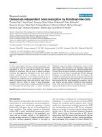
Figure 1
Naïve and memory CD4
+
and CD8
+
T-cell subpopulations in patients with rheumatoid arthritis (RA), patients with systemic lupus erythematosus
(SLE), and healthy control individuals. (a) Representative flow cytometry showing naïve and memory subpopulations of CD4
+
and CD8
+
T cells
from one RA patient and a sex- and age-matched control individual. (b) Pooled data showing naïve and memory subpopulations of CD4
+
and
CD8
+
T cells from RA patients (n =8) and control individuals (n =8). Horizontal lines indicate means. (c) Pooled data showing naïve and memory
subpopulations of CD4
+
and CD8
+
T cells from SLE patients (n =12) and control individuals (n =12). Horizontal lines indicate means. (d) The
correlation between age and CD45RA
+
CD62L
–
terminally differentiated CD8
+
T cells from control individuals (n =13), RA patients (n =8), and
SLE patients (n =12) is shown. The P values were calculated using Mann–Whitney U test and Student’s t-test for panel b and linear regression for
panel d.
individuals and in SLE patients, indicating that this pertur-
bation in homeostasis of T cells is a specific feature of RA.
Although the naïve/memory phenotype of T cells has previ-
ously been investigated in RA in numerous studies using
CD45RA and CD45RO expression as markers of naïve
and memory cells, respectively, that approach has suf-
fered from the limitation that large numbers of CD45RA
+
CD8
+
T cells are actually effector memory cells [10,13].
The CD45RA/CD45RO oversimplification has also
resulted in rather confusing conclusions regarding T-cell
homeostasis, such as defects in primary T-cell homeosta-
sis based on reduced T-cell receptor excision circle
(TREC) levels in naïve CD4
+
T cells (defined as
CD45RO
–
) in RA patients [14]. Our findings suggest that
reduced TREC levels in the CD45RO
–
CD4
+
T-cell popu-
lation may not be due to a reduction in TRECs in naïve
cells but rather to reduced TRECs in the
CD45RA
+
CD45RO
–
CD62L
–
effector memory CD4
+
T cells. It should be noted that previous studies have
reported ‘false naïve’ CD45RA
+
populations of CD4
+
and
CD8
+
T cells in peripheral blood of RA patients [15];
however, the nature of these cells, the exact phenotype,
and the significance was not known at that time.
Our finding that peripheral blood CD8
+
T cells exhibit
increased central memory phenotype and decreased ter-
minally differentiated effector memory phenotype suggests
that the peripheral blood homeostasis of CD8
+
T cells is
perturbed in RA. Perturbations in CD8
+
T-cell maturation
have been shown for HIV-specific CD8
+
T cells, in which
there is an accumulation of preterminally differentiated
CD45RA
–
CD62L
–
CD8
+
T cells [12,16], and such a lack
of differentiation may result in functional or homing
defects. In RA we found a decrease in terminally differenti-
ated CD45RA
+
CD62L
–
CD8
+
T cells with a concomitant
increase in the CD45RA
–
CD62L
+
central memory popula-
tion. If one accepts the linear model of differentiation [10],
which we note has been challenged [12], then our find-
ings indicate that in RA there may be an accelerated dif-
ferentiation of naïve cells into central memory CD4
+
and
CD8
+
T cells. This accelerated differentiation may be due
to a non-antigen-specific effect in RA that differentiates all
peripheral T cells irrespective of their specificity, or it may
actually reflect an antigen-specific expansion of T cells
potentially driven by autoantigen.
The decrease in CD45RA
+
CD62L
–
effector memory
CD8
+
T cells in peripheral blood we found in RA patients
may reflect a decrease in the survival of these cells. It
should be noted, however, that peripheral blood T cells
from RA patients do not exhibit an increase in apoptosis in
in vitro cultures, which is in contrast to synovial membrane
T cells [17,18]. This may suggest that the skewed pheno-
type of the CD45RA
+
CD62L
–
effector memory CD8
+
T cells is more likely due to an increase in the migration of
these cells into sites of inflammation. However, a blockade
of the differentiation of central memory CD45RA
–
CD62L
+
CD8
+
T cells into effector memory CD8
+
T cells would
also result in an increase in the central memory population
with a concomitant decrease in the effector T cells, as
observed in the present study.
Studies of the phenotype of CD8
+
T cells in the synovial
membrane and fluid may shed light as to whether this
skewed phenotype is also found in these sites or whether
there is an enrichment for CD45RA
+
CD62L
–
CD8
+
T cells, indicating increased recruitment into the inflamed
synovium in RA. Inflammation and production of
chemokines such as macrophage inflammatory protein-1α
and RANTES [7,8] in the synovium may result in preferen-
tial recruitment of such effector memory CD8
+
T cells
(which are important contributors to IFN-γ production) and
subsequent macrophage activation, because terminally
differentiated CD45RA
+
CD62L
–
CD8
+
T cells have been
shown to express higher levels of perforin and may be
more potent effector cells [10]. The question arises of
whether the observed skewed differentiation of CD8
+
T cells in RA patients is due to medication, especially
steroids. As shown in Table 1, 38% of the RA patients and
58% of the SLE patients were receiving steroid treatment.
However, the skewed memory phenotype was only
observed in the RA patients, suggesting that this treat-
ment is not responsible for the differences in CD4
+
and
CD8
+
T-cell phenotypes.
Findings from the present preliminary study show that
peripheral blood CD8
+
T cells in RA exhibit a skewed
effector memory phenotype. This skewed phenotype was
not found in CD4
+
T cells in RA and was not seen in age-
matched healthy control individuals or in SLE patients. The
skewed phenotype may be a result of accelerated differen-
Available online />R95
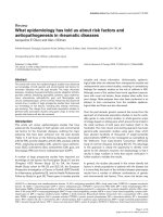
Figure 2
Representation of skewed CD8
+
T-cell phenotype in patients with
rheumatoid arthritis (RA) as compared with sex- and age-matched
healthy control individuals, indicating the relative sizes of the different
naïve and memory populations of CD8
+
T cells. Percentages refer to
the proportions of different naïve/memory population of total CD8
+
T cells.
tiation and migration into sites of inflammation. An under-
standing of the mechanisms that are involved in this
skewed differentiation of effector memory CD8
+
T cells
may prove valuable in elucidating the pathogenesis of RA.
Conclusion
In peripheral blood of RA patients a skewed homeostasis
of CD8
+
T cells was found, with an increase in central
memory and a decrease in terminally differentiated effector
memory T cells (Fig. 2). This skewed T-cell phenotype was
not found in healthy age- and sex-matched control individ-
uals or in patients with SLE. Reduction in peripheral blood
effector memory CD8
+
T cells in RA may indicate an
increase in the migration of these cells into sites of inflam-
mation, and therefore may contribute to ongoing synovial
inflammation.
Competing interests
None declared.
Acknowledgment
This work was supported by National Institutes of Health grants R01
AI46719 and R01 AI52005 to PDK.
References
1. Morita Y, Yamamura M, Kawashima M, Harada S, Tsuji K, Shibuya
K, Maruyama K, Makino H: Flow cytometric single-cell analysis
of cytokine production by CD4
+
T cells in synovial tissue and
peripheral blood from patients with rheumatoid arthritis.
Arthritis Rheum 1998, 41:1669-1676.
2. Kang YM, Zhang X, Wagner UG, Yang H, Beckenbaugh RD,
Kurtin PJ, Goronzy JJ, Weyand CM: CD8 T cells are required for
the formation of ectopic germinal centers in rheumatoid syn-
ovitis. J Exp Med 2002, 195:1325-1336.
3. Fitzgerald JE, Ricalton NS, Meyer AC, West SG, Kaplan H,
Behrendt C, Kotzin BL: Analysis of clonal CD8+ T cell expan-
sions in normal individuals and patients with rheumatoid
arthritis. J Immunol 1995, 154:3538-3547.
4. Wang EC, Lawson TM, Vedhara K, Moss PA, Lehner PJ,
Borysiewicz LK: CD8high+ (CD57+) T cells in patients with
rheumatoid arthritis. Arthritis Rheum 1997, 40:237-248.
5. Masuko-Hongo K, Sekine T, Ueda S, Kobata T, Yamamoto K,
Nishioka K, Kato T: Long-term persistent accumulation of
CD8+ T cells in synovial fluid of rheumatoid arthritis. Ann
Rheum Dis 1997, 56:613-621.
6. Fazou C, Yang H, McMichael AJ, Callan MF: Epitope specificity
of clonally expanded populations of CD8+ T cells found within
the joints of patients with inflammatory arthritis. Arthritis
Rheum 2001, 44:2038-2045.
7. Patel DD, Zachariah JP, Whichard LP: CXCR3 and CCR5 ligands
in rheumatoid arthritis synovium. Clin Immunol 2001, 98:39-
45.
8. Brennan FM, Cope AP, Katsikis P, Gibbons DL, Maini RN, Feld-
mann M: Selective immunosuppression of tumour necrosis
factor-alpha in rheumatoid arthritis. Chem Immunol 1995, 60:
48-60.
9. Hamann D, Baars PA, Rep MH, Hooibrink B, Kerkhof-Garde SR,
Klein MR, van Lier RA: Phenotypic and functional separation of
memory and effector human CD8+ T cells. J Exp Med 1997,
186:1407-1418.
10. Sallusto F, Lenig D, Forster R, Lipp M, Lanzavecchia A: Two
subsets of memory T lymphocytes with distinct homing
potentials and effector functions. Nature 1999, 401:708-712.
11. Tussey L, Speller S, Gallimore A, Vessey R: Functionally distinct
CD8+ memory T cell subsets in persistent EBV infection are
differentiated by migratory receptor expression. Eur J Immunol
2000, 30:1823-1829.
12. Champagne P, Ogg GS, King AS, Knabenhans C, Ellefsen K,
Nobile M, Appay V, Rizzardi GP, Fleury S, Lipp M, Förster R,
Rowland-Jones S, Sékaly R-P, McMichael AJ, Pantaleo G:
Skewed maturation of memory HIV-specific CD8 T lympho-
cytes. Nature 2001, 410:106-110.
13. Roederer M, Dubs JG, Anderson MT, Raju PA, Herzenberg LA:
CD8 naive T cell counts decrease progressively in HIV-
infected adults. J Clin Invest 1995, 95:2061-2066.
14. Koetz K, Bryl E, Spickschen K, O’Fallon WM, Goronzy JJ, Weyand
CM: T cell homeostasis in patients with rheumatoid arthritis.
Proc Natl Acad Sci USA 2000, 97:9203-9208.
15. Neidhart M, Pataki F, Schonbachler J, Bruhlmann P: Flow cyto-
metric characterisation of the ‘false naïve’ (CD45RA+,
CD45RO-, CD29 bright+) peripheral blood T-lymphocytes in
health and in rheumatoid arthritis. Rheumatol Int 1996, 16:77-
87.
16. Mueller YM, De Rosa SC, Hutton JA, Witek J, Roederer M, Altman
JD, Katsikis PD: Increased CD95/Fas-induced apoptosis of
HIV-specific CD8(+) T cells. Immunity 2001, 15:871-882.
17. Hasunuma T, Hoa TT, Aono H, Asahara H, Yonehara S,
Yamamoto K, Sumida T, Gay S, Nishioka K: Induction of Fas-
dependent apoptosis in synovial infiltrating cells in rheuma-
toid arthritis. Int Immunol 1996, 8:1595-1602.
18. Hoa TT, Hasunuma T, Aono H, Masuko K, Kobata T, Yamamoto K,
Sumida T, Nishioka K: Novel mechanisms of selective apopto-
sis in synovial T cells of patients with rheumatoid arthritis. J
Rheumatol 1996, 23:1332-1337.
Correspondence
Peter D Katsikis, MD, PhD, Department of Microbiology and Immunol-
ogy, Drexel University College of Medicine, Drexel University, 2900
Queen Lane, Philadelphia, PA 19129, USA. E-mail:
Arthritis Research & Therapy Vol 5 No 2 Maldonado et al.
R96