Báo cáo y học: "The VH gene repertoire of splenic B cells and somatic hypermutation in systemic lupus erythematosus" potx
Bạn đang xem bản rút gọn của tài liệu. Xem và tải ngay bản đầy đủ của tài liệu tại đây (131.18 KB, 8 trang )
R114
Introduction
Systemic lupus erythematosus (SLE) is characterized by
polyclonal B-lymphocyte activation, which leads to pro-
duction of autoantibodies with various specificities, princi-
pally against nuclear antigens including double stranded
(ds)DNA, ribonucleoprotein particles, histones and nonhi-
stone chromatin proteins. Other antibodies bind cell
surface structures and cytoplasmic antigens. Of these,
serum high-affinity IgG antibodies that are specific for
native dsDNA are believed to be the principal pathogenic
agents and are used as a diagnostic indicator [1]. These
autoantibodies differ from anti-DNA antibodies found in
the sera of healthy individuals in that they bind to dsDNA
with high affinity, they are often cationic in charge and they
do not usually cross-react with unrelated antigens [2].
Despite intensive study, the factors that lead to the pro-
duction of such autoantibodies remains in dispute,
although a number of hypotheses have been suggested.
Previous studies, using serum antibodies, hybridomas
generated from peripheral blood lymphocytes (PBLs) and
mouse models, concluded that autoantibodies produced
in SLE are associated with particular properties. These
include expression of characteristic idiotypes, clonal
restriction of anti-DNA and anti-Sm antibodies, somatic
hypermutation, V gene bias, and the presence of positively
charged complementarity determining region (CDR)
residues or sequence motifs in anti-dsDNA antibodies [3].
V(D)J rearrangement of immunoglobulin genes has the
capacity to generate an immense repertoire of immune
receptors that are able to recognize virtually any foreign
substance via somatic hypermutation, but because of the
nature of this process a number of immune receptors with
specificity for self molecules are also generated. These
self-reactive B cells are normally eliminated in the bone
marrow, but self-reactivity can also be generated in the
periphery by somatic mutation. For example, mutation of a
single amino acid at position 35 on the heavy chain culmi-
nates in a switch from anti-phosphoryl choline (a bacterial
hapten) to anti-dsDNA [4]. This supports the hypothesis
CDR = complementarity determining region; ds = double stranded; FDC = follicular dendritic cell; FR = framework region; GC = germinal centre;
PBL = peripheral blood lymphocyte; PCR = polymerase chain reaction; R/S ratio = replacement : silent ratio; SLE = systemic lupus erythematosus.
Arthritis Research & Therapy Vol 5 No 2 Fraser et al.
Research article
The V
H
gene repertoire of splenic B cells and somatic
hypermutation in systemic lupus erythematosus
Nicola L W Fraser
1
, Gary Rowley
1
, Max Field
2
and David I Stott
1
1
Division of Immunology, Infection and Inflammation, University of Glasgow, Western Infirmary, Glasgow, Scotland, UK
2
Department of Rheumatic Diseases, Glasgow Royal Infirmary, Glasgow, Scotland, UK
Corresponding author: Nicola Fraser (e-mail: )
Received: 27 September 2002 Revisions received: 18 December 2002 Accepted: 5 January 2003 Published: 3 February 2003
Arthritis Res Ther 2003, 5:R114-R121 (DOI 10.1186/ar627)
© 2003 Fraser et al., licensee BioMed Central Ltd (Print ISSN 1478-6354; Online ISSN 1478-6362). This is an Open Access article: verbatim
copying and redistribution of this article are permitted in all media for any non-commercial purpose, provided this notice is preserved along with the
article's original URL.
Abstract
In systemic lupus erythematosus (SLE) it has been
hypothesized that self-reactive B cells arise from virgin B cells
that express low-affinity, nonpathogenic germline V genes that
are cross-reactive for self and microbial antigens, which
convert to high-affinity autoantibodies via somatic
hypermutation. The aim of the present study was to determine
whether the V
H
family repertoire and pattern of somatic
hypermutation in germinal centre (GC) B cells deviates from
normal in SLE. Rearranged immunoglobulin V
H
genes were
cloned and sequenced from GCs of a SLE patient’s spleen.
From these data the GC V gene repertoire and the pattern of
somatic mutation during the proliferation of B-cell clones were
determined. The results highlighted a bias in V
H
5 gene family
usage, previously unreported in SLE, and under-representation
of the V
H
1 family, which is expressed in 20–30% of IgM
+
B cells of healthy adults and confirmed a defect in negative
selection. This is the first study of the splenic GC response in
human SLE.
Keywords: spleen, systemic lupus erythematosus
Open Access
Available online />R115
that the aetiological stimulant of the autoimmune response
observed in SLE may be of bacterial or viral origin, and this
is further supported by the observation that the anti-DNA
response is clonally restricted in both mouse models and
SLE patients [5,6]. This hypothesis suggests that self-
reactive B cells may arise from B cells that express low-
affinity V genes, which are cross-reactive for self and
microbial antigens, by somatic hypermutation to generate
high-affinity autoantibodies.
There have been only a limited number of studies on the
immunoglobulin V gene repertoire and somatic hypermuta-
tion in SLE, with the majority of those investigations per-
formed in PBLs. PBLs comprise a population of
recirculating memory cells that have encountered a vast
range of antigens, including many environmental antigens,
over a prolonged period of time, whereas germinal centres
(GCs) in the spleen or lymph nodes provide a profile of
B cells that respond to antigen at a given time point. In an
earlier investigation, Ravirajan and coworkers [7,8]
demonstrated the presence of autoantibody-producing
B cells in the spleen of an SLE patient by analysis of
hybridomas generated from splenic B cells. The aims of
the present study were to identify the immunoglobulin
V genes used by proliferating B cell clones in GCs of a
SLE spleen, and to determine whether there are abnormal-
ities in the pattern of somatic hypermutation and antigen
selection. To our knowledge, this is the first detailed study
of the repertoire of the splenic GC response in SLE.
Materials and methods
Spleen sections
The spleen used for this investigation was removed from a
female SLE patient (M) because of hypersplenism sec-
ondary to persistent haemolysis and thrombocytopaenia.
Patient consent was obtained using standard practice pro-
cedures at the time. The patient fulfilled the American
Rheumatism Association criteria for SLE [1], with the pres-
ence of arthritis, photosensitive skin rash, an autoimmune
haemolytic anaemia, lymphopaenia, thrombocytopaenia,
and homogeneous antinuclear antibodies characterized as
anti-DNA antibodies. At the time of splenectomy the patient
had detectable antibodies against DNA (Crithidia nega-
tive), and IgA and IgM antibodies against cardiolipin.
The spleen was cut into small pieces and snap frozen. Serial
frozen sections (6–8 µm thick) of the spleen, which had been
stored at –70°C, were cut with a cryostat and mounted on
slides precoated with 2% 3-amino-propyltriethoxy silane
(Sigma, Poole, UK). Sections were air dried, fixed in acetone
for 10 min and stored at –70°C with desiccant.
Immunohistochemical staining of tissue sections
Frozen sections were stained using mouse monoclonal
antibodies for B cells (anti-CD20; DAKO A/S, Cam-
bridgeshire, UK), T cells (anti-CD3; DAKO A/S), proliferat-
ing cells (anti-Ki67; DAKO A/S), follicular dendritic cells
(FDCs; Wue2) and plasma cells (Wue1). (The latter two
were both kindly donated by Dr A Greiner, University of
Würzburg, Germany.) This was followed by incubation
with rabbit anti-mouse IgG (DAKO A/S), and an alkaline
phosphatase/anti-alkaline phosphatase complex (DAKO
A/S). Immune complexes containing alkaline phos-
phatase/anti-alkaline phosphatase were detected by incu-
bation with new fuschin substrate, and the sections were
counter-stained with Mayer’s haematoxylin (Sigma).
Microdissection of germinal centres and DNA preparation
GCs were identified by staining with anti-CD20 and anti-
FDC. They were microdissected under sterile ultrapure
water using sterile blood lancets attached to Narishige
micromanipulators (Nikon, Telford, UK), linked to an
inverted microscope (Nikon). Excised tissue was digested
in 30 µl proteinase K (0.7 mg/ml; Boehringer Mannheim,
Mannheim, Germany) at 50°C for 1 hour, which was then
inactivated at 95°C for 10 min. This DNA preparation was
used as a template for subsequent primary PCR reactions.
Amplification and cloning of rearranged
immunoglobulin V genes
To avoid contamination with amplified immunoglobulin
V genes, all procedures prior to primary PCR amplification
were performed in a separate clean laboratory to that
where all steps after amplification were carried out. Nested
PCR was performed using a mixture of primers for the
leader sequences of all of the V
H
families with a universal
J
H
primer, in the primary amplification. All primers used are
described in detail elsewhere [9]. For primary amplification,
the conditions used for 35 cycles were 94°C for 1 min,
61°C for 1 min and 72°C for 2 min, followed by one cycle
at 72°C for 15 min. The Taq polymerase used was of high-
fidelity quality (Expand Easy; Roche, Mannheim, Germany),
which has a very low error rate. Secondary amplification
used individual primers for each of the first framework
regions of each V
H
family in conjunction with a J
H
primer
mix. Cycle conditions for secondary amplification were
94°C for 1 min; 61°C for 1 min in the case of V
H
1–3 and
65°C for 1 min in the case of V
H
4–6; and 72°C for 2 min
for 40 cycles; followed by 72°C for 15 min. This method
ensures that only rearranged V (D) J genes are amplified.
Successful secondary amplifications were identified as a
band corresponding to a product of approximately 400
base pairs on an agarose gel. The bands were excised and
subsequently purified using QIAquick cleanup columns
(Qiagen, Sussex, UK). The amplified DNA was then ligated
with TA-cloning vector pCRII, and transformed into IFN-αF
cells (Invitrogen, Paisley, UK) and cloned.
Sequencing and analysis of rearranged V genes
Plasmid DNA from clones containing gene inserts was
prepared using QIAprep spin mini-prep kits (Qiagen,
Arthritis Research & Therapy Vol 5 No 2 Fraser et al.
R116
Sussex, UK), according to the manufacturer’s instructions,
and precipitated, washed thoroughly and resuspended in
10 mmol/l Tris-HCl (pH 8.5). Cloned, rearranged
immunoglobulin V genes were sequenced in an ABI Prism
377 DNA sequencer (Applied Biosystems, Warwickshire,
UK). Germline immunoglobulin V genes providing the best
match to the cloned DNA sequence were identified by
blast searching the Vbase Sequence Directory of human
germline immunoglobulin V genes [10]. Sequences were
aligned and compared using the DNA plot 1.4 programme
(W. Müller, Institut für Genetik, Köln, Germany). The
nomenclature for the V, D and J gene segments adopted
here and the definitions of the CDRs were previously
described [11–13]. Family trees were constructed by
analysis of mutations shared by sequences with the same
V-D-J rearrangement. Replacement : silent ratios (R/S
ratios) were calculated by analyzing whether a mutation
resulted in an amino acid change (replacement mutation)
or not (silent mutation).
Results
Structural and immunochemical characterisation of
germinal centres within an SLE spleen
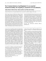
Clusters of B cells and FDCs, which resembled GCs,
were identified in frozen sections of the SLE spleen by
staining with anti-CD20 and anti-FDC, respectively. Two
GCs (GC A and GC B) were excised and the rearranged
V
H
genes amplified (as described under Materials and
methods). Both GCs were located within the same
0.5 cm
3
portion of spleen tissue. GC A can be seen in
Fig. 1, and has two distinct areas of staining for FDC and
one large area of B cells encompassing both FDC
regions. Very few T cells could be seen within GC A
and B, and no plasma cells could be seen at all within the
GC structure itself. No distinct mantle zone was observed
surrounding either GC.
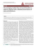
Two other areas, shown in Fig. 1 (e and f) appeared to be
made up entirely of B cells because there was no positive
staining for any of the other cell markers. These were
referred to as B-cell clusters C and D.
Repertoire of immunoglobulin V
H
genes isolated from
SLE splenic germinal centres
In total, 15 rearranged V
H
sequences were analyzed
(including seven independent V-D-J rearrangements) for
GC A and 16 (six V-D-J rearrangements) for GC B. From
B-cell cluster C, 37 functional and six nonfunctional V
H
sequences were analyzed (16 V-D-J rearrangements) and
cluster D yielded eight functional and 16 nonfunctional
sequences (seven V-D-J rearrangements).
The best matching germline V gene sequences corre-
sponding to each of these rearranged sequences were
identified (Table 1 and Fig. 2). The V
H
locus in humans
consists of 51 functional V
H
segments, which are classi-
fied as families V
H
1 through to V
H
7. V
H
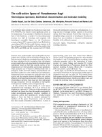
gene family usage
of the combined GCs and B-cell clusters differs signifi-
cantly from the expected frequencies, assuming that each
germline gene is equally likely to form a viable rearrange-
ment (P < 0.001). In particular V
H
1 was completely absent,
whereas it is normally expressed in 20–30% of PBL B
cells (P = 0.0066), and V
H
5 was observed in 16.6% of the
V
H
sequences in the present study as compared with the
3.92% that is theoretically expected (P = 0.046). The
D gene family use was skewed towards D2 but the small
number of possible D genes makes it difficult to determine
the significance of these findings. The J
H
family expression
exhibited by the SLE spleen studied here differed signifi-
cantly from the expected values (P = 0.0015). For
example, J4 was over-expressed, and J1 and J2 were
under-represented.
From these data we can see that GC A and GC B do not
share any common B-cell clones. We can also see that by
far the most common V
H
family present is V
H
3 for both
GCs. Analysis of V gene sequences indicated that a
Figure 1
Immunohistochemistry of sections from a patient with systemic lupus
erythematosus. (a) Anti-FDC (follicular dendritic cell) staining within
the area known as germinal centre (GC) A. (b) Staining for CD20.
FDC staining suggests two possible GCs within close proximity, but
B-cell staining shows no clear demarcation. (c) Anti-FDC staining for
GC B. (d) staining with anti-CD20. Panels (a) and (b) are consecutive
sections, as are (c) and (d). Panels (e) and (f) show B-cell clusters
identified using anti-CD20 on serial sections: (e) B-cell cluster C; (f) is
B-cell cluster D. The arrows indicate the areas of positive staining for
the marker in question. All images: 100×.
number of B cells shared common V, D, J and junctional
sequences for which mutational analysis was carried out
(described below).
The V
H
3 family is again highly represented in B-cell clus-
ters C and D, specifically V
H
3-23, which is known to be
among the most highly expressed genes in normal individ-
uals. However, V
H
5-51, one of only two functional
members of the V
H
5 family, is also found in large numbers
here. In fact, 10 of the V
H
5 sequences were shown to use
the same V-D-J rearrangement. From a total of 36 different
rearrangements (all four areas combined), six indepen-
dently rearranged groups of genes used the V
H
5-51 gene.
Available online />R117
Table 1
Variable region heavy chain genes identified from spleen
tissue
Number
of sequences
GC/cluster V
H
gene D
H
gene J
H
gene isolated
A V
H
2-5*02 3-10*01 5*02 1
V
H
3-15*01 2-21*02 4*02 3
V
H
3-23*01 1-26*01 5*02 3
V
H
3-30*03 3-22*01 4*02 1
V
H
3-43*02 1
V
H
3-74*02 2-15*01 6*02 2
V
H
5-51 3-10 4*02 4
BV
H
2-5*02 1-26*01 5*01 3
V
H
3-21*01 2
V
H
3-30*03 2-8*01 6*02 1
V
H
3-30*03 2-8*01 4*02 1
V
H
3-30.3*01 3-3*01 6*02 8
V
H
4-30.1 2-2*01 4*02 1
C V
H
2-5*02 5*02 2(1)
V
H
3-15*01 1
V
H
3-21*01 1
V
H
3-23*01 2*01 1
V
H
3-23*01 3-10*02 6*03 9
V
H
3-30*03 3*02 1
V
H
3-30*03 2
V
H
3-33*01 5-24*01 3*02 1
V
H
3-74*01 (1)
V
H
4-4*03 4*02 1
V
H
4-30.4*01 2-2*01 6*02 5(3)
V
H
4-34*01 5*02 1
V
H
4-59 1-26*01 4*02 1
V
H
5-51*01 2-2*01 4*02 11
V
H
5-51*01 2-2*01 4*02 3(1)
V
H
5-51*01 4-4*01 3*02 2
DV
H
3-07 2-15 3*02 13(10)
V
H
3-23 3-22 6*03 3(1)
V
H
3-33 6-13*01 4*02 2
V
H
3-72*01 5-5*01 4*02 1
V
H
4-59*02 5-24*01 4*02 1
V
H
5-51*01 2-2*01 1*01 3(2)
V
H
5-51*01 6-13*01 4*02 1
The best matching germline sequences identified from blast searching
the Vbase database from which the rearranged V-D-J sequences
identified from germinal centre (GC) A and GC B, and B-cell clusters
C and D were derived. Groups of sequences were deemed to be
clonally related when they used the same V, D, J and CDR3, and
differed only by base substitutions. The total number of members of
each clone is given in the right-most column, and the number of these
members that are nonfunctional is indicated in brackets. Identical
sequences are only counted once.
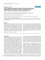
Figure 2
V
H
gene family usage. (a) Comparison of V
H
gene family usage of
functional rearrangements found in the combined germinal centres
(GCs) and B-cell clusters. V
H
gene family usage differed significantly
from the expected frequencies, assuming each germline gene is
equally likely to form a viable rearrangement (P < 0.01). V
H
1 was
significantly under-expressed (P = 0.0066) and V
H
5 over-expressed
(P = 0.046). (b) D gene family use. (c) J
H
family usage differed
significantly from expected (P = 0.0015).
0
10
20
30
40
50
60
70
VH1 VH2 VH3 VH4 VH5 VH6 VH7
VH gene family
% of functional independent V-genes
expected
observed
(a)
0
5
10
15
20
25
30
35
40
45
D1 D2 D3 D4 D5 D6 D7
D gene
% of functional independent V-genes
0
10
20
30
40
50
J1 J2 J3 J4 J5 J6
JH gene
% of functional independent V-genes
(b)
(c)
Somatic mutations in germinal centre V
H
genes
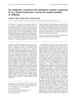
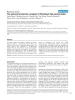
The distribution of the frequency of mutations for all four
areas is shown in Fig. 3. The data represent a mean
average number of mutations per sequence of 7.6 for
GC A, 15.8 for GC B, and 15 and 14.9 for clusters
C and D, respectively. All data sets, excluding B-cell
cluster D, contained at least one sequence in the 0–2
range, and the majority of GC A V
H
genes contained 3–10
mutations. The location of the mutations observed was
categorized as being within the framework region (FR),
CDR1 or CDR2. CDR3 was ignored for mutational analy-
sis because of difficulty in distinguishing between point
mutations and junctional variation in this region of the vari-
able gene because of recombination events.
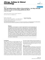
Although mutations were seen in higher numbers in the
FRs, this can be attributed to the fact that these segments
are longer and there is therefore increased likelihood of
random mutations occurring. In order to correct for this,
the number of mutations in each segment is expressed as
a percentage of the total number of bases in that segment
(Fig. 4). The graph illustrates the fact that there is a higher
frequency of mutation in the CDRs than in the FRs, which
is typical of an antigen-driven response.
R/S mutation ratios were calculated for the total V
H
segment, FRs and CDRs (Table 2). The ratio for each indi-
vidual clone is shown, as well as the R/S value of all the
clones together, disregarding all individual sequences.
The R/S ratio of framework regions of V
H
genes from
GC A and cluster D was remarkably high in comparison
with that seen in other studies of mutations in V
H
genes of
both autoimmune and healthy patients, whereas that for
the CDRs of GC A was not significantly above random.
The R/S ratios calculated for GC B were more in accord
with those previously reported. The R/S ratios for the CDR
are much higher than random, with the framework ratio
being slightly less than random. This indicates that affinity
selection of B cells with replacement mutations by antigen
is taking place.
Clonal genealogies
As Table 1 shows, there are a number of B cells within all
four areas that share the same germline genes. These
sequences are deemed to be clonally related if they share
the same V, D and J germline genes, as well as having
common junctional sequences. Genealogical trees were
constructed for all clones containing three or more
members. The most dominant V gene amplified from GC B
was V
H
3-30.3*01 followed by V
H
2-5*02. Genealogical
trees constructed from these clonally related sets, as well
as V
H
5-51 from GC A and V
H
3-23*01 from cluster C, are
shown in Fig. 5. This provides clear evidence that these
clones of B cells are proliferating and mutating in the
splenic GCs and B-cell clusters.
Serine codon usage
There is evidence for a bias toward serine codon usage
within immunoglobulin variable genes [14], especially
within the CDRs. From all of the sequences amplified from
all four sites, 70% of the serines present within CDR1 and
CDR2 were AGC or AGT. This was not the case for the
FR, in which TCC was the most frequent serine codon
present. Only AGC and AGT produced replacement
mutations within the CDRs, of which the most prevalent
was from serine to asparagine; other mutations included
serine to threonine, tyrosine, aspartate, methionine and
proline.
Discussion
The immunohistology of the spleen from the SLE patient
produced a picture similar to the cellular architecture of
healthy spleens in mice [15] and humans [16], which is
Arthritis Research & Therapy Vol 5 No 2 Fraser et al.
R118
Figure 3
The distribution of mutations between V
H
gene sequences in germinal
centre and B-cell clusters.
0
1
2
3
4
5
6
7
8
9
10
(0–2) (3–10) (11–20) (21–40)
Number of mutations
Number of sequences
GCA
GCB
C
D
Figure 4
Graph showing variable region mutations as a percentage of the total
length of each region of the V gene segment. Although there are fewer
mutations in the complementarity determining regions (CDRs), the
segment is also much smaller so the mutational frequency is higher
than in the framework regions (FRs).
0
5
10
15
20
25
30
35
40
GCA GCB C D
No. of mutations (% of length)
FR
CDR1
CDR2
known to be interspersed with GCs. In the present study,
however, a mantle surrounding the GC was not identified.
Each mature GC is generally derived from one to three
B-cell clones, which manage to survive a significant reduc-
tion in clonal diversity and then go on to endure V(D)J
hypermutation. The GC is of most interest because it is
the site of antigen driven V(D)J hypermutation and selec-
tion [15] where antigen-specific B cells acquire point
mutations in the V regions of transcriptionally active
rearranged immunoglobulin genes. These mutations accu-
mulate steadily during expansion of B-lymphocyte clones
in the dark zone of the GC. This clonal evolution occurs
independently in each GC, because little trafficking of
B cells between GCs has been observed [17].
The cellular components necessary for a GC response
were present in both GC A and GC B (i.e. B cells, FDCs
capable of presenting antigen and T cells). However, none
of the GCs and B-cell clusters had a discernible mantle
zone. No plasma cells were located within the GCs them-
selves but they were loosely distributed in the surrounding
tissue within close proximity to the GCs (data not shown).
The immunohistochemistry also demonstrates that both
GCs are of approximately equal size, which validates com-
parison of the data produced from each.
A recent study in Science [18] identified autoreactive
B cells in MRL.Fas
lpr
mice proliferating in the T-cell zone of
lymphoid tissues. This was thought to be due to their defi-
ciency of the Fas receptor, because these cells would nor-
mally be deleted through the Fas receptor/Fas
ligand-mediated pathway of apoptosis. A similar explana-
tion may account for the GCs identified in this study not
exhibiting a traditional mantle, and explain the lack of neg-
ative selection illustrated by the low number of nonfunc-
tional genes.
Available online />R119
Table 2
The replacement : silent ratios of sequences from each of the
systemic lupus erythematosus splenic germinal centres and B-
cell clusters
Location
Total V
H
CDR1
GC/cluster Segment Framework and CDR2
A
R/S ratio of all sequences 3.11 7.6 2.9
Clone V
H
5-51 2.5 5 1.4
Clone V
H
3-15 2 2 1
Clone V
H
3-23 2 1:0 1:0
R/S of all clones 2.75 5 1.4
B
R/S ratio of all sequences 3.6 2.6 8.1
Clone V
H
2-5 4.7 4:0 4.5
Clone V
H
3-3.30 2.8 1.5 12
R/S of all clones 31.79
C
R/S ratio of all sequences 2.9 3 3.2
V
H
5-51-J
H
44.581
V
H
3-23 2.7 2.4 4
V
H
5-51-D2-2 23.5 22 3:0
V
H
4-30 3 2 6
R/S of all clones 66.44.6
D
R/S ratio of all sequences 2. 9 2.9 2.9
V
H
3-07 2.4 1.7 7
V
H
3-23 4.5 3.5 2:0
V
H
5-51 2.75 2 3:0
R/S of all clones 2.8 2.1 12
Replacement : silent ratios (R/S ratios) were calculated for the
complete V
H
segment as well as individual complementarity
determining regions (CDRs) and framework regions. The ratios for
each individual clone are shown as well as the R/S value of all the
clones together, disregarding individual sequences. The colons
represent instances where there are no silent replacements, therefore
no figure can be given since it is not possible to divide by zero.
Figure 5
Clonal genealogical trees constructed from sequence data from
germinal centre (GC) A, GC B and cluster C. Numbers in bold indicate
the minimum number of mutations required between each sequence.
The bracketed figures represent silent mutations. The dashed circle
shown in cluster C represents a hypothetical intermediate that was not
actually found among the sequences. GC A contained two highly
mutated B cell clones represented as (a) and (b).
GC A GC B
(a)
10 7 0
1
2 13
(b)
Cluster C
11
9 17
25
2 2
V
H
5-51
a b
c
d
V
H
2-5*02
a
b c
V
H
3-30-3*01
a
b c
d e
g
h
f
V
H
3-23*01
a b
c d
ef
h
i
g
(1)
3
(1)
1
(1)
Of the total number of rearranged V
H
genes, 19% were
found to contain stop codons and out-of-frame rearrange-
ments, and were therefore deemed to be nonfunctional,
the majority being found in cluster D. This correlates with a
similar study conducted by Jacobi et al. [19], who found
13% of PBL V
H
gene sequences amplified from an SLE
patient to be nonfunctional, as compared with 53% of
genes amplified from PBLs of a healthy individual. Those
investigators observed a similar R/S ratio in the productive
rearrangements to that seen in the nonproductive
rearrangements. It was therefore suggested that there may
be some abnormality in selection in SLE related to an
intrinsic failure of B-cell apoptosis or enhanced B-cell acti-
vation by T cells, which overwhelms protective mecha-
nisms that are effective in normal individuals.
The most abundantly expressed V
H
gene family was V
H
3,
which is not surprising because it is the largest family and
has been found to be the most dominant in the normal
repertoire [20]. What is perhaps more interesting is the
large number of V
H
5 sequences (16.6%) as compared
with the 3.9% expected to be produced randomly in the
normal human repertoire. V
H
5-51 has also been isolated
from breast tumours (Nzula et al., unpublished data) and
thymic GCs from a patient with myasthenia gravis [9] pre-
viously in our laboratory, but not from GCs from the sali-
vary glands of two patients with Sjögren’s syndrome,
using the same primers [21]. It is therefore unlikely that the
V
H
5 family primers used here preferentially amplify
V
H
5-51. V
H
5 sequences have been consistently associ-
ated with IgE antibodies, and it has been suggested that
these antibodies may be associated with an unidentified
superantigen [22]. Two studies analyzing anti-DNA anti-
bodies in SLE both identified a heavy chain clone compa-
rable to V
H
5-51 [23,24]. Comparison of our V-D-J
rearrangements with the two anti-DNA specific antibodies
revealed very few somatic mutations in common, however.
One hotspot highlighted at position 77 (in the tip of the
FR3 loop) [25] was found to be present in the anti-DNA
specific antibodies as well as in most of the analyzed
V
H
5-51 sequences from this study, but this was not often
the same amino acid substitution.
V
H
5-51 was also used by two human IgG monoclonal anti-
bodies that bind phospholipid, derived from the PBL of a
SLE patient. In this instance the J segment used was J
H
6b,
which was not found in combination with V
H
5-51 in the
present study. It may be significant that patient M pro-
duced anticardiolipin antibodies, but not anti-dsDNA,
although there is no direct evidence that the cardiolipin
antibodies used V
H
5-51 because no information about the
specificity of these antibodies was obtained.
A complete absence of V
H
1 family genes was observed in
the present study of splenic GC, which contrasts with the
theoretically expected value of 20–30%. This was also
found to be the case in another study of SLE patients con-
ducted by Hansen et al. [26]. They found that 13% of
functional PBL V genes from healthy control individuals
were from the V
H
1 family, but only about 1% of the V
H
genes expressed by PBLs of a SLE patient used this
family. de Wildt et al. [27], on the other hand, found very
little difference between expression of V
H
1 in healthy
control individuals and SLE patients. The PBL B-cell
repertoire may not reflect the repertoire of splenic B cells.
Using high-fidelity Taq polymerase and the same primers
as used here, the PCR error rate for V
H
genes was less
than one base per four V
H
genes [9]. The average
numbers of single base mutations in the V
H
genes from
GC A and GC B were 7.6 and 15.8, respectively, and for
clusters C and D they were 15 and 14.9, respectively.
This is significantly higher than the Taq polymerase error
rate, demonstrating somatic hypermutation in vivo. The dif-
ference in mutation rates between the two GCs may indi-
cate that they are in different states of maturity or that
GC B might have been founded by a memory B cell that
was already mutated (e.g. the founder cell of clone B-b;
Fig. 5). Hypermutation within a GC is closely linked to
antigen-induced B-cell proliferation; thus, from the data
presented here, this appears to be the case in both GCs
and the B-cell clusters. The second phase of this process,
during an immune response against a xenoantigen, is
selection of B cells that express high-affinity antigen
receptors resulting from rare mutations, by competition for
binding to antigen on the surface of FDCs. This process is
generally believed to result in an increase in the ratio of
replacement to silent mutations, especially within CDRs,
and often in selection against replacement mutations in
the FRs. The evidence presented here is supportive of
antigen selection in GC B and clusters C and D, in which
the R/S ratio is higher than random in the CDRs. Selection
for replacement mutations in the FRs has previously been
observed, for example in two anti-hen egg lysozyme anti-
bodies (HyHEL-5 and HyHEL-10) [28,29], in which both
had contact residues in the FRs. It is also possible that the
high R/S ratio in the FR region of the sequences in this
study are the result of antigen selection, but this was not
confirmed because of the absence of data on specificity.
A serine codon bias was also observed (not shown), with
70% of the serines in CDR1 and CDR2 represented by
AGC or AGT, both of which are recognized targets of the
hypermutation machinery (for review [30]). Only AGC and
AGT serine codons produced replacement mutations in
the CDRs. Of these, the mutation from serine to
asparagine was the most prevalent, which is in accor-
dance with mutational analysis performed on patients with
myasthaenia gravis [9].
The clonal genealogies show that the groups of
rearranged V
H
genes included sequences that could be
Arthritis Research & Therapy Vol 5 No 2 Fraser et al.
R120
Available online />R121
assigned to parental and daughter cells on the basis of
shared mutations and junctional sequences. By far the
most dominant sequences were V
H
3-30*01 and
V
H
5-51*01 (approximately 16% each). In studies of PBLs
in SLE, the most common V
H
segment observed was
V
H
3-23 (12%) [27], which we have seen in similar
numbers (11%).
Conclusion
The present study highlights a possible bias toward
expression of V
H
5 immunoglobulin V genes by splenic
GC B cells in SLE, as well as normal high levels of V
H
3
and an under-representation of V
H
1. It also confirms that
there is a defect in the negative selection process in SLE
patients.
Competing interests
None declared.
Acknowledgements
This project was funded by the Arthritis Research Campaign. We
would like to thank Rod Ferrier (Department of Pathology, University of
Glasgow) for cutting frozen sections, and Dr Ian McKay for assistance
with statistical analysis.
References
1. Tan EM: Autoantibodies to nuclear antigens (ANA): their
immunobiology and medicine. Adv Immunol 1982, 33:167-
240.
2. Tan EM: Antinuclear antibodies: diagnostic markers for
autoimmune diseases and probes for cell biology. Adv
Immunol 1989, 44:93-151.
3. Khalil M, Spatz L, Diamond B: Anti-DNA antibodies. In Systemic
Lupus Erythematosus. Edited by Lahita RG. New York: Academic
Press; 1999:197-217.
4. Diamond B, Scharff MD: Somatic mutation of the T15 heavy
chain gives rise to an antibody with autoantibody specificity.
Proc Natl Acad Sci USA 1984, 81:5841-5844.
5. Stott DI: Lessons about autoantibody specificity in systemic
lupus erythematosus from animal models. Clin Exp Immunol
1990, 81:1-4.
6. Stott DI: Spectrotypes of anti-DNA antibodies show that anti-
DNA-secreting B-cell clones of SLE patients are restricted in
number, stable and long lived. Autoimmunity 1992, 12:249-
258.
7. Ravirajan CT, Harmer I, McNally T, Hohmann A, Mackworth-Young
CG, Isenberg DA: Phospholipid binding specificities and idio-
type expression of hybridoma derived monoclonal autoanti-
bodies from splenic cells of patients with systemic lupus
erythematosus. Ann Rheum Dis 1995, 54:471-476.
8. Ravirajan CT, Kalsi J, Wiloch HW, Barakat S, Tuaillon N, Irvine W,
Cockayne A, Harris A, Williams DG, Williams W: Antigen-
binding diversity of human hybridoma autoantibodies derived
from splenocytes of patients with SLE. Lupus 1992, 1:157-
165.
9. Sims GP, Shiono H, Willcox N, Stott DI: Somatic hypermutation
and selection of B cells in thymic germinal centers respond-
ing to acteylcholine receptor in myasthenia gravis. J Immunol
2001, 167:1935-1944.
10. Tomlinson IM, Williams SC, Ignatovich O, Corbett SJ, WinterG:
Human VBase Directory of Immunoglobulin Genes. Cambridge,
UK: MRC Centre for Protein Engineering; 1997.
11. Chothia C, Lesk AM, Gherardi E, Tomlinson IM, Walter G, Marks
JD, Llewelyn MB, Winter G: Structural repertoire of the human
VH segments. J Mol Biol 1992, 227:799-817.
12. Mattila PS, Schugk J, Wu H, Makela O: Extensive allelic
sequence variation in the J region of the human immunoglob-
ulin heavy chain gene locus. Eur J.Immunol 1995, 25:2578-
2582.
13. Corbett SJ, Tomlinson IM, Sonnhammer EL, Buck D, Winter G:
Sequence of the human immunoglobulin diversity (D)
segment locus: a systematic analysis provides no evidence
for the use of DIR segments, inverted D segments, ‘minor’ D
segments or D-D recombination. J Mol Biol 1997, 270:587-
597.
14. Jolly CJ, Wagner SD, Rada C, Klix N: The targeting of somatic
hypermutation. Semin Immunol 1996, 8:159-168.
15. Camacho SA, Kosco-Vilbois MH, Berek C: The dynamic struc-
ture of the germinal center. Immunol Today 1998, 19:511-514.
16. Lydyard PM, Grossi CE: Cells, tissues and organs of the
immune system. In Immunology. Edited by Roitt I, Brostoff J,
Male D. Edinburgh: Mosby; 2001:37.
17. Jacob J, Kelsoe G, In situ studies of the primary immune
response to (4-hydroxy-3- nitrophenyl)acetyl. II. A common
clonal origin for periarteriolar lymphoid sheath-associated
foci and germinal centers. J Exp Med 1992, 176:679-687.
18. William J, Euler C, Christensen S, Shlomchik MJ, Evolution of
autoantibody response via somatic hypermutation outside of
germinal centers. Science 2002, 297:2066-2070.
19. Jacobi AM, Hansen A, Burmester GR, Dorner T, Lipsky PE:
Enhanced mutational activity and disturbed selection of muta-
tions in V(H) gene rearrangements in a patient with systemic
lupus erythematosus. Autoimmunity 2000, 33:61-76.
20. Brezinschek HP, Foster SJ, Brezinschek RI, Dorner T, Domiati-
Saad R, Lipsky PE: Analysis of the human VH gene repertoire.
Differential effects of selection and somatic hypermutation on
human peripheral CD5(+)/IgM+ and CD5(-)/IgM+ B cells. J
Clin Invest 1997, 99:2488-2501.
21. Stott DI, Hiepe F, Hummel M, Steinhauser G, Berek C: Antigen-
driven clonal proliferation of B-cells within the target tissues
of an autoimmune disease. J Clin Invest 1998, 102:938-946.
22. Snow RE, Chapman CJ, Frew AJ, Holgate ST, Stevenson FK:
Pattern of usage and somatic hypermutation in the V(H)5
gene segments of a patient with asthma: implications for IgE.
Eur J Immunol 1997, 27:162-170.
23. Roben P, Barbas SM, Sandoval L, Lecerf J-M, Stollar BD,
Solomen A: Repertoire cloning of lupus anti-DNA antibodies. J
Clin Invest 1996, 98:2827-2837.
24. Seal SN, Hoet RM, Raats JMA, Radic MZ: Analysis of autoim-
mune bone marrow by antibody-phage display. Arthritis
Rheum 2000, 43:2132-2138.
25. Cai J, Humphries C, Richardson A, Tucker PW: Extensive and
selective mutation of a rearranged VH5 gene in human B cell
chronic lymphocytic leukemia. J Exp Med 1992, 176:1073-
1081.
26. Hansen A, Dorner T, Lipsky PE: Use of immunoglobulin vari-
able-region genes by normal subjects and patients with sys-
temic lupus erythematosus. Int Arch Allergy Immunol 2000,
123:36-45.
27. de Wildt RMT, Tomlinson IM, van Venrooij WJ, Winter Ga, Hoet
RMA: Comparable heavy and light chain pairings in normal
and systemic lupus erythematosus IgG+ B cells. Eur J
Immunol 2000, 30:254-261.
28. Smith-Gill SJ, Wilson AC, Potter M, Prager EM, Feldmann RJ,
Mainhart CR, Mapping the antigenic epitope for a monoclonal
antibody against lysozyme. J Immunol 1982, 128:314-322.
29. Padlan EA, Silverton EW, Sheriff S, Cohen GH, Smith-Gill SJ,
Davies DR: Structure of an antibody-antigen complex: crystal
structure of the HyHEL-10 Fab-lysozyme complex. Immunol-
ogy 1989, 86:5938-5942.
30. Jacobs H, Bross L: Towards an understanding of somatic
hypermutation. Curr Opin Immunol 2001, 13:208-218.
Correspondence
Dr Nicola Fraser, Division of Immunology, Infection and Inflammation,
University of Glasgow, Western Infirmary, Glasgow G11 6NT, UK. Tel:
+44 (0)141 211 2152; fax: +44 (0)141 337 3217; e-mail: