Báo cáo y học: "Developments in the scientific and clinical understanding of autoinflammatory disorders" doc
Bạn đang xem bản rút gọn của tài liệu. Xem và tải ngay bản đầy đủ của tài liệu tại đây (1.09 MB, 10 trang )
Page 1 of 10
(page number not for citation purposes)
Available online />Abstract
The autoinflammatory diseases, also known as periodic fever
syndromes, are disorders of innate immunity which can be
inherited or acquired and which cause recurrent, self-limiting,
seemingly spontaneous episodes of systemic inflammation and
fever in the absence of autoantibody production or infection. There
has been much recent progress in elucidating their aetiologies and
treatment. With the exception of familial Mediterranean fever, which
is common in certain populations, autoinflammatory diseases are
mostly rare but should not be overlooked in the differential
diagnosis of recurrent fevers since DNA diagnosis and effective
therapies are available for many of them.
Introduction
The autoinflammatory conditions are a group of multisystem
disorders of innate immunity characterised by fluctuating or
irregularly recurring episodes of fever and systemic inflam-
mation, affecting the skin, eyes, joints, and serosal surfaces.
They include the hereditary syndromes familial Mediterranean
fever (FMF), tumour necrosis factor (TNF) receptor-asso-
ciated periodic syndrome (TRAPS), the hyper-IgD and
periodic fever syndrome (HIDS), and the cryopyrin-associated
periodic syndrome (CAPS) and acquired diseases of adult-
hood, including urate arthropathy and Schnitzler syndrome.
Despite some similarities in symptoms, there are major
distinctions in the aetiology, inheritance, duration and
frequency of ‘attacks’, and the overall clinical picture of the
various disorders (Table 1). These diseases are generally
compatible with normal life expectancy, bar the significant risk
of developing AA amyloidosis. Recent insights into their
molecular pathogenesis with identification of susceptibility
genes and characterisation of new proteins and pathways
have led to improved diagnosis and development of rational
therapies and have shed fascinating new light on aspects of
the innate immune system.
The inherited fever syndromes
Familial Mediterranean fever
This was first described in New York in 1945 by Sheppard
Siegal, although the term familial Mediterranean fever was not
coined until 1958 [1].
Genetics and pathophysiology
The gene associated with FMF, MEFV on chromosome 16,
encodes a protein called pyrin and was identified through
positional cloning in 1997 [2,3]. MEFV is constitutively
expressed in neutrophils, eosinophils, monocytes, dendritic
cells, and synovial fibroblasts and is upregulated in response
to inflammatory activators such as interferon-γ and TNF-α [4].
The more than 40 MEFV mutations associated with FMF
encode either single amino acid substitutions or deletions
(Infevers registry database [5]). Disease-causing mutations
occur mostly in exon 10 but also occur in exons 1, 2, 3, 5,
and 9. Mutations in each of the two MEFV alleles are found in
85% of patients with FMF, whilst the great majority of
individuals with a single mutated allele are healthy carriers [6].
The methionine residue at position 694 may be especially
important for pyrin’s function; three different mutations
involving M694 have been identified, and homozygosity for
M694V is associated with a severe phenotype. Interestingly,
simple heterozygous deletion of this residue has been
associated with autosomal dominant FMF in northern
Review
Developments in the scientific and clinical understanding of
autoinflammatory disorders
Helen J Lachmann and Philip N Hawkins
National Amyloidosis Centre and Centre for Acute Phase Proteins, Department of Medicine, University College London Medical School,
Hampstead Campus, Rowland Hill Street, London NW3 2PF, UK
Corresponding author: Helen J Lachmann,
Published: 30 January 2009 Arthritis Research & Therapy 2009, 11:212 (doi:10.1186/ar2579)
This article is online at />© 2009 BioMed Central Ltd
CAPS = cryopyrin-associated periodic syndrome; CB2BP1 = CD2-binding protein-1; CINCA = chronic infantile neurological, cutaneous, and artic-
ular syndrome; CPPD = calcium pyrophosphate dihydrate; FCAS = familial cold autoinflammatory syndrome; FMF = familial Mediterranean fever;
HIDS = hyper-IgD and periodic fever syndrome; IL = interleukin; LRR = leucine-rich repeat; MSU = monosodium urate; MVA = mevalonic aciduria;
MVK = mevalonate kinase; MWS = Muckle-Wells syndrome; NF-κB = nuclear factor-kappa-B; NOMID = neonatal onset multisystem inflammatory
disease; PAMP = pathogen associated molecular patterns; PAPA = pyogenic sterile arthritis, pyoderma gangrenosum, and acne; PYD = pyrin
domain; SAA = serum amyloid A protein; TNF = tumour necrosis factor; TNFR1 = tumour necrosis factor receptor 1; TNFRSF1A = tumour necro-
sis factor receptor superfamily 1A; TRAPS = tumour necrosis factor receptor-associated periodic syndrome.
Page 2 of 10
(page number not for citation purposes)
Arthritis Research & Therapy Vol 11 No 1 Lachmann and Hawkins
Table 1
The autoinflammatory conditions of known genetic aetiology
Periodic Predominant Potential Distinctive Typical Typical Characteristic
fever Mode of ethnic Usual age precipitants clinical duration frequency laboratory
syndrome Gene inheritance groups at onset of attacks features of attacks of attacks abnormalities Treatment
FMF MEFV Autosomal Eastern Childhood/ Usually none Short severe 1 to 3 days Variable Marked acute- Colchicine
Chromosome recessive Mediterranean early adult and occasionally attacks, phase response
16 (dominant in menstruation, colchicine- during attacks
rare families) fasting, stress, responsive, and
and trauma erysipelas-like
erythema
TRAPS TNFRSF1A Autosomal Northern Childhood/ Usually none Prolonged More than a Variable and Marked acute- Etanercept and
Chromosome dominant European early adult symptoms week and may may be phase response high-dose
12 and can be but reported be very continuous during attacks corticosteroids
de novo in many prolonged and low levels
ethnic groups of soluble TNFR1
when well
HIDS MVK Autosomal Northern Infancy Immunisations Diarrhoea and 3 to 7 days 1 to 2 monthly Elevated IgD Anti-TNF and
Chromosome recessive European lymphadenopathy and IgA, acute- anti-IL-1
12 phase response, therapies
and mevalonate
aciduria during
attacks
FCAS NLRP3 Autosomal Northern Childhood Exposure to Cold-induced 24 to 48 hours Depends on Acute-phase Cold avoidance
Chromosome 1 dominant European cold fever, arthralgia, environmental response during and anti-IL-1
environment rash, and factors attacks and to a therapies
conjunctivitis lesser extent
when well
MWS NLRP3 Autosomal Northern Neonatal/ Marked diurnal Urticarial rash, Continuous, Often daily Varying but Anti-IL-1
Chromosome 1 dominant European infancy variation and conjunctivitis, often worse in marked acute- therapies
cold environment and sensorineural the evenings phase response
but less marked deafness most of the time
than in FCAS
CINCA/ NLRP3 Sporadic Northern Infancy None Urticarial rash, Continuous Continuous Varying but Anti-IL-1
NOMID Chromosome 1 European aseptic meningitis, marked acute- therapies
deforming arthropathy, phase response
ensorineural deafness, most of the time
and mental retardation
PAPA PSTPIP1 Autosomal Northern Childhood None Pyogenic arthritis, Intermittent Variable and Acute-phase Anti-TNF
(CD2BP1) dominant European pyoderma attacks with may be response during therapy
Chromosome (only 3 families gangrenosum, migratory arthritis continuous attacks
15 reported) and cystic acne
Blau NOD2 Autosomal None Childhood None Granulomatous Continuous Continuous Sustained Corticosteroids
syndrome (CARD15) dominant polyarthritis, iritis, modest acute-
Chromosome 16 and dermatitis phase response
CINCA, chronic infantile neurological, cutaneous, and articular syndrome; FCAS, familial cold autoinflammatory syndrome; FMF, familial Mediterranean fever; IL, interleukin; MVK, mevalonate
kinase; MWS, Muckle-Wells syndrome; NOMID, neonatal onset multisystem inflammatory disease; PAPA, pyogenic sterile arthritis, pyoderma gangrenosum, and acne; TNF, tumour necrosis
factor; TNFR1, tumour necrosis factor receptor 1; TNFRSF1A, tumour necrosis factor receptor superfamily 1A; TRAPS, tumour necrosis factor receptor-associated periodic syndrome.
Page 3 of 10
(page number not for citation purposes)
Europeans [7]. Greater disruption of a single MEFV allele by
two or more mutations can also cause dominant inheritance,
although FMF affecting more than one generation in typical
populations usually represents pseudodominant inheritance
due to consanguinity or a high prevalence of carriers.
One particular pyrin variant, E148Q encoded in exon 2, has
allele frequencies of 10% to 20% in Asian populations and
up to 1% to 2% in Caucasians. Whilst pyrin E148Q can
cause FMF when coupled with an exon 10 mutation,
homozygosity for E148Q alone is not associated with the
disease in the vast majority of cases. There is some evidence
that FMF carriers, perhaps especially those with pyrin
E148Q, may have an augmented response to some types of
non-FMF inflammation [8,9].
Neither the structure nor the function of pyrin has yet been
fully characterised, although subtle abnormalities of leukocyte
function have been reported in FMF patients and upregulated
MEFV expression has been identified in critically ill children
with multiple organ failure [10]. The putative 781-amino acid
protein has sequence homologies with a number of proteins
of apparently disparate function and cellular localisation. Pyrin
is thought to interact with a variety of proteins within the
cytoplasm and to play a key role in the modulation of
inflammation and apoptosis [11]. Many of its interactions
appear to involve its 90-amino acid N-terminal death domain,
which is now classified generically as a pyrin domain (PYD) in
other proteins that have homology with pyrin’s N-terminal
sequence [12]. Members of the death domain superfamily are
involved in the assembly and activation of apoptotic and
inflammatory complexes through homotypic protein-protein
interactions [13]. Proteins with PYDs play important roles in the
regulation of caspase-1 and thus modulate production of inter-
leukin-1 (IL-1). In this regard, pyrin is thought to interact with
another member of the superfamily, apoptosis-associated
speck-like protein with a caspase recruitment domain (ASC).
Recent work also suggests that pyrin may itself be a substrate
for cleavage by caspase-1 and that pyrin variants may serve as a
more efficient substrate than the wild-type protein [14]. Another
postulated mechanism by which variant pyrin could promote
inflammation is translocation of the resulting N-terminal PYD
cleavage fragments to the nucleus, where they could potentiate
activation of nuclear factor-kappa-B (NF-κB) [15].
Clinical features
FMF is the most common in Middle Eastern populations but
occurs worldwide [16]. The prevalence of FMF is estimated
to be 1/250 to 1/500 among Sephardic Jews and 1/1,000 in
the Turkish population. Carrier frequency exceeds 1 in 4 in
some eastern Mediterranean populations, prompting specu-
lation that the FMF trait may have conferred survival benefit,
possibly through enhanced resistance to microbial infection
mediated via an upregulated innate immune response
[17,18]. Males and females are affected equally and the
disease usually presents in childhood.
Attacks of FMF occur irregularly and apparently sponta-
neously although some may be precipitated by minor physical
or emotional stress, the menstrual cycle, or diet. Attacks
evolve rapidly and symptoms resolve within 72 hours. Fever
with serositis are the cardinal features, and these can vary
from mild to incapacitating. Peritonitis that can mimic an
acute surgical abdomen occurs in 85% of cases, and indeed
40% of patients will undergo exploratory surgery before FMF
is diagnosed. Pleuritic chest pain occurs in 40% of patients,
characteristically unilaterally, either alone or with peritonitis.
Headache with features of meningism has been reported in
children in particular, but the nervous system is not usually
involved. Orchitis occurs in less than 5% of patients, most
commonly in early childhood, and can be confused with
testicular torsion. Joint involvement usually affects the lower
limbs: arthralgia is common in acute attacks and usually
subsides within a couple of days, but a chronic destructive
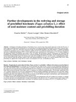
arthritis can rarely occur. A characteristic erysipelas-like rash
occurs in 20% of patients, usually around the ankles
(Figure 1). A degree of myalgia can occur during acute
attacks, but up to a fifth of patients complain of persistent
muscle pain on exertion, usually affecting the calves. Pro-
tracted febrile myalgia is rare and is characterised by severe
pain in the lower limbs or abdominal musculature which may
persist for weeks and can be accompanied by a vasculitic
rash; it usually responds to corticosteroids therapy.
Acute attacks are accompanied by a neutrophilic leuko-
cytosis, raised erythrocyte sedimentation rate, and a dramatic
acute-phase response. Investigations may be required to
exclude other diagnoses but imaging by x-ray, ultrasound, or
echocardiography during attacks is usually unrewarding.
Diagnosis is supported by DNA analysis but essentially
remains clinical and centres on the history of recurrent self-
limiting idiopathic attacks of fever and serositis that can be
prevented by prophylatic colchicine treatment. Genetic
results must be interpreted cautiously given that certain
individuals with paired pathogenic MEFV mutations never
develop FMF and that others with heterozygous carrier status
can do so. Furthermore, most diagnostic laboratories offer
only limited analysis of the large 10-exon MEFV gene.
Treatment
Supportive measures, including analgesia, are often required
during acute attacks, but the mainstay of management is
long-term prophylactic treatment with low-dose colchicine.
This was discovered serendipitously in 1972 by Goldfinger
[19] and has entirely transformed the outlook of this pre-
viously disabling disease. Continuous treatment with
colchicine at a dose of 1 to 2 mg daily in adults prevents or
substantially reduces symptoms of FMF in at least 95% of
cases and almost completely eliminates the risk of AA
amyloidosis (see below). The mechanism of action of
colchicine remains incompletely understood, but colchicine
binds to tubulin and evidently modulates neutrophil adhesion,
Available online />mobility, and cytokine release in a presumably rather specific
manner in patients with defective pyrin variants [20,21].
Long-term colchicine is advisable in every patient with FMF and
mandatory in those who already have AA amyloidosis. Although
colchicine is very toxic in acute overdose, the low daily doses
required for treatment of FMF are generally very well tolerated.
Diarrhoea is the most common side effect and usually can be
avoided by gradual introduction of the drug. Despite theoretical
concerns, there is no evidence that colchicine causes infertility
or birth defects and it can be taken safely by nursing mothers
[22]. Colchicine is a purely prophylactic treatment in FMF, and
introduction or dose escalation during an acute FMF attack is
not generally effective.
Genuine resistance to colchicine is probably very rare, although
issues of compliance are surprisingly common. Anecdotal
reports of benefit from treatment with etanercept or anakinra in
‘refractory’ patients are beginning to emerge [23,24].
Tumour necrosis factor receptor-associated periodic
syndrome
TRAPS is the second most common inherited fever
syndrome, although with an estimated prevalence of about 1
per million in the UK, it is very rare.
Genetics and pathophysiology
TRAPS is an autosomal dominant disease associated with
mutations in the gene for TNF receptor superfamily 1A
(TNFRSF1A), a 10-exon gene located on chromosome
12p13 [25]. TNF is a key mediator of inflammation with pleio-
tropic actions, including pyrexia, cachexia, leukocyte activa-
tion, induction of cytokine secretion, expression of adhesion
molecules, and resistance to intracellular pathogens. TNF
receptor 1 (TNFR1) is a member of the death domain
superfamily and comprises an extracellular motif containing
four cysteine-rich domains, a transmembrane domain, and an
intracellular death domain. Binding of soluble circulating TNF
causes trimerization of the receptor and activation of NF-κB,
with downstream induction of inflammation and inhibition of
apoptosis via production of cellular caspase-8-like inhibitory
protein (cFLIP). Events following endocytosis of the activated
TNFR1 complex result in apoptosis. The mechanism(s) by
which heterozygous TRFRSF1A mutations cause TRAPS
remain unclear and may well differ between mutations. Most
TRAPS-associated mutations lie within exons 2 to 4, of which
about half are missense substitutions affecting highly
conserved cysteine residues that disrupt structurally impor-
tant cysteine-cysteine disulphide bonds in the extracellular
domain. Under normal circumstances, TNF signalling is
terminated by metalloproteinase-dependent cleavage of a
proximal region of the extracellular domain, which releases
soluble TNFR1 that competitively inhibits binding of circu-
lating TNF to cell surface receptors. Whilst cleavage of
certain TNFR1 variants is impaired producing a ‘shedding
defect’, this is not the case with other TRAPS-causing
mutations, which must exert their pathogenic effect by
different means. It is thought that mutant misfolded receptors
may give rise to enhanced or prolonged signalling, possibly
through retention within the endoplasmic reticulum [26-29].
Despite initial hopes to the contrary, the mechanisms and
downstream effects by which TNFR1 mutations result in
TRAPS remain far from clear.
Clinical features
The clinical entity now known as TRAPS was described in
1982 as familial Hibernian fever [30], reflecting the Irish/
Scottish ancestry of patients in early reports, but TRAPS has
subsequently been reported in many ethnic groups, including
Jews, Arabs, and Central Americans. Males and females are
affected equally and presentation is usually before 4 years of
age. Most mutations are associated with high penetrance, but
two variants, P46L and R92Q, that can be associated with
TRAPS are present in approximately 10% of healthy West
Africans [31] and 1% of healthy Caucasians, respectively.
Attacks in TRAPS are far less distinct than in FMF. Febrile
episodes typically last 1 to 4 weeks and symptoms are nearly
continuous in a third of patients. Approximately half of
patients give no clear family history, many of whom have the
P46L or R92Q variants, which are also associated with
milder disease and later onset [32]. The clinical picture
varies: more than 95% of patients experience fever, and 80%
have arthralgia or myalgia that typically follows a centripetal
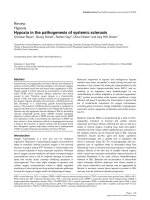
migratory path; abdominal pain occurs in 80%; and skin
manifestations, including erythematous rash (Figure 2),
oedematous plaques (often overlying areas of mylagic pain),
and discrete reticulate or serpiginous lesions, occur in 70%
of patients. Other features include headache, pleuritic pain,
lymphadenopathy, conjunctivitis, and periorbital oedema.
There are also reports of central nervous system manifes-
tations and imaging findings resembling multiple sclerosis
Arthritis Research & Therapy Vol 11 No 1 Lachmann and Hawkins
Page 4 of 10
(page number not for citation purposes)
Figure 1
Erysipelas-like erythema around the ankle, the characteristic painful
rash seen in attacks of familial Mediterranean fever.
[33]. Symptoms are almost universally accompanied by a
marked acute-phase response. During quiescent periods, the
plasma concentration of soluble TNFR1 may be abnormally
low in patients with decreased receptor shedding. Genetic
testing is central to diagnosis.
Treatment
Despite high initial hopes for response to anti-TNF biologics,
treatment of TRAPS often remains disappointing. Acute
attacks do respond to high-dose corticosteroids, and
etanercept (but interestingly not infliximab) is useful in some
patients, although response may gradually decline [34]. A
recent report suggested that IL-1 blockade with anakinra can
be very effective in some patients [35].
The hyper IgD and periodic fever syndrome
Genetics and pathophysiology
Hyper IgD and periodic fever syndrome (HIDS) is an auto-
somal recessive disease caused by mutations in the mevalo-
nate kinase (MVK) gene on the long arm of chromosome 12
[36]. About 60 mutations have been described, spanning the
11-exon gene, the most common of which encode MVK
variants V377I and I268T. MVK is the enzyme following HMG
CoA (or 3-hydroxy-3-methylglutaryl-coenzyme A) reductase in
the pathway involved in cholesterol, farnasyl, and isoprenoid
biosynthesis. Most HIDS-causing MVK mutations are
missense variants that reduce enzyme activity by 90% to
99% [37]. Other mutations resulting in near-complete
absence of enzyme activity cause a much more severe
inflammatory disease known as mevalonic aciduria (MVA),
features of which include stillbirth, congenital malformations,
severe psychomotor retardation, ataxia, myopathy, failure to
thrive, and early death.
It is not yet known how MVK deficiency causes inflammation
or increased IgD production, although reduction in preny-
lation due to failure of flux through the isoprenoid pathway
currently seems more likely to be responsible than accumu-
lation of the enzyme’s substrate [38,39]. The relationship of
the isoprenoid pathway to inflammation is of all the more
interest given the anti-inflammatory properties of statin drugs
that are widely used to inhibit cholesterol synthesis. Whilst
various effects of statins on caspase-1 activation and IL-1
secretion have been postulated, a clinical study of simvastatin
of six patients with HIDS suggested only minor benefit [40];
rather worryingly, two other children with MVA were reported
to develop severe flares of inflammatory disease following
statin treatment [41].
Clinical features
HIDS is extremely rare and is predominantly a Dutch disease,
probably through a founder effect. It was described in The
Netherlands in 1984 and the international registry in
Nijmegen has data on just over 200 patients [42]. The
carriage rate of MVK V337I is 1 in 350 in the Dutch popu-
lation [43], but HIDS has been reported in many other
countries and other ethnic groups, including Arabs and
Southeast Asians. The disease occurs equally in males and
females and usually presents in the first year of life [44].
Attacks are irregular, typically lasting 4 to 7 days, and are
characteristically provoked by vaccination, minor trauma,
surgery, or stress, perhaps triggered by a reduction in MVK
enzyme associated with increased body temperature [45].
Attacks of HIDS typically comprise fever, cervical lymph-
adenopathy, splenomegaly, and abdominal pain with vomiting
and diarrhoea. Headache, arthralgia, large-joint arthritis,
erythematous macules and papules, and aphthous ulcers are
also common. HIDS typically ameliorates in adult life and
older patients may remain well for years.
Diagnosis of HIDS is supported by a high serum IgD
concentration, although this is not specific and is not always
present [46]. More accessibly, serum IgA concentration is
Available online />Page 5 of 10
(page number not for citation purposes)
Figure 2
Erythematous rash complicating an acute attack in tumour necrosis
factor receptor-associated periodic syndrome (TRAPS).
also elevated in 80% of patients. Attacks are accompanied
by an acute-phase response, leukocytosis, and the transient
presence of mevalonic acid in the urine. A mutation in both
alleles of the MVK gene can be identified in most patients,
including the MVK V337I variant in 50% to 80% of cases.
Treatment
Treatment is largely supportive, including nonsteroidal anti-
inflammatory drugs, although responses to etanercept
[47,48] and anakinra have lately been reported. A cautious
therapeutic trial of statin therapy may be worthwhile.
Cryopyrin-associated periodic syndrome
CAPS comprises a much-overlapping spectrum of three
hitherto separately described diseases, ranging from mild to
severe, respectively: familial cold urticaria, now known as
familial cold autoinflammatory syndrome (FCAS); Muckle-
Wells syndrome (MWS); and chronic infantile neurological,
cutaneous, and articular syndrome (CINCA), which is known
in the US as neonatal onset multisystem inflammatory disease
(NOMID).
Genetics and pathophysiology
CAPS is associated with various mutations in NLRP3/CIAS1
on chromosome 1q44, a gene that encodes the death
domain protein known variously as NLRP3, NALP3, and
cryopyrin [49]. Dominant inheritance is evident in about 75%
of patients with FCAS and MWS, whereas CINCA, at the
most severe end of the clinical spectrum, is usually due to de
novo mutation. More than 60, mostly missense, mutations
have been reported and all but three of them are in exon 3.
The genotype-phenotype relationship can differ markedly
between individuals, even within a family.
NLRP3 is expressed in granulocytes, dendritic cells, B and
T lymphocytes, epithelial cells of the oral and genital tracts,
and chondrocytes. It encodes a protein that has a PYD, a
nucleotide-binding site domain, and a leucine-rich repeat
(LRR) motif. Signalling through a variety of danger signals,
including intracellular pathogen associated molecular
patterns (PAMP) and uric acid, results in the association of
NLRP3 via its LRR with other members of the death domain
superfamily to form a multimeric cytosolic protein complex,
known collectively as the inflammasome [50,51]. This results
in activation of caspase-1, which cleaves pro-IL-1 to produce
active IL-1-β and IL-1-α; it also upregulates NF-κB expression
and thereby increases IL-1 gene expression. IL-1 is a major
proinflammatory cytokine that mediates a multitude of local
and systemic responses to infection and tissue injury and, as
proved by the complete response of CAPS to IL-1 receptor
blockade, is pivotal in causing the clinical features of this
disease [52].
Clinical features
Most reported patients with CAPS have European ancestry
but cases have been described from South Asia and
elsewhere [53]. Onset of disease is usually in early infancy,
often from birth, and there is no gender bias. FCAS is the
most common in North America and was described in 1940
as recurrent episodes of cold-induced fever, arthralgia,
conjunctivitis, and rash (Figure 3). MWS was described in
1962 [54] as a syndrome with often daily attacks of urticarial
rash, conjunctivitis, arthralgia, and fever, complicated by
progressive sensorineural deafness in 40% of patients, and a
high risk of AA amyloidosis. CINCA is a sporadic severe
inflammatory disorder that presents in the neonatal period
with multisystem involvement including the skin, skeletal
system, and central nervous system [55]. Bony overgrowth
and premature ossification may occur particularly in the skull
and knees (Figure 4); chronic aseptic meningitis results in
developmental retardation; and blindness due to optic
atrophy and deafness are also common. The relationship
between these three overlapping syndromes, essentially
encompassing a spectrum of severity, was recognised in only
the past few years after their common genetic aetiology was
discovered.
Clinical disease is accompanied by an acute-phase response
and often leukocytosis and thrombocytosis and anaemia of
chronic disease. Sensorineural hearing loss should be sought
with audiometry, and characteristic bony abnormalities may
be evident radiologically. Fundoscopy and brain imaging may
show features consistent with elevated intracranial pressure.
A mutation in NLRP3 can be identified in almost all patients
with clinical FCAS or MWS, although mutations are found in
only about 50% of children with classic CINCA; it is possible
that ‘mutation-negative’ cases of FCAS and MWS may also
exist but are simply not being recognised.
Treatment
Daily injections of anakinra (recombinant IL-1 receptor
antagonist) produce rapid and complete clinical and sero-
logical remission in CAPS [52]. It is hoped that early anti-IL-1
therapy may prevent developmental abnormalities in children
with disease toward the severe end of the spectrum [56].
Various new longer acting IL-1 inhibitors are also proving to
be very effective [57] and early safety and efficacy data look
encouraging [58].
Pyogenic sterile arthritis, pyoderma gangrenosum, and
acne (PAPA) syndrome
This exceptionally rare autosomal dominant disease is caused
by mutations in the proline serine threonine phosphatase-
interacting protein-1 (PTSTPIP) gene encoding a protein also
known as CD2-binding protein-1 (CB2BP1) [59]. Stimulated
macrophages isolated from patients demonstrate increased
IL-1β release, suggesting that mutations result in increased
activation of caspase-1. The underlying pathogenesis remains
poorly understood, although there is evidence that CD2BP1,
which interacts with actin and is an important component of
cytoskeletal organisation, interacts with pyrin [60]. This
interaction is significantly increased by tyrosine phosphory-
Arthritis Research & Therapy Vol 11 No 1 Lachmann and Hawkins
Page 6 of 10
(page number not for citation purposes)
lation of native CD2BP1. Disease-associated mutations have
also been shown to potentiate the pyrin-CD2BP1 interaction.
There is some evidence that this may result in unmasking of
pyrin’s PYD domain and thus a possible mechanism by which
mutations could result in caspase-1 activation [61]. PAPA is
characterised clinically by severe acne and recurrent pustular
sterile arthritis that typically occurs after minor trauma. Early
reports suggest that therapy with anakinra may be effective.
Blau syndrome or early-onset sarcoidosis
This sarcoid-like syndrome was described in 1985 as an
autosomal dominant syndrome of granulomatous infiltration of
the joints causing camptodactyly, skin, and sometimes viscera
associated with uveitis [62]. Another syndrome, early-onset
sarcoidosis, is probably the same disease and both have been
shown to be associated with missense mutations in NOD2/
CARD15. This is another member of the death domain super-
family [63] and is thought to serve as an intracellular receptor
for PAMPs leading to NF-κB activation. NOD2 mutations have
also been implicated in familial Crohn disease, another
granulomatous disease. Treatment is with corticosteroids.
Acquired autoinflammatory conditions
Schnitzler syndrome
Schnitzler syndrome is a disorder of unknown pathogenesis
characterised by relapsing urticarial rashes, periodic fevers,
arthralgias/arthritis, lymphadenopathy, and IgM parapro-
teinaemia, which can be of a very low level. Fewer than 100
patients have been reported. Onset is in adulthood, reflecting
susceptibility with increasing age to paraproteinaemia. Long-
term outcomes appear good, with 15-year survival exceeding
90%, although overt lymphoproliferative disease evolves in
more than 15% of patients. Chemotherapy directed toward
the underlying clonal B-cell disorder is effective in some but
not all patients, possibly due to the low proportion in whom
complete suppression of the IgM paraproteinaemia can be
achieved. A pivotal role of IL-1 in the pathogenesis of this
acquired disorder has lately been suggested by remarkable
therapeutic efficacy of anakinra in a number of patients [64].
Gout and pseudogout
A place for these acute inflammatory arthritides in the
umbrella of autoinflammatory disorders has recently been
Available online />Page 7 of 10
(page number not for citation purposes)
Figure 3
Characteristic urticarial lesions that develop almost every afternoon in
this patient with cryopyrin-associated periodic syndrome (CAPS)
accompanied by fever, generalised myalgia, and conjunctivitis.
Figure 4
Severe cryopyrin-associated periodic syndrome (CAPS), toward the
chronic infantile neurological, cutaneous, and articular syndrome
(CINCA) end of the spectrum, is frequently associated with arthropathy
as shown here. The knees are enlarged with deformed femora without
synovitis. Short stature and finger clubbing are also well-recognised
features of the syndrome.
suggested by observations that monosodium urate (MSU)
and/or calcium pyrophosphate dihydrate (CPPD) crystals can
activate the NLRP3 inflammasome, resulting in the produc-
tion of active IL1-β and IL-18 [65]. Macrophages from mice
with knockouts of a variety of inflammasome components
produce significantly less IL-1β compared with wild-type
animals following challenge with MSU or CPPD crystals.
Involvement of IL-1β in crystal arthritis has recently been
confirmed clinically in an open-label study of anakinra in 10
patients with acute gout [66].
Long-term outcomes
Although CINCA/NOMID can be sufficiently severe to cause
death within the first few decades, life expectancy among
many patients with autoinflammatory disorders is typically
near normal and is expected to be excellent in those for
whom there is now effective therapy. The most serious and
life-threatening complication of these diseases generally is
AA amyloidosis.
AA amyloidosis
Reactive systemic (AA) amyloidosis is an often fatal
disorder, predominantly affecting the kidneys, which occurs
in a small proportion of patients with one of a wide range of
chronic inflammatory diseases [67]. AA amyloid fibrils are
derived from the circulating acute-phase reactant serum
amyloid A protein (SAA), and their accumulation in tissues
throughout the body progressively damages the structure
and function of vital organs. SAA is synthesised by the liver
under transcriptional regulation of IL-1, interleukin 6 (IL-6)
and TNF-α, and its plasma concentration, which in health is
less than 3 mg/l, may rise a thousand fold in the presence of
inflammation [68]. Whilst the lifetime incidence of AA
amyloidosis is about 1% to 5% in patients with chronic
inflammatory diseases generally, it is much more common
among patients with inherited periodic fever syndromes,
although the factors that determine susceptibility to its
development, other than the presence of an acute-phase
response for a long period, are not known. The median
duration of inflammatory disease in patients who develop
amyloidosis is about 20 years, and the life-long nature of
inherited periodic fever syndromes is presumably a factor in
the high prevalence of amyloid in these diseases; another
factor may be the unusually high plasma concentrations of
SAA that typically occur in inherited periodic fever
syndromes. Up to 60% of patients with FMF died of renal
failure due to AA amyloidosis before prophylactic colchicine
was widely prescribed, and even recently it was reported in
13% of a large Turkish series. The incidence of AA
amyloidosis in TRAPS and CAPS is approximately 25% but
is less than 5% in HIDS, perhaps because the disease often
ameliorates spontaneously in early adulthood. The natural
history of untreated AA amyloidosis is of renal failure and
early death, but this can be prevented by treatment of the
underlying inflammatory disorder that substantially sup-
presses SAA production.
Conclusions
Recent progress in elucidating the pathogenesis of many
autoinflammatory diseases has led to major advances in their
treatment, most remarkably the introduction of IL-1 inhibition
in CAPS. The clinical significance of low-penetrance muta-
tions/polymorphisms in the inherited period fever syndrome
genes remains unclear, although there is early evidence that
they may potentiate inflammation more generally [69,70]. The
multitude of studies currently in progress, both in rare
hereditary autoinflammatory diseases and in more common
acquired ones (including Crohn disease, systemic onset
juvenile arthritis, and Behçet syndrome), are expected to shed
important further light on aspects of the innate immune
system and inflammation generally over the next few years.
Competing interests
The authors declare that they have no competing interests.
Acknowledgements
Written consent for publication of their photographs was obtained from
all patients featured in this article.
References
1. Heller H, Sohar E, Sherf L: Familial Mediterranean fever. AMA
Arch Intern Med 1958, 102:50-71.
2. Consortium TFF: A candidate gene for familial Mediterranean
fever. Nat Genet 1997, 17:25-31.
3. Consortium TIF: Ancient missense mutations in a new member
of the RoRet gene family are likely to cause familial Mediter-
ranean fever. Cell 1997, 90:797-807.
4. Centola M, Wood G, Frucht DM, Galon J, Aringer M, Farrell C,
Kingma DW, Horwitz ME, Mansfield E, Holland SM, O’Shea JJ,
Rosenberg HF, Malech HL, Kastner DL: The gene for familial
Mediterranean fever, MEFV, is expressed in early leukocyte
development and is regulated in response to inflammatory
mediators. Blood 2000, 95:3223-3231.
5. Infevers homepage [ />6. Lachmann HJ, Sengül B, Yavuzsen TU, Booth DR, Booth SE,
Bybee A, Gallimore JR, Soytürk M, Akar S, Tunca M, Hawkins PN:
Clinical and subclinical inflammation in patients with familial
Mediterranean fever and in heterozygous carriers of MEFV
mutations. Rheumatology (Oxford) 2006, 45:746-750.
7. Booth DR, Gillmore JD, Lachmann HJ, Booth SE, Bybee A,
Soytürk M, Akar S, Pepys MB, Tunca M, Hawkins PN: The
genetic basis of autosomal dominant familial Mediterranean
fever. QJM 2000, 93:217-221.
Arthritis Research & Therapy Vol 11 No 1 Lachmann and Hawkins
Page 8 of 10
(page number not for citation purposes)
This article is part of a special collection of reviews, The
Scientific Basis of Rheumatology: A Decade of
Progress, published to mark Arthritis Research &
Therapy’s 10th anniversary.
Other articles in this series can be found at:
/>The Scientific Basis
of Rheumatology:
A Decade of Progress
8. Cañete JD, Arostegui JI, Queiró R, Gratacós J, Hernández MV,
Larrosa M, Alperí M, Moll C, Rius J, Sanmartí R, Yagüe J: An
unexpectedly high frequency of MEFV mutations in patients
with anti-citrullinated protein antibody-negative palindromic
rheumatism. Arthritis Rheum 2007, 56:2784-2788.
9. Kalyoncu M, Acar BC, Cakar N, Bakkaloglu A, Ozturk S, Dereli E,
Tunca M, Kasapcopur O, Yalcinkaya F, Ozen S: Are carriers for
MEFV mutations “healthy”? Clin Exp Rheumatol 2006, 24:
S120-122.
10. Hall MW, Gavrilin MA, Knatz NL, Duncan MD, Fernandez SA,
Wewers MD: Monocyte mRNA phenotype and adverse out-
comes from pediatric multiple organ dysfunction syndrome.
Pediatr Res 2007, 62:597-603.
11. Tidow N, Chen X, Müller C, Kawano S, Gombart AF, Fischel-
Ghodsian N, Koeffler HP: Hematopoietic-specific expression of
MEFV, the gene mutated in familial Mediterranean fever, and
subcellular localization of its corresponding protein, pyrin.
Blood 2000, 95:1451-1455.
12. Martinon F, Hofmann K, Tschopp J: The pyrin domain: a possi-
ble member of the death domain-fold family implicated in
apoptosis and inflammation. Curr Biol 2001, 10:R118-R120.
13. Park HH, Lo YC, Lin SC, Wang L, Yang JK, Wu H: The death
domain superfamily in intracellular signaling of apoptosis and
inflammation. Annu Rev Immunol 2007, 25:561-586.
14. Chae JJ, Wood G, Masters SL, Richard K, Park G, Smith BJ,
Kastner DL: The B30.2 domain of pyrin, the familial Mediter-
ranean fever protein, interacts directly with caspase-1 to mod-
ulate IL-1beta production. Proc Natl Acad Sci U S A 2006, 103:
9982-9987.
15. Chae JJ, Wood G, Richard K, Jaffe H, Colburn NT, Masters SL,
Gumucio DL, Shoham NG, Kastner DL: The familial Mediter-
ranean fever protein, pyrin, is cleaved by caspase-1 and acti-
vates NF-kappaB through its N-terminal fragment. Blood
2008, 112:1794-1803.
16. Mikula M, Buller A, Sun W, Strom CM: Prevalence of known
mutations in the familial Mediterranean fever gene (MEFV) in
various carrier screening populations. Genet Med 2008, 10:
349-352.
17. Ross JJ: Goats, germs, and fever: are the pyrin mutations
responsible for familial Mediterranean fever protective
against Brucellosis? Med Hypotheses 2007, 68:499-501.
18. Schaner P, Richards N, Wadhwa A, Aksentijevich I, Kastner D,
Tucker P, Gumucio D: Episodic evolution of pyrin in primates:
human mutations recapitulate ancestral amino acid states.
Nat Genet 2001, 27:318-321.
19. Goldfinger SE: Colchicine for familial Mediterranean fever. N
Engl J Med 1972, 287:1302.
20. Rigante D, La Torraca I, Avallone L, Pugliese AL, Gaspari S,
Stabile A: The pharmacologic basis of treatment with
colchicine in children with familial Mediterranean fever. Eur
Rev Med Pharmacol Sci 2006, 10:173-178.
21. Chia EW, Grainger R, Harper JL: Colchicine suppresses neu-
trophil superoxide production in a murine model of gouty
arthritis: a rationale for use of low-dose colchicine. Br J Phar-
macol 2008, 153:1288-1295.
22. Ben-Chetrit E, Levy M: Reproductive system in familial Mediter-
ranean fever: an overview. Ann Rheum Dis 2003, 62:916-919.
23. Mor A, Pillinger MH, Kishimoto M, Abeles AM, Livneh A: Familial
Mediterranean fever successfully treated with etanercept. J
Clin Rheumatol 2007, 13:38-40.
24. Roldan R, Ruiz AM, Miranda MD, Collantes E: Anakinra: new
therapeutic approach in children with Familial Mediterranean
Fever resistant to colchicine. Joint Bone Spine 2008, 75:504-
505.
25. McDermott MF, Aksentijevich I, Galon J, McDermott EM, Ogunko-
lade BW, Centola M, Mansfield E, Gadina M, Karenko L, Petters-
son T, McCarthy J, Frucht DM, Aringer M, Torosyan Y, Teppo AM,
Wilson M, Karaarslan HM, Wan Y, Todd I, Wood G, Schlimgen R,
Kumarajeewa TR, Cooper SM, Vella JP, Amos CI, Mulley J, Quane
KA, Molloy MG, Ranki A, Powell RJ, et al.: Germline mutations in
the extracellular domains of the 55 kDa TNF receptor, TNFR1,
define a family of dominantly inherited autoinflammatory syn-
dromes. Cell 1999, 97:133-144.
26. D’Osualdo A, Ferlito F, Prigione I, Obici L, Meini A, Zulian F, Pon-
tillo A, Corona F, Barcellona R, Di Duca M, Santamaria G, Tra-
verso F, Picco P, Baldi M, Plebani A, Ravazzolo R, Ceccherini I,
Martini A, Gattorno M: Neutrophils from patients with
TNFRSF1A mutations display resistance to tumor necrosis
factor-induced apoptosis: pathogenetic and clinical implica-
tions. Arthritis Rheum 2006, 54:998-1008.
27. Rebelo SL, Bainbridge SE, Amel-Kashipaz MR, Radford PM,
Powell RJ, Todd I, Tighe PJ: Modeling of tumor necrosis factor
receptor superfamily 1A mutants associated with tumor
necrosis factor receptor-associated periodic syndrome indi-
cates misfolding consistent with abnormal function. Arthritis
Rheum 2006, 54:2674-2687.
28. Todd I, Radford PM, Daffa N, Bainbridge SE, Powell RJ, Tighe PJ:
Mutant tumor necrosis factor receptor associated with tumor
necrosis factor receptor-associated periodic syndrome is
altered antigenically and is retained within patients’ leuko-
cytes. Arthritis Rheum 2007, 56:2765-2773.
29. Nedjai B, Hitman GA, Yousaf N, Chernajovsky Y, Stjernberg-
Salmela S, Pettersson T, Ranki A, Hawkins PN, Arkwright PD,
McDermott MF, Turner MD: Abnormal tumor necrosis factor
receptor I cell surface expression and NF-
κκ
B activation in
tumor necrosis factor receptor-associated periodic syndrome.
Arthritis Rheum 2008, 58:273-283.
30. Williamson LM, Hull D, Mehta R, Reeves WG, Robinson BH,
Toghill PJ: Familial Hibernian fever. QJM 1982, 51:469-480.
31. Tchernitchko D, Chiminqgi M, Galactéros F, Préhu C, Segbena Y,
Coulibaly H, Rebaya N, Loric S: Unexpected high frequency of
P46L TNFRSF1A allele in sub-Saharan West African popula-
tions. Eur J Hum Genet 2005, 13:513-515.
32. Ravet N, Rouaghe S, Dodé C, Bienvenu J, Stirnemann J, Lévy P,
Delpech M, Grateau G: Clinical significance of P46L and R92Q
substitutions in the tumour necrosis factor superfamily 1A
gene. Ann Rheum Dis 2006, 65:1158-1162.
33. Hoffmann LA, Lohse P, Konig FB, Feneberg W, Hohlfeld R,
Kumpfel T: TNFRSF1A R92Q mutation in association with a
multiple sclerosis-like demyelinating syndrome. Neurology
2008, 70:1155-1156.
34. Nowlan ML, Drewe E, Bulsara H, Esposito N, Robins RA, Tighe
PJ, Powell RJ, Todd I: Systemic cytokine levels and the effects
of etanercept in TNF receptor-associated periodic syndrome
(TRAPS) involving a C33Y mutation in TNFRSF1A. Rheumatol-
ogy (Oxford) 2006, 45:31-37.
35. Gattorno M, Pelagatti MA, Meini A, Obici L, Barcellona R, Federici
S, Buoncompagni A, Plebani A, Merlini G, Martini A: Persistent
efficacy of anakinra in patients with tumor necrosis factor
receptor-associated periodic syndrome. Arthritis Rheum 2008,
58:1516-1520.
36. van der Meer JW, Vossen JM, Radl J, van Nieuwkoop JA, Meyer
CJ, Lobatto S, van Furth R: Hyperimmunoglobulinaemia D and
periodic fever: a new syndrome. Lancet 1984, 1:1087-1090.
37. Cuisset L, Drenth JP, Simon A, Vincent MF, van der Velde Visser
S, van der Meer JW, Grateau G, Delpech M; International Hyper-
IgD Study Group: Molecular analysis of MVK mutations and
enzymatic activity in hyper-IgD and periodic fever syndrome.
Eur J Hum Genet 2001, 9:260-266.
38. Houten SM, Frenkel J, Waterham HR: Isoprenoid biosynthesis
in hereditary periodic fever syndromes and inflammation. Cell
Mol Life Sci 2003, 60:1118-1134.
39. Schneiders MS, Houten SM, Turkenburg M, Wanders RJ, Water-
ham HR: Manipulation of isoprenoid biosynthesis as a possi-
ble therapeutic option in mevalonate kinase deficiency.
Arthritis Rheum 2006, 54:2306-2313.
40. Simon A, Drewe E, van der Meer JW, Powell RJ, Kelley RI, Stalen-
hoef AF, Drenth JP: Simvastatin treatment for inflammatory
attacks of the hyperimmunoglobulinemia D and periodic fever
syndrome. Clin Pharmacol Ther 2004, 75:476-483.
41. Hoffmann GF, Charpentier C, Mayatepek E, Mancini J, Leichsen-
ring M, Gibson KM, Divry P, Hrebicek M, Lehnert W, Sartor K,
Trefz FK, Rating D, Bremer HJ, Nyhan WL: Clinical and biochem-
ical phenotype in 11 patients with mevalonic aciduria. Pedi-
atrics 1993, 91:915-921.
42. Hyper-IgD and periodic fever syndrome (HIDS) homepage
[].
43. Houten SM, van Woerden CS, Wijburg FA, Wanders RJ, Water-
ham HR: Carrier frequency of the V377I (1129G>A) MVK
mutation, associated with Hyper-IgD and periodic fever syn-
drome, in the Netherlands. Eur J Hum Genet 2003, 11:196-
200.
44. Drenth JP, Haagsma CJ, van der Meer JW: Hyperimmunoglobu-
linemia D and periodic fever syndrome. The clinical spectrum
Available online />Page 9 of 10
(page number not for citation purposes)
in a series of 50 patients. International Hyper-IgD Study
Group. Medicine (Baltimore) 1994, 73:133-144.
45. Houten SM, Frenkel J, Rijkers GT, Wanders RJ, Kuis W, Water-
ham HR: Temperature dependence of mutant mevalonate
kinase activity as a pathogenic factor in hyper-IgD and peri-
odic fever syndrome. Hum Mol Genet 2002, 11:3115-3124.
46. Ammouri W, Cuisset L, Rouaghe S, Rolland MO, Delpech M,
Grateau G, Ravet N: Diagnostic value of serum immunoglobu-
linaemia D level in patients with a clinical suspicion of hyper
IgD syndrome. Rheumatology (Oxford) 2007, 46:1597-1600.
47. Takada K, Aksentijevich I, Mahadevan V, Dean JA, Kelley RI,
Kastner DL: Favorable preliminary experience with etanercept
in two patients with the hyperimmunoglobulinemia D and
periodic fever syndrome. Arthritis Rheum 2003, 48:2645-2651.
48. Lachmann HJ, Goodman HJ, Andrews PA, Gallagher H, Marsh J,
Breuer S, Rowczenio DM, Bybee A, Hawkins PN: AA amyloido-
sis complicating hyperimmunoglobulinemia D with periodic
fever syndrome: a report of two cases. Arthritis Rheum 2006,
54:2010-2014.
49. Hoffman HM, Wright FA, Broide DH, Wanderer AA, Kolodner RD:
Identification of a locus on chromosome 1q44 for familial cold
urticaria. Am J Hum Genet 2000, 66:1693-1698.
50. Tschopp J, Martinon F, Burns K: NALPs: a novel protein family
involved in inflammation. Nat Rev Mol Cell Biol 2003, 4:95-
104.
51. Martinon F, Agostini L, Meylan E, Tschopp J: Identification of
bacterial muramyl dipeptide as activator of the NALP3/cry-
opyrin inflammasome. Curr Biol 2004, 14:1929-1934.
52. Hawkins PN, Lachmann HJ, McDermott MF: Interleukin-1-recep-
tor antagonist in the Muckle-Wells syndrome. N Engl J Med
2003, 348:2583-2584.
53. Leslie KS, Lachmann HJ, Bruning E, McGrath JA, Bybee A, Gal-
limore JR, Roberts PF, Woo P, Grattan CE, Hawkins PN: Pheno-
type, genotype, and sustained response to anakinra in 22
patients with autoinflammatory disease associated with
CIAS-1/NALP3 mutations. Arch Dermatol 2006, 142:1591-
1597.
54. Muckle TJ, Wells MV: Urticaria, deafness and amyloidosis: a
new heredo-familial syndrome. QJM 1962, 31:235-248.
55. Prieur AM, Griscelli C, Lampert F, Truckenbrodt H, Guggenheim
MA, Lovell DJ, Pelkonnen P, Chevrant-Breton J, Ansell BM: A
chronic, infantile, neurological, cutaneous and articular
(CINCA) syndrome. A specific entity analysed in 30 patients.
Scand J Rheumatol Suppl 1987, 66:57-68.
56. Goldbach-Mansky R, Dailey NJ, Canna SW, Gelabert A, Jones J,
Rubin BI, Kim HJ, Brewer C, Zalewski C, Wiggs E, Hill S, Turner
ML, Karp BI, Aksentijevich I, Pucino F, Penzak SR, Haverkamp
MH, Stein L, Adams BS, Moore TL, Fuhlbrigge RC, Shaham B,
Jarvis JN, O’Neil K, Vehe RK, Beitz LO, Gardner G, Hannan WP,
Warren RW, Horn W, et al.: Neonatal-onset multisystem
inflammatory disease responsive to interleukin-1b inhibition.
N Engl J Med 2006, 355:581-592.
57. Hoffman HM, Throne ML, Amar NJ, Sebai M, Kivitz AJ, Kavanaugh
A, Weinstein SP, Belomestnov P, Yancopoulos GD, Stahl N,
Mellis SJ: Efficacy and safety of rilonacept (interleukin-1 trap)
in patients with cryopyrin-associated periodic syndromes:
results from two sequential placebo-controlled studies. Arthri-
tis Rheum 2008, 58:2443-2452.
58. Goldbach-Mansky R, Shroff SD, Wilson M, Snyder C, Plehn S,
Barham B, Pham TH, Pucino F, Wesley RA, Papadopoulos JH,
Weinstein SP, Mellis SJ, Kastner DL: A pilot study to evaluate
the safety and efficacy of the long-acting interleukin-1
inhibitor rilonacept (interleukin-1 Trap) in patients with famil-
ial cold autoinflammatory syndrome. Arthritis Rheum 2008, 58:
2432-2442.
59. Wise CA, Gillum JD, Seidman CE, Lindor NM, Veile R, Bashiardes
S, Lovett M: Mutations in CD2BP1 disrupt binding to PTP
PEST and are responsible for PAPA syndrome, an autoinflam-
matory disorder. Hum Mol Genet 2002, 11:961-969.
60. Shoham NG, Centola M, Mansfield E, Hull KM, Wood G, Wise
CA, Kastner DL: Pyrin binds the PSTPIP1/CD2BP1 protein,
defining familial Mediterranean fever and PAPA syndrome as
disorders in the same pathway. Proc Natl Acad Sci U S A
2003, 100:13501-13506.
61. Yu JW, Fernandes-Alnemri T, Datta P, Wu J, Juliana C, Solorzano
L, McCormick M, Zhang Z, Alnemri ES: Pyrin activates the ASC
pyroptosome in response to engagement by autoinflamma-
tory PSTPIP1 mutants. Mol Cell 2007, 28:214-227.
62. Blau EB: Familial granulomatous arthritis, iritis, and rash. J
Pediatr 1985, 107:689-693.
63. Miceli-Richard C, Lesage S, Rybojad M, Prieur AM, Manouvrier-
Hanu S, Häfner R, Chamaillard M, Zouali H, Thomas G, Hugot JP:
CARD15 mutations in Blau syndrome. Nat Genet 2001, 29:19-
20.
64. de Koning HD, Bodar EJ, van der Meer JW, Simon A: Schnitzler
syndrome: beyond the case reports: review and follow-up of
94 patients with an emphasis on prognosis and treatment.
Semin Arthritis Rheum 2007, 37:137-148.
65. Martinon F, Petrilli V, Mayor A, Tardivel A, Tschopp J: Gout-asso-
ciated uric acid crystals activate the NALP3 inflammasome.
Nature 2006, 440:237-241.
66. So A, De Smedt T, Revaz S, Tschopp J: A pilot study of IL-1
inhibition by anakinra in acute gout. Arthritis Res Ther 2007, 9:
R28.
67. Lachmann HJ, Goodman HJ, Gilbertson JA, Gallimore JR, Sabin
CA, Gillmore JD, Hawkins PN: Natural history and outcome in
systemic AA amyloidosis. N Engl J Med 2007, 356:2361-2371.
68. Ledue TB, Weiner DL, Sipe JD, Poulin SE, Collins MF, Rifai N:
Analytical evaluation of particle-enhanced immunonephelo-
metric assays for C-reactive protein, serum amyloid A and
mannose-binding protein in human serum. Ann Clin Biochem
1998, 35:745-753.
69. Aganna E, Hawkins PN, Ozen S, Pettersson T, Bybee A, McKee
SA, Lachmann HJ, Karenko L, Ranki A, Bakkaloglu A, Besbas N,
Topaloglu R, Hoffman HM, Hitman GA, Woo P, McDermott MF:
Allelic variants in genes associated with hereditary periodic
fever syndromes as susceptibility factors for reactive sys-
temic AA amyloidosis. Genes Immun 2004, 5:289-293.
70. Aksentijevich I, Galon J, Soares M, Mansfield E, Hull K, Oh HH,
Goldbach-Mansky R, Dean J, Athreya B, Reginato AJ, Henrickson
M, Pons-Estel B, O’Shea JJ, Kastner DL: The tumor-necrosis-
factor receptor-associated periodic syndrome: new mutations
in TNFRSF1A, ancestral origins, genotype-phenotype studies,
and evidence for further genetic heterogeneity of periodic
fevers. Am J Hum Genet 2001, 69:301-314.
Arthritis Research & Therapy Vol 11 No 1 Lachmann and Hawkins
Page 10 of 10
(page number not for citation purposes)