Báo cáo y học: "Increased expression of lipocalin-type prostaglandin D2 synthase in osteoarthritic cartilage" docx
Bạn đang xem bản rút gọn của tài liệu. Xem và tải ngay bản đầy đủ của tài liệu tại đây (2.43 MB, 12 trang )
Open Access
Available online />Page 1 of 12
(page number not for citation purposes)
Vol 10 No 6
Research article
Increased expression of lipocalin-type prostaglandin D
2
synthase
in osteoarthritic cartilage
Nadia Zayed
1
, Xinfang Li
1
, Nadir Chabane
1
, Mohamed Benderdour
2
, Johanne Martel-Pelletier
1
,
Jean-Pierre Pelletier
1
, Nicolas Duval
3
and Hassan Fahmi
1
1
Osteoarthritis Research Unit, Research Centre of the University of Montreal Hospital Center (CR-CHUM), Notre-Dame Hospital, 1560 Sherbrooke
Street East, J.A. DeSève Pavilion, Y-2628, and Department of Medicine, University of Montreal, Montreal, QC, H2L 4M1, Canada
2
Research Centre, Sacré-Coeur Hospital, 5400, Gouin Boulevard West, Montreal, QC, H4J 1C5, Canada
3
Centre de Convalescence, de Charmilles Pavillon, 1487 des Laurentides Boulevard, Montreal, QC, H7M 2Y3, Canada
Corresponding author: Hassan Fahmi,
Received: 12 Sep 2008 Revisions requested: 17 Oct 2008 Revisions received: 2 Dec 2008 Accepted: 18 Dec 2008 Published: 18 Dec 2008
Arthritis Research & Therapy 2008, 10:R146 (doi:10.1186/ar2581)
This article is online at: />© 2008 Zayed et al.; licensee BioMed Central Ltd.
This is an open access article distributed under the terms of the Creative Commons Attribution License ( />),
which permits unrestricted use, distribution, and reproduction in any medium, provided the original work is properly cited.
Abstract
Introduction Prostaglandin D synthase (PGDS) is responsible
for the biosynthesis of PGD and J series, which have been
shown to exhibit anti-inflammatory and anticatabolic effects.
Two isoforms have been identified: hematopoietic- and lipocalin-
type PGDS (H-PGDS and , respectively). The aims of this study
were to investigate the expressions of H-PGDS and L-PGDS in
cartilage from healthy donors and from patients with
osteoarthritis (OA) and to characterize their regulation by
interleukin-1-beta (IL-1β) in cultured OA chondrocytes.
Methods The expressions of H-PGDS and L-PGDS mRNA and
protein in cartilage were analyzed by real-time reverse
transcriptase-polymerase chain reaction (RT-PCR) and
immunohistochemistry, respectively. Chondrocytes were
stimulated with IL-1β, and the expression of L-PGDS was
evaluated by real-time RT-PCR and Western blotting. The roles
of de novo protein synthesis and of the signalling pathways
mitogen-activated protein kinases (MAPKs), nuclear factor-
kappa-B (NF-κB), and Notch were evaluated using specific
pharmacological inhibitors.
Results L-PGDS and H-PGDS mRNAs were present in both
healthy and OA cartilage, with higher levels of L-PGDS than H-
PGDS (> 20-fold). The levels of L-PGDS mRNA and protein
were increased in OA compared with healthy cartilage.
Treatment of chondrocytes with IL-1β upregulated L-PGDS
mRNA and protein expressions as well as PGD
2
production in a
dose- and time-dependent manner. The upregulation of L-PGDS
by IL-1β was blocked by the translational inhibitor
cycloheximide, indicating that this effect is indirect, requiring de
novo protein synthesis. Specific inhibitors of the MAPK p38 (SB
203580) and c-jun N-terminal kinase (JNK) (SP600125) and of
the NF-κB (SN-50) and Notch (DAPT) signalling pathways
suppressed IL-1β-induced upregulation of L-PGDS expression.
In contrast, an inhibitor of the extracellular signal-regulated
kinase (ERK/MAPK) (PD98059) demonstrated no significant
influence. We also found that PGD
2
prevented IL-1β-induced
upregulation of L-PGDS expression.
Conclusions This is the first report demonstrating increased
levels of L-PGDS in OA cartilage. IL-1β may be responsible for
this upregulation through activation of the JNK and p38 MAPK
and NF-κB signalling pathways. These data suggest that L-
PGDS might have an important role in the pathophysiology of
OA.
15d-PGJ
2
: 15-deoxy-delta12,14-PGJ
2
; AA: arachidonic acid; AP-1: activation protein-1; CHX: cycloheximide; COX: cyclooxygenase; CRTH2: che-
moattractant-receptor-like molecule expressed on Th2 cells; C
T
: threshold cycle; DAPT: N-[N-(3,5-diflurophenylacetate)-L-alanyl]-(S)-phenylglycine t-
butyl ester; DMEM: Dulbecco's modified Eagle's medium; DP: D prostanoid receptor; ERK: extracellular signal-regulated kinase; FCS: foetal calf
serum; GAPDH: glyceraldehyde-3-phosphate dehydrogenase; H-PGDS: hematopoietic-type prostaglandin D synthase; IL-1β: interleukin-1-beta;
JNK: c-jun N-terminal kinase; L-PGDS: lipocalin-type prostaglandin D synthase; MAPK: mitogen-activated protein kinase; MMP: matrix metalloprotei-
nase; mPGES-1: microsomal prostaglandin E synthase-1; NF-κB: nuclear factor-kappa-B; OA: osteoarthritis; PBS: phosphate-buffered saline; PCR:
polymerase chain reaction; PG: prostaglandin; PGDS: prostaglandin D synthase; PPARγ: peroxisome proliferator-activated receptor-gamma; RT:
reverse transcriptase; RT-PCR: reverse transcriptase-polymerase chain reaction; SD: standard deviation; SEM: standard error of the mean; UNG:
uracil-N-glycosylase.
Arthritis Research & Therapy Vol 10 No 6 Zayed et al.
Page 2 of 12
(page number not for citation purposes)
Introduction
Osteoarthritis (OA) is the most common joint disorder and is a
leading cause of disability throughout the world [1]. It can
cause pain, stiffness, swelling, and loss of function in the
joints. Pathologically, OA is characterized by progressive
degeneration of articular cartilage, synovial inflammation, and
subchondral bone remodeling. These processes are thought
to be largely mediated through excess production of proinflam-
matory and catabolic mediators. Among these mediators,
interleukin-1-beta (IL-1β) has been demonstrated to be pre-
dominantly involved in the initiation and progression of the dis-
ease [2-4]. One mechanism through which IL-1β exerts its
effects is by inducing connective tissue cells, including
chondrocytes, to produce matrix metalloproteinases (MMPs),
aggrecanases, reactive oxygen species, and prostaglandins
(PGs) [2].
The biosynthesis of PGs involves multiple enzymatically regu-
lated reactions. The process is initiated through the release of
arachidonic acid (AA) from the cell membrane by phospholi-
pases. Subsequently, AA is converted to an intermediate sub-
strate PGH
2
by the actions of cyclooxygenase (COX). Two
distinct isoforms have been identified: COX-1 is constitutively
expressed, whereas COX-2 is induced by various stimuli such
as proinflammatory cytokines and growth factors [5]. Once
formed by COX-1 or COX-2, the unstable PGH
2
intermediate
is metabolized by specific PG synthase enzymes to generate
the classical bioactive PGs, including PGE
2
, PGD
2
, PGF
2
α,
PGI
2
, and thromboxane [6].
There is a growing body of evidence suggesting that PGD
2
may have protective effects in OA and possibly other chronic
articular diseases. For instance, treatment with PGD
2
enhances the expression of the cartilage-specific matrix com-
ponents collagen type II and aggrecan [7] and prevents
chondrocyte apoptosis [8]. In addition, we have recently
shown that PGD
2
inhibits the induction of MMP-1 and MMP-
13, which play an important role in cartilage damage [9]. Thus,
PGD
2
can mediate its chondroprotective effects not only
through chondrogenesis enhancement, but also through inhi-
bition of catabolic events. PGD
2
was also shown to exhibit
anti-inflammatory properties. Indeed, increased levels of PGD
2
are observed during the resolution phase of inflammation and
the inflammation is exacerbated by COX inhibitors [10,11].
The anti-inflammatory role of PGD
2
is supported by studies
using PGD
2
synthase-deficient and transgenic mice. The
knockout animals show impaired resolution of inflammation,
and transgenic animals have little detectable inflammation
[12]. In addition, retroviral delivery of PGD
2
synthase sup-
presses inflammatory responses in a murine air-pouch model
of monosodium urate monohydrate crystal-induced inflamma-
tion [13]. Some effects of PGD
2
can be mediated by its dehy-
dration end product, 15d-PGJ
2
(15-deoxy-delta12,14-PGJ
2
),
which has been shown to exhibit potent anti-inflammatory and
anticatabolic properties [14]. PGD
2
exerts its effects princi-
pally by binding and activating two plasma membrane recep-
tors, the D prostanoid receptor (DP) 1 [15] and
chemoattractant-receptor-like molecule expressed on Th2
cells (CRTH2), also known as DP2 [16]. The effects of the
PGD
2
metabolite 15d-PGJ
2
are mediated through mecha-
nisms independent of and dependent on nuclear peroxisome
proliferator-activated receptor-gamma (PPARγ) [14,17,18].
The biosynthesis of PGD
2
from its precursor PGH
2
is cata-
lyzed by two PGD synthases (PGDSs): one is gluthatione-
independent, the lipocaline-type PGDS (L-PGDS), and the
other is glutathione-requiring, the hematopoietic PGDS (H-
PGDS) [19]. L-PGDS (also called β-trace) is expressed abun-
dantly in the central nervous system [20,21], the heart [22],
the retina [23], and the genital organs [24]. H-PGDS is
expressed mainly in mast cells [25], megakaryocytes [26], and
T-helper 2 lymphocytes [27]. So far, little is known about the
expression and regulation of L-PGDS and H-PGDS in carti-
lage. To better understand the role of PGD
2
in the joint, we
investigated the expressions of H-PGDS and L-PGDS in
healthy and OA cartilage. Moreover, we explored the effect of
IL-1β, a key cytokine in the pathogenesis of OA, on L-PGDS
expression in cultured chondrocytes.
Materials and methods
Reagents
Recombinant human IL-1β was obtained from Genzyme (Cam-
bridge, MA, USA). Cycloheximide (CHX) was purchased from
Sigma-Aldrich Canada (Oakville, ON, Canada). SB203580,
SP600125, PD98059, SN-50, and N-[N-(3,5-diflurophenyla-
cetate)-L-alanyl]-(S)-phenylglycine t-butyl ester (DAPT) were
from Calbiochem (now part of EMD Biosciences, Inc., San
Diego, CA, USA). PGD
2
was from Cayman Chemical Com-
pany (Ann Arbor, MI, USA). Dulbecco's modified Eagle's
medium (DMEM), penicillin and streptomycin, foetal calf serum
(FCS), and TRIzol
®
reagent were from Invitrogen (Burlington,
ON, Canada). All other chemicals were purchased from either
Bio-Rad Laboratories (Mississauga, ON, Canada) or Sigma-
Aldrich Canada.
Specimen selection and chondrocyte culture
Healthy cartilage and synovial fluids were obtained at
necropsy, within 12 hours of death, from donors with no his-
tory of arthritic diseases (n = 13, mean ± standard deviation
[SD] age of 64 ± 17 years). To ensure that only healthy tissue
was used, cartilage specimens were thoroughly examined
both macroscopically and microscopically. OA cartilage and
synovial fluids were obtained from patients undergoing total
knee replacement (n = 32, mean ± SD age of 67 ± 16 years).
All OA patients were diagnosed on criteria developed by the
American College of Rheumatology Diagnostic Subcommittee
for OA [28]. At the time of surgery, the patients had sympto-
matic disease requiring medical treatment in the form of nons-
teroidal anti-inflammatory drugs or selective COX-2 inhibitors.
Patients who had received intra-articular injections of steroids
Available online />Page 3 of 12
(page number not for citation purposes)
were excluded. The Clinical Research Ethics Committee of
Notre-Dame Hospital (Montreal, QC, Canada) approved the
study protocol and the informed consent form.
Chondrocytes were released from cartilage by sequential
enzymatic digestion as previously described [29]. Briefly, this
consisted of 2 mg/mL pronase for 1 hour followed by 1 mg/mL
collagenase for 6 hours (type IV; Sigma-Aldrich Canada) at
37°C in DMEM and antibiotics (100 U/mL penicillin and 100
μg/mL streptomycin). The digested tissue was briefly centri-
fuged and the pellet was washed. The isolated chondrocytes
were seeded at high density in tissue culture flasks and cul-
tured in DMEM supplemented with 10% heat-inactivated
FCS. At confluence, the chondrocytes were detached,
seeded at high density, and allowed to grow in DMEM, supple-
mented as above. The culture medium was changed every
second day, and 24 hours before the experiment, the cells
were incubated in fresh medium containing 0.5% FCS. Only
first-passaged chondrocytes were used.
RNA extraction and reverse transcriptase-polymerase
chain reaction
Total RNA from homogenized cartilage or stimulated chondro-
cytes was isolated using the TRIzol
®
reagent (Invitrogen) in
accordance with the manufacturer's instructions. To remove
contaminating DNA, isolated RNA was treated with RNase-
free DNase I (Ambion, Inc., Austin, TX, USA). The RNA was
quantitated using the RiboGreen RNA quantitation kit (Molec-
ular Probes, Inc., now part of Invitrogen Corporation, Carlsbad,
CA, USA), dissolved in diethylpyrocarbonate (DEPC)-treated
H
2
O, and stored at -80°C until use. One microgram of total
RNA was reverse-transcribed using Moloney murine leukemia
virus reverse transcriptase (RT) (Fermentas, Burlington, ON,
Canada), as detailed in the manufacturer's guidelines. One fif-
tieth of the RT reaction was analyzed by real-time quantitative
polymerase chain reaction (PCR) as described below. The fol-
lowing primers were used: L-PGDS [GeneBank: NM000954
],
sense 5'-AACCAGTGTGAGACCCGAAC-3', antisense 5'-
AGGCGGTGAATTTCTCCTTT-3'; H-PGDS [GeneBank:
NM014485
], sense 5'-CCCCATTTTGGAAGTTGATG-3',
antisense 5'-TGAGGCGCATTATACGTGAG-3; and glyceral-
dehyde-3-phosphate dehydrogenase (GAPDH) [GeneBank:
NM002046
], sense 5'-CAGAACATCATCCCTGCCTCT-3',
antisense 5'-GCTTGACAAAGTGGTCGTTGAG-3'.
Quantitative PCR analysis was performed in a total volume of
50 μL containing template DNA, 200 nM of sense and anti-
sense primers, 25 μL of SYBR
®
Green master mix (Qiagen,
Mississauga, ON, Canada), and uracil-N-glycosylase (UNG)
(0.5 units; Epicentre Biotechnologies, Madison, WI, USA).
After incubation at 50°C for 2 minutes (UNG reaction) and at
95°C for 10 minutes (UNG inactivation and activation of the
AmpliTaq Gold enzyme; Qiagen), the mixtures were subjected
to 40 amplification cycles (15 seconds at 95°C for denatura-
tion and 1 minute for annealing and extension at 60°C). Incor-
poration of SYBR
®
Green dye into PCR products was
monitored in real time using a GeneAmp 5700 Sequence
detection system (Applied Biosystems, Foster City, CA, USA),
allowing the determination of the threshold cycle (C
T
) at which
exponential amplification of PCR products begins. After PCR,
dissociation curves were generated with one peak, indicating
the specificity of the amplification. A C
T
value was obtained
from each amplification curve using the software provided by
the manufacturer (Applied Biosystems).
Relative amounts of mRNA in healthy and OA cartilage were
determined using the standard curve method. Serial dilutions
of internal standards (plasmids containing cDNA of target
genes) were included in each PCR run, and standard curves
for the target gene and for GAPDH were generated by linear
regression using log (C
T
) versus log (cDNA relative dilution).
The C
T
values were then converted to number of molecules.
Relative mRNA expression in cultured chondrocytes was
determined using the ΔΔC
T
method, as detailed in the guide-
lines of the manufacturer (Applied Biosystems). A ΔC
T
value
was first calculated by subtracting the C
T
value for the house-
keeping gene GAPDH from the C
T
value for each sample. A
ΔΔC
T
value was then calculated by subtracting the ΔC
T
value
of the control (unstimulated cells) from the ΔC
T
value of each
treatment. Fold changes compared with the control were then
determined by raising 2 to the -ΔΔC
T
power. Each PCR gen-
erated only the expected specific amplicon as shown by the
melting-temperature profiles of the final product and by gel
electrophoresis of test PCRs. Each PCR was performed in
triplicate on two separate occasions for each independent
experiment.
Immunohistochemistry
Cartilage specimens were processed for immunohistochemis-
try as previously described [29]. The specimens were fixed in
4% paraformaldehyde and embedded in paraffin. Sections (5
μm) of paraffin-embedded specimens were deparaffinized in
toluene and were dehydrated in a graded series of ethanol.
The specimens were then preincubated with chondroitinase
ABC (0.25 U/mL in phosphate-buffered saline [PBS] pH 8.0)
for 60 minutes at 37°C, followed by a 30-minute incubation
with Triton X-100 (0.3%) at room temperature. Slides were
then washed in PBS followed by 2% hydrogen peroxide/meth-
anol for 15 minutes. They were further incubated for 60 min-
utes with 2% healthy serum (Vector Laboratories, Burlingame,
CA, USA) and overlaid with primary antibody for 18 hours at
4°C in a humidified chamber. The antibody was a rabbit poly-
clonal anti-human L-PGDS (United States Biological Inc.,
Swampscott, MA, USA), used at 10 μg/mL. Each slide was
washed three times in PBS pH 7.4 and stained using the avi-
din-biotin complex method (Vectastain ABC kit; Vector Labo-
ratories). The colour was developed with 3,3'-
diaminobenzidine (DAB) (Vector Laboratories) containing
hydrogen peroxide. The slides were counterstained with eosin.
The specificity of staining was evaluated by using antibody
Arthritis Research & Therapy Vol 10 No 6 Zayed et al.
Page 4 of 12
(page number not for citation purposes)
that had been preadsorbed (1 hour at 37°C) with a 20-fold
molar excess of recombinant human L-PGDS (Cayman Chem-
ical Company) and by substituting the primary antibody with
nonimmune rabbit IgG (Chemicon International, Temecula,
CA, USA), used at the same concentration as the primary anti-
body. The evaluation of positive-staining chondrocytes was
performed using our previously published method [29]. For
each specimen, six microscopic fields were examined under ×
40 magnification. The total number of chondrocytes and the
number of chondrocytes staining positive were evaluated, and
the results were expressed as the percentage of chondrocytes
staining positive (cell score).
Western blot analysis
Chondrocytes were lysed in ice-cold lysis buffer (50 mM Tris-
HCl, pH 7.4, 150 mM NaCl, 2 mM EDTA [ethylenediamine-
tetraacetic acid], 1 mM PMSF [phenylmethylsulphonyl fluo-
ride], 10 μg/mL each of aprotinin, leupeptin, and pepstatin,
1% NP-40, 1 mM Na
3
VO
4
, and 1 mM NaF). Lysates were son-
icated on ice and centrifuged at 12,000 revolutions per minute
for 15 minutes. The protein concentration of the supernatant
was determined using the bicinchoninic acid method (Pierce,
Rockford, IL, USA). Twenty micrograms of total cell lysate was
subjected to SDS-PAGE and electrotransferred to a nitrocel-
lulose membrane (Bio-Rad Laboratories). After blocking in 20
mM Tris-HCl pH 7.5 containing 150 mM NaCl, 0.1% Tween
20, and 5% (wt/vol) nonfat dry milk, blots were incubated over-
night at 4°C with the primary antibody and washed with a Tris
buffer (Tris-buffered saline pH 7.5 with 0.1% Tween 20). The
blots were then incubated with horseradish peroxidase-conju-
gated secondary antibody (Pierce), washed again, incubated
with SuperSignal Ultra Chemiluminescent reagent (Pierce),
and, finally, exposed to Kodak X-Omat film (Eastman Kodak
Company, Rochester, NY, USA). Bands on the films were
scanned using the imaging system Chemilmager 4000 (Alpha
Innotech Corporation, San Leandro, CA, USA), and the inten-
sity of the L-PGDS bands was normalized by dividing them by
the intensity of the β-actin band of the corresponding sample.
11β-PGF
2
α and PGD
2
assays
The levels of 11β-PGF2α in hyaluronidase-treated synovial flu-
ids and of PGD
2
in chondrocyte supernatants were deter-
mined using competitive enzyme immunoassays from Cayman
Chemical Company. Assays were performed according to the
manufacturer's recommendation.
Statistical analysis
Data are expressed as the mean ± standard error of the mean
(SEM). Statistical significance was assessed by the two-tailed
Student t test. P values of less than 0.05 were considered sig-
nificant.
Results
Expressions of L-PGDS and H-PGDS in healthy and
osteoarthritis cartilage
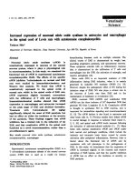
We first analyzed the levels of L-PGDS and H-PGDS mRNAs
in healthy and OA cartilage using real-time quantitative RT-
PCR. As shown in Figure 1, cartilage predominantly expresses
L-PGDS mRNA, and its levels of expression were approxi-
mately threefold higher in OA cartilage compared with healthy
cartilage. In contrast to L-PGDS, there was no statistically sig-
nificant difference in the levels of H-PGDS mRNA between
OA and healthy cartilage (Figure 1). In preliminary experi-
ments, we showed that the amplification efficiencies of tested
genes and GAPDH were similar. The efficiencies for the ampli-
fication of each gene and the reference were approximately
equal, ranging between 1.95 and 2.
Next, we used immunohistohemistry to analyze the localization
and the expression level of L-PGDS and H-PGDS proteins in
healthy and OA cartilage. As shown in Figures 2a and 2b, the
immunostaining for L-PGDS was located in the superficial and
upper intermediate layers of cartilage. Statistical evaluation for
the cell score revealed a clear and significant increase in the
number of chondrocytes staining positive for L-PGDS in OA
cartilage (43% ± 6%, mean ± SEM) compared with healthy
cartilage (20% ± 4%, mean ± SEM). The specificity of the
staining was confirmed using antibody that had been pread-
sorbed (1 hour at 37°C) with a 20-fold molar excess of the
Figure 1
Lipocalin-type prostaglandin D synthase (L-PGDS) and hematopoietic-type PGDS (H-PGDS) mRNA levels in healthy and osteoarthritis (OA) human cartilageLipocalin-type prostaglandin D synthase (L-PGDS) and hematopoi-
etic-type PGDS (H-PGDS) mRNA levels in healthy and osteoarthri-
tis (OA) human cartilage. RNA was extracted from healthy (n = 9) and
OA (n = 9) cartilage, reverse-transcribed into cDNA, and processed for
real-time polymerase chain reaction. The threshold cycle values were
converted to the number of molecules, as described in Materials and
methods. Data are expressed as copies of the gene's mRNA detected
per 10,000 GAPDH copies. *P < 0.05 versus healthy samples.
GAPDH, glyceraldehyde-3-phosphate dehydrogenase.
Available online />Page 5 of 12
(page number not for citation purposes)
recombinant protein (Figure 2c) or nonimmune control IgG
(data not shown). Using several commercially available anti-
bodies directed against human H-PGDS, we were unable to
detect H-PGDS protein expression in OA or healthy cartilage.
Together, these data indicate that the expression level of L-
PGDS is increased in OA cartilage.
To assess the level of PGD
2
in synovial fluids from OA and
healthy donors, we quantified its major stable metabolite, 11β-
PGF
2
α. We measured this metabolite because PGD
2
is unsta-
ble in vivo [30] and quantification of PGD
2
in synovial fluid can
be unreliable. We found a higher level of 11β-PGF
2
α in OA
synovial fluid when compared with healthy synovial fluid (Fig-
ure 3), indicating that the production of PGD
2
is higher in OA
synovial fluids. Together, these data indicate increased
expression and activity of L-PGDS in OA tissues.
Figure 2
Expression of lipocalin-type prostaglandin D synthase (L-PGDS) protein in healthy and osteoarthritis (OA) cartilageExpression of lipocalin-type prostaglandin D synthase (L-PGDS) protein in healthy and osteoarthritis (OA) cartilage. Representative immu-
nostaining of human healthy (a) and OA (b) cartilage for L-PGDS protein. (c) OA specimens treated with anti-L-PGDS antibody that was pread-
sorbed with a 20-fold molar excess of recombinant human L-PGDS (control for staining specificity). (d) Percentage of chondrocytes expressing L-
PGDS in healthy and OA cartilage. Results are expressed as the mean ± standard error of the mean of nine healthy and nine OA specimens. *P <
0.05 versus healthy cartilage.
Arthritis Research & Therapy Vol 10 No 6 Zayed et al.
Page 6 of 12
(page number not for citation purposes)
Interleukin-1-beta induces L-PGDS expression in
chondrocytes
IL-1β plays a major role in the cartilage physiology and in the
pathogenesis of OA [2]; therefore, we examined its effects on
the expression of L-PGDS in cultured OA chondrocytes. Cells
were treated with IL-1β (100 pg/mL) for different time periods,
and the levels of L-PGDS mRNA were quantified using real-
time RT-PCR. IL-1β-induced changes in gene expression were
evaluated as fold over control (untreated cells) after normaliza-
tion to the internal control gene, GAPDH. As shown in Figure
4a, treatment with IL-1β (100 pg/mL) enhanced L-PGDS
mRNA expression in a time-dependent manner. L-PGDS
mRNA expression started to gradually increase 24 hours post-
stimulation with IL-1β and remained elevated until 72 hours.
The induction of L-PGDS mRNA by IL-1β was also dose-
dependent. A significant increase at concentrations as low as
10 pg/mL was observed and the maximal effect was reached
at 100 pg/mL (Figure 4b). To determine whether changes in
mRNA levels were paralleled by changes in L-PGDS protein
levels, we performed Western blot analysis. Consistent with
its effects on L-PGDS mRNA, treatment with IL-1β led to a
dose- and time-dependent increase in the L-PGDS protein
expression (Figure 4c, d). To establish whether the IL-1β-
induced increase in L-PGDS expression corresponded with
an increase in PGDS activity, we measured PGD
2
levels in
conditioned media. As shown in Figures 4e and 4f, the
increased expression of L-PGDS protein was accompanied by
a time- and dose-dependent increase in PGD
2
production.
The upregulation of L-PGDS mRNA expression in
chondrocytes requires de novo protein synthesis
The lag period required for IL-1β to induce L-PGDS mRNA in
chondrocytes contrasts with those required for other IL-1β-
inducible genes, the expression of which starts as early as 2 to
6 hours and reaches a maximum at 8 to 18 hours. This sug-
gests that de novo protein synthesis is required for IL-1β-
induced L-PGDS expression. To evaluate this possibility, we
examined the impact of the protein synthesis inhibitor CHX.
Chondrocytes were stimulated with IL-1β in the absence or
presence of CHX, and the levels of L-PGDS mRNA were ana-
lyzed by real-time PCR. As shown in Figure 5, treatment with
CHX prevented IL-1β-mediated upregulation of L-PGDS
mRNA expression. This suggests that, to upregulate L-PGDS
expression in chondrocytes, IL-1β must induce the synthesis
of one or more proteins.
JNK and p38 MAPKs and NF-κB pathways contribute to
interleukin-1-beta-induced upregulation of L-PGDS
IL-1β exerts its effects acting through activation of the
mitogen-activated protein kinase (MAPK) (extracellular signal-
regulated kinase [ERK], c-jun N-terminal kinase [JNK], and
p38) and nuclear factor-kappa-B (NF-κB) signalling cascades
[31-35]. To evaluate the potential contribution of these path-
ways in IL-1β-induced L-PGDS expression, we used specific
pharmacological inhibitors. Chondrocytes were pretreated for
30 minutes with selective inhibitors for the above pathways
and then stimulated or not with IL-1β for 48 hours. As shown
in Figure 6a, pretreatment with the p38 MAPK inhibitor
SB203580 (1 μM), the JNK MAPK inhibitor SP600125 (10
μM), or the NF-κB inhibitor SN-50 (1 μM) suppressed IL-1β-
induced upregulation of L-PGDS expression. In contrast, pre-
treatment with the p42/44 MAPK inhibitor PD98059 (10 μM)
had no effect on IL-1β-induced upregulation of L-PGDS. The
concentration of the MAPK and NF-κB inhibitors used for
these experiments had no significant effect on cell viability as
indicated by the results of the MTT (3- [4,5-dimethylthiazol-2-
yl]-2,5-diphenyltetrazolium bromide) assay (data not shown).
These results suggest that the activation of JNK and p38
MAPK as well as NF-κB is essential to the induction of L-
PGDS by IL-1β in chondrocytes.
The Notch signalling pathway regulates diverse cellular proc-
esses, including proliferation, differentiation, and apoptosis
[36], and was reported to contribute to the regulation of L-
PGDS expression [37]. To determine whether this pathway
participates in IL-1β-induced L-PGDS expression in human
chondrocytes, we assessed the effect of DAPT. DAPT is a γ-
secretase inhibitor, which blocks cleavage of the intracellular
domain of all Notch proteins, and is widely used to evaluate
the effect of Notch inhibition [36]. As shown in Figure 6b, pre-
treatment with DAPT dose-dependently prevented IL-1β-
induced L-PGDS protein expression, indicating the involve-
ment of Notch signalling in this process. Notch inhibition was
confirmed by transcriptional inhibition of its direct target gene,
Hes1 (data not shown).
PGD
2
downregulated L-PGDS expression
To further characterize the regulation of L-PGDS expression in
cartilage, we examined the effect of PGD
2
, the end product of
Figure 3
Synovial levels of the prostaglandin D
2
(PGD
2
) metabolite 11β-PGF
2
αSynovial levels of the prostaglandin D
2
(PGD
2
) metabolite 11β-
PGF
2
α. 11β-PGF
2
α levels were measured in synovial fluids from
healthy subjects and patients with osteoarthritis (OA). The results are
expressed as picograms per milligram of proteins and are the mean ±
standard error of the mean of 7 healthy subjects and 11 OA patients.
*P < 0.05 versus healthy subjects.
Available online />Page 7 of 12
(page number not for citation purposes)
Figure 4
Effect of interleukin-1-beta (IL-1β) on lipocalin-type prostaglandin D synthase (L-PGDS) expression in osteoarthritis chondrocytesEffect of interleukin-1-beta (IL-1β) on lipocalin-type prostaglandin D synthase (L-PGDS) expression in osteoarthritis chondrocytes.
Chondrocytes were treated with 100 pg/mL IL-1β for the indicated time periods or with increasing concentrations of IL-1β for 48 hours. (a, b) Total
RNA was isolated and reverse-transcribed into cDNA, and L-PGDS and GAPDH mRNAs were quantified using real-time polymerase chain reaction.
All experiments were performed in triplicate, and negative controls without template RNA were included in each experiment. Results are expressed
as fold changes, considering 1 as the value of untreated cells, and represent the mean ± standard error of the mean (SEM) of four independent
experiments. *P < 0.05 compared with unstimulated cells. (c, d) Cell lysates were prepared and analyzed for L-PGDS and β-actin proteins by West-
ern blotting. Representative Western blots are shown in the upper panels. In the lower panels, the bands were scanned, and the L-PGDS band
intensity values were normalized to the corresponding β-actin band intensity value. Data are expressed as fold induction, considering 1 as the value
of unstimulated cells, and represent the mean ± SEM of four independent experiments. *P < 0.05 compared with unstimulated cells. (e, f) Condi-
tioned media was collected and analyzed for prostaglandin D
2
(PGD
2
) content. Results are expressed as the mean ± SEM of four independent
experiments. *P < 0.05 compared with unstimulated cells. GAPDH, glyceraldehyde-3-phosphate dehydrogenase.
Arthritis Research & Therapy Vol 10 No 6 Zayed et al.
Page 8 of 12
(page number not for citation purposes)
L-PGDS. Chondrocytes were stimulated with IL-1β in the
absence or presence of increasing concentrations of PGD
2
for
48 hours, and the expression of L-PGDS was evaluated by
Western blotting. As shown in Figure 7, treatment with PGD
2
dose-dependently reduced IL-1β-induced L-PGDS expres-
sion.
Discussion
This is the first report to demonstrate the presence of L-PGDS
in human cartilage and to show that its levels are elevated in
OA cartilage compared with healthy cartilage. The proinflam-
matory cytokine IL-1β upregulated, whereas PGD
2
downregu-
lated, the expression of L-PGDS in cultured chondrocytes.
These findings suggest that L-PGDS may be implicated in the
pathogenesis of OA.
In healthy cartilage, L-PGDS immunostaining was located in
only a few cells in the superficial and middle zones. By con-
trast, in OA cartilage, the cell score was significantly higher,
particularly in cartilage areas showing significant damage
(fibrillation). Given the anti-inflammatory and anticatabolic
roles of PGD
2
, it is reasonable to speculate that the upregula-
tion of L-PGDS may act as a sort of chondroprotective mech-
anism. Increased expression of L-PGDS was described in
other diseases such as atherosclerosis [22], multiple sclerosis
[38], diabetes [39] essential hypertension [40], and Tay-
Figure 5
The interleukin-1-beta (IL-1β)-induced upregulation of lipocalin-type prostaglandin D synthase (L-PGDS) mRNA expression requires de novo protein synthesisThe interleukin-1-beta (IL-1β)-induced upregulation of lipocalin-
type prostaglandin D synthase (L-PGDS) mRNA expression
requires de novo protein synthesis. Chondrocytes were incubated
with cycloheximide (CHX) (10 μg/mL) for 30 minutes prior to stimula-
tion with 100 pg/mL IL-1β for 48 hours. Total RNA was isolated and
reverse-transcribed into cDNA, and L-PGDS mRNA was quantified
using real-time polymerase chain reaction. Results are expressed as
fold changes, considering 1 as the value of untreated cells, and repre-
sent the mean ± standard error of the mean of four independent experi-
ments. *P < 0.05 compared with cells treated with IL-1β alone.
Figure 6
Effect of mitogen-activated protein kinase, nuclear factor-kappa-B, and Notch inhibitors on interleukin-1-beta (IL-1β)-induced upregulation of lipocalin-type prostaglandin D synthase (L-PGDS) expressionEffect of mitogen-activated protein kinase, nuclear factor-kappa-B,
and Notch inhibitors on interleukin-1-beta (IL-1β)-induced upregu-
lation of lipocalin-type prostaglandin D synthase (L-PGDS) expres-
sion. Osteoarthritis chondrocytes were pretreated with SB203580 (1
μM), SP600125 (10 μM), PD98059 (10 μM), or SN-50 (1 μM) for 30
minutes (a) or with increasing concentrations (1, 5, and 10 mM) of
DAPT for 48 hours (b) prior to stimulation with IL-1β (100 pg/mL). After
48 hours, cell lysates were prepared and analyzed for L-PGDS and β-
actin protein expression by Western blotting. Representative Western
blots are shown in the upper panels. In the lower panels, the bands
were scanned, and the L-PGDS band intensity values were normalized
to the corresponding β-actin band intensity value. Data are expressed
as fold induction, considering 1 as the value of unstimulated cells, and
represent the mean ± standard error of the mean of four independent
experiments. *P < 0.05 compared with cells treated with IL-1β alone.
DAPT, N-[N-(3,5-diflurophenylacetate)-L-alanyl]-(S)-phenylglycine t-
butyl ester.
Available online />Page 9 of 12
(page number not for citation purposes)
Sachs and Sandhoff diseases [41]. Thus, L-PGDS expression
is upregulated in many pathologies.
The enhanced expression of L-PGDS in the superficial and
middle zones of cartilage could potentially be due to the
increased level of the proinflammatory cytokine IL-1β in these
zones. Indeed, IL-1β, which plays pivotal roles in the initiation
and progression of OA, has been shown to accumulate in
these zones [42-46]. To prove this hypothesis, we performed
cell culture experiments. Our results revealed that exposure to
IL-1β led to a time- and concentration-dependent upregulation
of L-PGDS expression and PGD
2
production. The upregula-
tion of L-PGDS expression by IL-1β was blocked by CHX,
suggesting that this effect of IL-1β requires de novo protein
synthesis and would be consistent with an indirect stimulatory
mechanism.
The delayed induction of L-PGDS by IL-1β in chondrocytes is
consistent with the recently reported anti-inflammatory and
anticatabolic properties of PGD
2
. Indeed, the production of
PGD
2
is markedly elevated during the resolution of inflamma-
tion in carrageenan-induced pleurisy in rats, and exogenous
PGD
2
significantly reduces neutrophil levels in the inflamma-
tory exudates [10,11]. Enhanced production of PGD
2
was
also described during the resolution phase of the wound-heal-
ing process [47]. Cipollone and colleagues [48] examined the
expression of L-PGDS in atherosclerotic arteries and found
lower expression of L-PGDS and higher expression of micro-
somal prostaglandin E synthase-1 (mPGES-1) in symptomatic
plaques and found higher expression of L-PGDS and lower
expression of mPGES-1 in asymptomatic ones. This suggests
that the balance between PGD
2
and PGE
2
contributes to the
pathology of atherosclerosis and that a shift toward PGD
2
syn-
thesis may have an anti-inflammatory role. This is supported by
the observation that increased biosynthesis of PGD
2
is asso-
ciated with reduced production of PGE
2
in several in vitro
studies [49,50]. Recently, two separate studies demonstrated
anti-inflammatory properties of PGD
2
in an air-pouch model of
inflammation induced by monosodium urate monohydrate
crystals [13,51]. Moreover, H-PGDS knockout mice fail to
resolve a delayed-type hypersensitivity reaction [12]. In addi-
tion to its anti-inflammatory effects, PGD
2
was shown to
induce the expression of collagen type II and aggrecan [7], to
prevent apoptosis [8], and to inhibit the induction of MMP-1
and MMP-13 [52] in chondrocytes. Together, these data and
those from the present study favour the hypothesis that the
upregulation of L-PGDS expression in chondrocytes may be
part of a negative feedback control of inflammatory and cata-
bolic responses activated by IL-1β in the joint.
The production of PGD
2
by chondrocytes is of particular inter-
est since PGD
2
is readily converted to 15d-PGJ
2
, a potent
antiarthritic agent [14]. 15dPGJ
2
downregulates the expres-
sion of a number of inflammatory and catabolic mediators
involved in the pathogenesis of OA, including IL-1β, tumour
necrosis factor-alpha, inducible nitric-oxide synthase, and
MMPs [14]. Moreover, many in vivo studies support a protec-
tive effect of 15d-PGJ
2
and other PPARγ ligands in experimen-
tal animal models of OA [53,54]. Thus, the increased
expression of L-PGDS can lead to the production of a PPARγ
ligand in the joint. In contrast to classical PGs, which induce
their effects through binding to cell surface G protein-coupled
receptors, 15d-PGJ
2
induces most of its effects through the
nuclear receptor PPARγ. We have previously shown that
PPARγ expression is reduced in OA cartilage and that IL-1β
downregulates its expression in chondrocytes [29], which may
interfere with the protective effect of the PGD
2
metabolite
15d-PGJ
2
. Therefore, the increased expression of L-PGDS
observed in our study may represent a compensatory mecha-
nism to counter the reduced expression of PPARγ in OA and
to limit local inflammatory and catabolic responses. Also, it
should be noted that 15d-PGJ
2
can induce many of its effects
independently of PPARγ [14,17,18]. In addition, PGD
2
can
directly exert protective effects in OA before being metabo-
lized into 15d-PGJ
2
. Indeed, we have recently demonstrated
Figure 7
Effect of prostaglandin D
2
(PGD
2
) on interleukin-1-beta (IL-1β)-induced upregulation of lipocalin-type prostaglandin D synthase (L-PGDS) expressionEffect of prostaglandin D
2
(PGD
2
) on interleukin-1-beta (IL-1β)-
induced upregulation of lipocalin-type prostaglandin D synthase
(L-PGDS) expression. Osteoarthritis chondrocytes were pretreated
with increasing concentrations of PGD
2
for 30 minutes prior to stimula-
tion with IL-1β (100 pg/mL). After 48 hours, cell lysates were prepared
and analyzed for L-PGDS and β-actin protein expression by Western
blotting. A representative Western blot is shown in the upper panel. In
the lower panel, the bands were scanned, and the L-PGDS band inten-
sity values were normalized to the corresponding β-actin band intensity
value. Data are expressed as fold induction, considering 1 as the value
of unstimulated cells, and represent the mean ± standard error of the
mean of four independent experiments. *P < 0.05 compared with cells
stimulated with IL-1β alone.
Arthritis Research & Therapy Vol 10 No 6 Zayed et al.
Page 10 of 12
(page number not for citation purposes)
that human chondrocytes express functional DP1 and CRTH-
2 and that PGD
2
downregulates MMP-1 and MMP-13 expres-
sions through activation of the DP1 pathway [9].
To elucidate the mechanisms by which IL-1β upregulates L-
PGDS expression, we evaluated the roles played by down-
stream signalling cascades using specific pharmacological
inhibitors. We found that JNK and p38 MAPK inhibitors
blocked IL-1β-induced L-PGDS upregulation, whereas an
inhibitor of the ERK MAPK was without effect. We also found
that NF-κB blockade caused a significant decrease in IL-1β-
induced upregulation of L-PGDS protein expression. These
findings support the hypothesis that the JNK and p38 MAPKs
as well as the NF-κB pathways are involved in the upregulation
of L-PGDS expression by IL-1β. Our results are concordant
with previous reports that implicate activation of MAPKs (JNK
and p38) and NF-κB in the upregulation of L-PGDS in lep-
tomeningel cells [55], endothelial cells [56], and macrophages
[57]. The activation of JNK and p38 MAPK and of NF-κB path-
ways in chondrocytes has been shown to cause activation of
their downstream transcription factors, including activation
protein-1 (AP-1) and NF-κB [31-35]. Interestingly, the pro-
moter region of the human L-PGDS contains binding sites for
NF-κB and AP-1 [55,56]. Therefore, one could speculate that
upregulation of L-PGDS expression by IL-1β could be medi-
ated by AP-1 and NF-κB. Our results also demonstrate that
the Notch signalling pathway positively contributes to IL-1β-
induced L-PGDS expression in chondrocytes because DAPT,
a Notch signalling inhibitor, blocked this process. These find-
ings contrast with previous data showing that the Notch path-
way downregulates L-PGDS expression in the brain-derived
TE671 cells [37]. The reasons for these discrepancies are
presently unclear but are most likely due to cell-type differ-
ences or to differences in experimental conditions.
We also found that PGD
2
inhibits IL-1β-induced L-PGDS
expression. These results suggest that PGD
2
may exert a neg-
ative feedback mechanism to downregulate L-PGDS expres-
sion and activity. Given that the levels of L-PGDS are elevated
in OA cartilage and that IL-1β upregulated its expression in
chondrocytes, it is possible that the IL-1β effect prevails over
that of PGD
2
in vivo during advanced stages of the disease.
Indeed, the OA cartilage specimens used in this study were
from donors with long-established OA. Further studies are
clearly warranted to determine the expression profile of L-
PGDS over the course of OA in animal models of the disease.
The concentrations of PGD
2
used to suppress IL-1β-induced
L-PGDS expression are likely to be much higher than those
produced in synovial fluids. However, it should be noted that,
like other eicosanoids, PGD
2
functions as an autocrine and
paracrine molecule and can readily reach pharmacological lev-
els in the microenvironment of cells that produce it.
Conclusion
Our study has demonstrated for the first time that L-PGDS is
upregulated in OA cartilage. The proinflammatory cytokine IL-
1β may be responsible for this upregulation via a mechanism
that seems to involve the activation of the JNK and p38 MAPK
and NF-κB signalling pathways. These results suggest that the
increased expression of L-PGDS may play a protective role
against articular inflammation and cartilage damage.
Competing interests
The authors declare that they have no competing interests.
Authors' contributions
NZ conceived the study and designed and carried out cell and
real-time RT-PCR experiments and some immunohistochemis-
try experiments. NC contributed to the study design and car-
ried out immunoassays and some cell experiments. XL carried
out some cell experiments and data analysis. MB participated
in the study design and data analysis. JM-P, J-PP, and ND
helped to obtain tissues and participated in the study design
and some immunohistochemistry experiments. HF conceived,
designed, and coordinated the study, carried out some cell
experiments, and drafted the manuscript. All authors read and
approved the final manuscript.
Acknowledgements
This work was supported by the Canadian Institutes of Health Research
(CIHR) (grant MOP-84282) and the Fonds de la Recherche du Centre
de Recherche du Centre Hospitalier de l'Université de Montréal
(CHUM). HF is a Research Scholar of the Fonds de Recherche en
Santé du Québec (FRSQ).
References
1. Lawrence RC, Helmick CG, Arnett FC, Deyo RA, Felson DT, Gian-
nini EH, Heyse SP, Hirsch R, Hochberg MC, Hunder GG, Liang
MH, Pillemer SR, Steen VD, Wolfe F: Estimates of the preva-
lence of arthritis and selected musculoskeletal disorders in
the United States. Arthritis Rheum 1998, 41:778-799.
2. Goldring MB: The role of cytokines as inflammatory mediators
in osteoarthritis: lessons from animal models. Connect Tissue
Res 1999, 40:1-11.
3. Pelletier JP, Martel-Pelletier J, Abramson SB: Osteoarthritis, an
inflammatory disease: potential implication for the selection of
new therapeutic targets. Arthritis Rheum 2001, 44:1237-1247.
4. Goldring MB, Berenbaum F: The regulation of chondrocyte
function by proinflammatory mediators: prostaglandins and
nitric oxide. Clin Orthop Relat Res 2004:S37-46.
5. Smith WL, Langenbach R: Why there are two cyclooxygenase
isozymes. J Clin Invest 2001, 107:1491-1495.
6. Martel-Pelletier J, Pelletier JP, Fahmi H: Cyclooxygenase-2 and
prostaglandins in articular tissues. Semin Arthritis Rheum
2003, 33:155-167.
7. Jakob M, Demarteau O, Suetterlin R, Heberer M, Martin I: Chon-
drogenesis of expanded adult human articular chondrocytes
is enhanced by specific prostaglandins. Rheumatology
(Oxford) 2004, 43:852-857.
8. Relic B, Benoit V, Franchimont N, Ribbens C, Kaiser MJ, Gillet P,
Merville MP, Bours V, Malaise MG: 15-deoxy-delta12,14-pros-
taglandin J2 inhibits Bay 11–7085-induced sustained extracel-
lular signal-regulated kinase phosphorylation and apoptosis
in human articular chondrocytes and synovial fibroblasts. J
Biol Chem 2004, 279:22399-22403.
9. Zayed N, Afif H, Chabane N, Mfuna-Endam L, Benderdour M, Mar-
tel-Pelletier J, Pelletier JP, Motiani RK, Trebak M, Duval N, Fahmi H:
Inhibition of interleukin-1beta-induced matrix metalloprotein-
Available online />Page 11 of 12
(page number not for citation purposes)
ases 1 and 13 production in human osteoarthritic chondro-
cytes by prostaglandin D(2). Arthritis Rheum 2008,
58:3530-3540.
10. Gilroy DW, Colville-Nash PR, Willis D, Chivers J, Paul-Clark MJ,
Willoughby DA: Inducible cyclooxygenase may have anti-
inflammatory properties. Nat Med 1999, 5:698-701.
11. Ianaro A, Ialenti A, Maffia P, Pisano B, Di Rosa M: Role of
cyclopentenone prostaglandins in rat carrageenin pleurisy.
FEBS Lett 2001, 508:61-66.
12. Trivedi SG, Newson J, Rajakariar R, Jacques TS, Hannon R,
Kanaoka Y, Eguchi N, Colville-Nash P, Gilroy DW: Essential role
for hematopoietic prostaglandin D
2
synthase in the control of
delayed type hypersensitivity. Proc Natl Acad Sci USA 2006,
103:5179-5184.
13. Murakami Y, Akahoshi T, Hayashi I, Endo H, Hashimoto A, Kono S,
Kondo H, Kawai S, Inoue M, Kitasato H: Inhibition of monoso-
dium urate monohydrate crystal-induced acute inflammation
by retrovirally transfected prostaglandin D synthase. Arthritis
Rheum 2003, 48:2931-2941.
14. Fahmi H, Pelletier JP, Martel-Pelletier J: PPARgamma ligands as
modulators of inflammatory and catabolic responses on
arthritis. An overview. J Rheumatol 2002, 29:3-14.
15. Hirata M, Kakizuka A, Aizawa M, Ushikubi F, Narumiya S: Molecu-
lar characterization of a mouse prostaglandin D receptor and
functional expression of the cloned gene. Proc Natl Acad Sci
USA 1994, 91:11192-11196.
16. Hirai H, Tanaka K, Yoshie O, Ogawa K, Kenmotsu K, Takamori Y,
Ichimasa M, Sugamura K, Nakamura M, Takano S, Nagata K: Pros-
taglandin D
2
selectively induces chemotaxis in T helper type 2
cells, eosinophils, and basophils via seven-transmembrane
receptor CRTH2. J Exp Med 2001, 193:255-261.
17. Boyault S, Bianchi A, Moulin D, Morin S, Francois M, Netter P, Ter-
lain B, Bordji K: 15-Deoxy-delta(12,14)-prostaglandin J(2)
inhibits IL-1beta-induced IKK enzymatic activity and Ikappa-
Balpha degradation in rat chondrocytes through a PPAR-
gamma-independent pathway. FEBS Lett 2004, 572:33-40.
18. Bianchi A, Moulin D, Sebillaud S, Koufany M, Galteau MM, Netter
P, Terlain B, Jouzeau JY: Contrasting effects of peroxisome-pro-
liferator-activated receptor (PPAR)gamma agonists on mem-
brane-associated prostaglandin E
2
synthase-1 in IL-1beta-
stimulated rat chondrocytes: evidence for PPARgamma-inde-
pendent inhibition by 15-deoxy-Delta12,14prostaglandin J
2
.
Arthritis Res Ther 2005, 7:R1325-1337.
19. Urade Y, Eguchi N: Lipocalin-type and hematopoietic prostag-
landin D synthases as a novel example of functional conver-
gence. Prostaglandins Other Lipid Mediat 2002, 68–
69:375-382.
20. Blodorn B, Mader M, Urade Y, Hayaishi O, Felgenhauer K, Bruck
W: Choroid plexus: the major site of mRNA expression for the
beta-trace protein (prostaglandin D synthase) in human brain.
Neurosci Lett 1996, 209:117-120.
21. Urade Y, Fujimoto N, Hayaishi O: Purification and characteriza-
tion of rat brain prostaglandin D synthetase. J Biol Chem 1985,
260:12410-12415.
22. Eguchi Y, Eguchi N, Oda H, Seiki K, Kijima Y, Matsu-ura Y, Urade
Y, Hayaishi O: Expression of lipocalin-type prostaglandin D
synthase (beta-trace) in human heart and its accumulation in
the coronary circulation of angina patients. Proc Natl Acad Sci
USA 1997, 94:14689-14694.
23. Beuckmann CT, Gordon WC, Kanaoka Y, Eguchi N, Marcheselli
VL, Gerashchenko DY, Urade Y, Hayaishi O, Bazan NG: Lipoca-
lin-type prostaglandin D synthase (beta-trace) is located in
pigment epithelial cells of rat retina and accumulates within
interphotoreceptor matrix. J Neurosci 1996, 16:6119-6124.
24. Gerena RL, Irikura D, Eguchi N, Urade Y, Killian GJ: Immunocyto-
chemical localization of lipocalin-type prostaglandin D syn-
thase in the bull testis and epididymis and on ejaculated
sperm. Biol Reprod 2000, 62:547-556.
25. Urade Y, Ujihara M, Horiguchi Y, Igarashi M, Nagata A, Ikai K, Hay-
aishi O: Mast cells contain spleen-type prostaglandin D syn-
thetase. J Biol Chem 1990, 265:371-375.
26. Fujimori K, Kanaoka Y, Sakaguchi Y, Urade Y: Transcriptional
activation of the human hematopoietic prostaglandin D syn-
thase gene in megakaryoblastic cells. Roles of the oct-1 ele-
ment in the 5'-flanking region and the AP-2 element in the
untranslated exon 1. J Biol Chem 2000, 275:40511-40516.
27. Tanaka K, Ogawa K, Sugamura K, Nakamura M, Takano S, Nagata
K: Cutting edge: differential production of prostaglandin D
2
by
human helper T cell subsets. J Immunol 2000, 164:2277-2280.
28. Altman RD: Criteria for the classification of osteoarthritis of the
knee and hip. Scand J Rheumatol Suppl 1987, 65:31-39.
29. Afif H, Benderdour M, Mfuna-Endam L, Martel-Pelletier J, Pelletier
JP, Duval N, Fahmi H: Peroxisome proliferator-activated recep-
tor gamma1 expression is diminished in human osteoarthritic
cartilage and is downregulated by interleukin-1beta in articu-
lar chondrocytes. Arthritis Res Ther 2007, 9:R31.
30. O'Sullivan S, Mueller MJ, Dahlen SE, Kumlin M: Analyses of pros-
taglandin D
2
metabolites in urine: comparison between
enzyme immunoassay and negative ion chemical ionisation
gas chromatography-mass spectrometry. Prostaglandins
Other Lipid Mediat 1999, 57:149-165.
31. Geng Y, Valbracht J, Lotz M: Selective activation of the mitogen-
activated protein kinase subgroups c-Jun NH2 terminal kinase
and p38 by IL-1 and TNF in human articular chondrocytes. J
Clin Invest 1996, 98:2425-2430.
32. Ding GJ, Fischer PA, Boltz RC, Schmidt JA, Colaianne JJ, Gough
A, Rubin RA, Miller DK: Characterization and quantitation of NF-
kappaB nuclear translocation induced by interleukin-1 and
tumor necrosis factor-alpha. Development and use of a high
capacity fluorescence cytometric system. J Biol Chem 1998,
273:28897-28905.
33. Mengshol JA, Vincenti MP, Coon CI, Barchowsky A, Brinckerhoff
CE: Interleukin-1 induction of collagenase 3 (matrix metallo-
proteinase 13) gene expression in chondrocytes requires p38,
c-Jun N-terminal kinase, and nuclear factor kappaB: differen-
tial regulation of collagenase 1 and collagenase 3. Arthritis
Rheum 2000, 43:801-811.
34. Mendes AF, Caramona MM, Carvalho AP, Lopes MC: Role of
mitogen-activated protein kinases and tyrosine kinases on IL-
1-Induced NF-kappaB activation and iNOS expression in
bovine articular chondrocytes. Nitric Oxide 2002, 6:35-44.
35. Fan Z, Bau B, Yang H, Aigner T: IL-1beta induction of IL-6 and
LIF in normal articular human chondrocytes involves the ERK,
p38 and NFkappaB signaling pathways. Cytokine 2004,
28:17-24.
36. Fiuza UM, Arias AM: Cell and molecular biology of Notch. J
Endocrinol 2007, 194:459-474.
37. Fujimori K, Kadoyama K, Urade Y: Protein kinase C activates
human lipocalin-type prostaglandin D synthase gene expres-
sion through de-repression of notch-HES signaling and
enhancement of AP-2 beta function in brain-derived TE671
cells. J Biol Chem 2005, 280:18452-18461.
38. Kagitani-Shimono K, Mohri I, Oda H, Ozono K, Suzuki K, Urade Y,
Taniike M: Lipocalin-type prostaglandin D synthase (beta-
trace) is upregulated in the alphaB-crystallin-positive oli-
godendrocytes and astrocytes in the chronic multiple sclero-
sis. Neuropathol Appl Neurobiol 2006, 32:64-73.
39. Hirawa N, Uehara Y, Ikeda T, Gomi T, Hamano K, Totsuka Y,
Yamakado M, Takagi M, Eguchi N, Oda H, Seiki K, Nakajima H,
Urade Y: Urinary prostaglandin D synthase (beta-trace) excre-
tion increases in the early stage of diabetes mellitus. Nephron
2001, 87:321-327.
40. Hirawa N, Uehara Y, Yamakado M, Toya Y, Gomi T, Ikeda T, Eguchi
Y, Takagi M, Oda H, Seiki K, Urade Y, Umemura S: Lipocalin-type
prostaglandin d synthase in essential hypertension. Hyperten-
sion 2002, 39:449-454.
41. Mohri I, Taniike M, Okazaki I, Kagitani-Shimono K, Aritake K,
Kanekiyo T, Yagi T, Takikita S, Kim HS, Urade Y, Suzuki K: Lipoc-
alin-type prostaglandin D synthase is up-regulated in oli-
godendrocytes in lysosomal storage diseases and binds
gangliosides. J Neurochem 2006, 97:641-651.
42. Pelletier JP, Lascau-Coman V, Jovanovic D, Fernandes JC, Man-
ning P, Connor JR, Currie MG, Martel-Pelletier J: Selective inhibi-
tion of inducible nitric oxide synthase in experimental
osteoarthritis is associated with reduction in tissue levels of
catabolic factors. J Rheumatol 1999, 26:2002-2014.
43. Tetlow LC, Adlam DJ, Woolley DE: Matrix metalloproteinase and
proinflammatory cytokine production by chondrocytes of
human osteoarthritic cartilage: associations with degenera-
tive changes. Arthritis Rheum 2001, 44:585-594.
44. Towle CA, Hung HH, Bonassar LJ, Treadwell BV, Mangham DC:
Detection of interleukin-1 in the cartilage of patients with oste-
Arthritis Research & Therapy Vol 10 No 6 Zayed et al.
Page 12 of 12
(page number not for citation purposes)
oarthritis: a possible autocrine/paracrine role in pathogene-
sis. Osteoarthritis Cartilage 1997, 5:293-300.
45. Melchiorri C, Meliconi R, Frizziero L, Silvestri T, Pulsatelli L, Maz-
zetti I, Borzi RM, Uguccioni M, Facchini A: Enhanced and coordi-
nated in vivo expression of inflammatory cytokines and nitric
oxide synthase by chondrocytes from patients with osteoar-
thritis. Arthritis Rheum 1998, 41:2165-2174.
46. Moos V, Fickert S, Muller B, Weber U, Sieper J: Immunohistolog-
ical analysis of cytokine expression in human osteoarthritic
and healthy cartilage. J Rheumatol 1999, 26:870-879.
47. Kapoor M, Kojima F, Yang L, Crofford LJ: Sequential induction of
pro- and anti-inflammatory prostaglandins and peroxisome
proliferators-activated receptor-gamma during normal wound
healing: a time course study. Prostaglandins Leukot Essent
Fatty Acids 2007, 76:103-112.
48. Cipollone F, Fazia M, Iezzi A, Ciabattoni G, Pini B, Cuccurullo C,
Ucchino S, Spigonardo F, De Luca M, Prontera C, Chiarelli F, Cuc-
curullo F, Mezzetti A: Balance between PGD synthase and PGE
synthase is a major determinant of atherosclerotic plaque
instability in humans. Arterioscler Thromb Vasc Biol 2004,
24:1259-1265.
49. Fournier T, Fadok V, Henson PM: Tumor necrosis factor-alpha
inversely regulates prostaglandin D
2
and prostaglandin E
2
pro-
duction in murine macrophages. Synergistic action of cyclic
AMP on cyclooxygenase-2 expression and prostaglandin E
2
synthesis. J Biol Chem 1997, 272:31065-31072.
50. Matsumoto H, Naraba H, Murakami M, Kudo I, Yamaki K, Ueno A,
Oh-ishi S: Concordant induction of prostaglandin E
2
synthase
with cyclooxygenase-2 leads to preferred production of pros-
taglandin E
2
over thromboxane and prostaglandin D
2
in
lipopolysaccharide-stimulated rat peritoneal macrophages.
Biochem Biophys Res Commun 1997, 230:110-114.
51. Jung SM, Schumacher HR, Kim H, Kim M, Lee SH, Pessler F:
Reduction of urate crystal-induced inflammation by root
extracts from traditional oriental medicinal plants: elevation of
prostaglandin D
2
levels. Arthritis Res Ther 2007, 9:R64.
52. Stewart MD, Li J, Wong J: Relationship between histone H3
lysine 9 methylation, transcription repression, and heterochro-
matin protein 1 recruitment. Mol Cell Biol 2005, 25:2525-2538.
53. Kobayashi T, Notoya K, Naito T, Unno S, Nakamura A, Martel-Pel-
letier J, Pelletier JP: Pioglitazone, a peroxisome proliferator-
activated receptor gamma agonist, reduces the progression of
experimental osteoarthritis in guinea pigs. Arthritis Rheum
2005, 52:479-487.
54. Boileau C, Martel-Pelletier J, Fahmi H, Mineau F, Boily M, Pelletier
JP: The peroxisome proliferator-activated receptor gamma
agonist pioglitazone reduces the development of cartilage
lesions in an experimental dog model of osteoarthritis: in vivo
protective effects mediated through the inhibition of key sign-
aling and catabolic pathways. Arthritis Rheum 2007,
56:2288-2298.
55. Fujimori K, Fujitani Y, Kadoyama K, Kumanogoh H, Ishikawa K,
Urade Y: Regulation of lipocalin-type prostaglandin D synthase
gene expression by Hes-1 through E-box and interleukin-1
beta via two NF-kappa B elements in rat leptomeningeal cells.
J Biol Chem 2003, 278:6018-6026.
56. Miyagi M, Miwa Y, Takahashi-Yanaga F, Morimoto S, Sasaguri T:
Activator protein-1 mediates shear stress-induced prostag-
landin d synthase gene expression in vascular endothelial
cells. Arterioscler Thromb Vasc Biol 2005, 25:970-975.
57. Joo M, Kwon M, Sadikot RT, Kingsley PJ, Marnett LJ, Blackwell TS,
Peebles RS Jr, Urade Y, Christman JW: Induction and function of
lipocalin prostaglandin D synthase in host immunity. J Immu-
nol 2007, 179:2565-2575.