Báo cáo y học: "Antibody engineering to develop new antirheumatic therapies" ppsx
Bạn đang xem bản rút gọn của tài liệu. Xem và tải ngay bản đầy đủ của tài liệu tại đây (1.17 MB, 11 trang )
Available online />Page 1 of 11
(page number not for citation purposes)
Abstract
There has been a therapeutic revolution in rheumatology over the
past 15 years, characterised by a move away from oral immuno-
suppressive drugs toward parenteral targeted biological therapies.
The potency and relative safety of the newer agents has facilitated
a more aggressive approach to treatment, with many more patients
achieving disease remission. There is even a prevailing sense that
disease ‘cure’ may be a realistic goal in the future. These develop-
ments were underpinned by an earlier revolution in molecular
biology and protein engineering as well as key advances in our
understanding of rheumatoid arthritis pathogenesis. This review will
focus on antibody engineering as the key driver behind our current
and developing range of antirheumatic treatments.
Antibody structure, function, and molecular
genetics: a primer
The biological therapy ‘revolution’ was made possible by
elucidation of the fine detail of the structure-function relation-
ship in immunoglobulin molecules and the ‘modular’ organisa-
tion of the underlying genes. Antibodies are essentially
multidomain adapter molecules used by the immune system
to neutralise and/or destroy invading microorganisms and
their products (antigens). They do this by connecting the
antigen with various effector mechanisms. At one end of the
antibody molecule (Figure 1), two identical variable (V)
regions have a molecular structure that, in three dimensions,
is highly complementary to the target antigen. Non-covalent
molecular interactions between antibody and antigen ensure
a tight fit. The constant (C) region, at the other end of the
antibody molecule, determines the fate of the bound antigen.
An antibody comprises four covalently linked polypeptide
chains: two identical heavy chains and two identical light
chains (Figure 1). The heavy chains usually contain four and
the light chain two distinct domains, where a domain is a
discrete, folded, functional unit (Figure 2a). The first domain
in each chain is the V domain, VH and VL on the heavy and
light chains, respectively. The rest of the heavy chain
comprises three (four for IgE) constant domains (CH1 to
CH3), whilst the light chains have one constant domain (CL).
There is a flexible peptide segment (the hinge) between the
CH1 and CH2 domains.
The antibody V region is composed of the VH and VL
domains. The C region is composed of the CL, CH1, CH2,
and CH3 domains. Digesting an antibody with papain
releases a single Fc (fragment crystallisable) fragment corres-
ponding to the CH2 and CH3 domains (Figure 2a). Two Fab
(fragment antigen-binding) fragments are also generated,
corresponding to the antibody binding arms (Figure 2b).
Within each VH and VL domain, three short polypeptide
segments form the hypervariable or complementarity-deter-
mining regions (CDRs) (Figure 1). These segments have a
highly variable sequence when compared with the rest of the
molecule and dictate the precise antigen-binding charac-
teristics of the antibody. The remainder of the V domain is
much less variable and forms a scaffold that supports the
CDRs. In the three-dimensional structure of an antibody
molecule, the three heavy-chain and three light-chain CDRs
are closely apposed to form the antigen-binding site. CDR3 is
the most variable of the CDRs and plays a dominant role in
antibody specificity. Antibody fragments such as Fab
fragments (Figure 2b), Fvs (non-covalently linked VH and VL
domains, Figure 2c), and single-chain Fvs (scFvs) (covalently
linked VH and VL domains, Figure 2d) generally have the
same specificity for antigen as the full-length antibody from
which they are derived.
Review
Antibody engineering to develop new antirheumatic therapies
John D Isaacs
Wilson Horne Immunotherapy Centre and Musculoskeletal Research Group, Institute of Cellular Medicine, Newcastle University, Framlington Place,
Newcastle-Upon-Tyne, NE2 4HH, UK
Corresponding author: John D Isaacs,
Published: 19 May 2009 Arthritis Research & Therapy 2009, 11:225 (doi:10.1186/ar2594)
This article is online at />© 2009 BioMed Central Ltd
BLyS = B-lymphocyte stimulator; C = constant; CDR = complementarity-determining region; CH = heavy chain C domain; CL = light chain C
domain; dAb = domain antibody; Fab = fragment antigen-binding; Fc = fragment crystallisable; FcγR = fragment crystallisable gamma receptor
(receptor for the constant region of IgG); Fvs = non-covalently linked heavy and light chain V domains; mAb = monoclonal antibody; PCR = poly-
merase chain reaction; RA = rheumatoid arthritis; scFvs = single-chain covalently linked heavy and light chain V domains; SLE = systemic lupus ery-
thematosus; SMIP = small modular immunopharmaceutical; TACI = transmembrane activator and calcium modulator and cyclophilin ligand
interactor; TNF = tumour necrosis factor; V = variable; VH = heavy chain V domain; VL = light chain V domain.
Arthritis Research & Therapy Vol 11 No 3 Isaacs
Page 2 of 11
(page number not for citation purposes)
The antibody C region determines the class and subclass of
the antibody. There are five human heavy-chain classes (IgM,
IgG, IgA, IgE, and IgD) and two light-chain classes (lambda
and kappa). IgG is the predominant class in blood and
tissues and comprises four subclasses, IgG1 to IgG4. Most
therapeutic antibodies are IgG molecules. Antibody class and
subclass determine the consequences of antibody binding to
antigen. IgM, IgG1, and IgG3 activate complement efficiently,
leading to chemotaxis and to opsonisation and lysis of the
target. IgG1 and IgG3 also have the highest affinity for Fc-
gamma receptors (FcγR I to III) on white blood cells, resulting
in activation of the cells followed by phagocytosis and cell-
mediated cytotoxicity. IgG2 and IgG4 are relatively poor at
harnessing effector function, and light-chain class (kappa or
lambda) has not been shown to contribute significantly. The
neonatal Fc receptor, FcRn, is an important and ubiquitously
expressed Fc receptor that, by rescuing IgG molecules from
lysosomal degradation, has an important influence on serum
half-life [1].
Specific amino acid residues in the C region of immuno-
globulin molecules, particularly in the CH2 domain, dictate
the capacity of certain subclasses to interact with effector
mechanisms. For example, residues 318, 320, and 322 are
critical for IgG binding to complement C1q and residues 234
to 237 are critical for FcγR binding [2-4]. An asparagine
residue at position 297 in IgG molecules is an N-linked
glycosylation site that also plays a critical role in effector
function [5].
The genetic organisation encoding antibody structure is
simultaneously simple and sophisticated, comprising a
number of blocks of genes. For a VH domain, these are as
follows:
• V segments, which code for most of the V domain,
including CDRs 1 and 2 and the first part of CDR3,
• D segments that code for the intermediate part of CDR3,
and
• J segments that code for the terminal part of CDR3.
In humans, there are about 51 heavy-chain V segments, 25 D
segments, and 6 J segments [6]. During B-cell development,
antibody-encoding DNA undergoes various rearrangements
(Figure 3). Essentially, any V segment can fuse to any D
segment and any fused VD segment to any J segment. A
similar process occurs in the light chain, where overall there
are 71 V segment and 9 J segment (but no D segment)
genes. This random pairing of segments (VDJ recombination)
leads to a very large number of possible CDR3 sequences,
explaining why CDR3 is the most variable CDR. In contrast,
the sequences of CDR1 and CDR2 are encoded within the
non-rearranged germline antibody sequence. The joins of V to
D and D to J are imprecise, with loss or addition of nucleo-
tides contributing to further CDR3 diversity. Further along the
chromosome from the J segments are the C-region genes in
the order Cμ (encodes IgM heavy chain), Cδ (encodes IgD
heavy chain), and then the genes for the subclasses of IgG
and IgA and for IgE. Following VDJ recombination, IgM or IgD
antibodies are produced initially, dependent upon RNA-
processing events (Figure 3).
After contact with antigen, affinity maturation occurs as a
consequence of further mutations within the rearranged
immunoglobulin gene. These somatic mutations are concen-
trated in the CDRs and occur during DNA replication such
that the progeny of a B cell produce antibody that is subtly
different from that of the parent in terms of affinity for antigen.
Those that produce antibody with a higher affinity have a
survival advantage over those that do not improve their affinity.
Antibody engineering
Following the description of monoclonal antibody (mAb)
generation by Kohler and Milstein in 1975 [7], increasing
knowledge of antibody structure-function relationships and of
immunoglobulin gene organisation rendered the production
of ‘man-made’ antibodies conceptually attractive and simple.
A number of strategies led up to the ‘bespoke’ process of
antibody design that we are now familiar with.
Chimeric antibodies
The first therapeutic antibodies were murine proteins pro-
duced from murine ‘hybridomas’ by conventional fusion tech-
nology [7]. In rheumatology practice, one of the earliest anti-
CD4 mAbs was murine [8]. A significant limitation to the use
of such ‘foreign’ molecules was their immunogenicity. For
example, OKT3, a murine mAb against human CD3, was
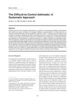
Figure 1
Basic antibody structure and the different types of therapeutic
antibody. (a) Basic antibody structure. (b) Basic structure of a murine,
chimeric, humanised, and human monoclonal antibody. Red indicates
murine sequence and black indicates human sequence. CDR,
complementarity-determining region.
effective for reversing allograft rejection. A subsequent
course of therapy was often ineffective, however, due to
neutralising anti-antibody – anti-globulin or human anti-murine
(HAMA) – responses [9]. A further potential limitation of
using murine mAbs was their interaction with human effector
functions. There are subtle differences in amino acid
sequence between murine and human Fc regions and
between murine and human FcγR. Consequently, the
interaction between a murine mAb and human FcγR will be
suboptimal, potentially limiting the cytotoxic potential of the
antibody in the therapeutic situation.
The modular design of immunoglobulins led to an obvious
solution to these issues in the form of chimeric mAbs.
Neuberger and colleagues [10] first demonstrated the
feasibility of linking a murine antibody V-region gene segment
to a human C-region gene segment. The resulting gene
construct encoded a chimeric, ‘half human/half mouse’, mAb
(Figure 1b). The chimeric C region did not interfere with
antigen binding but, as predicted, dictated the effector
function of the encoded mAb. The production of ‘matched
sets’ of chimeric mAbs confirmed the expected inter-class
and inter-subclass variation of effector function, enabling the
selection of the appropriate C region for a particular
therapeutic task and the birth of ‘designer’ mAbs [11,12].
Two chimeric mAbs are used in everyday rheumatological
practice: infliximab and rituximab (the nomenclature of mAbs
is explained in Table 1). Both possess a human IgG1 C
region and these highly effective drugs neutralise tumour
necrosis factor-alpha (TNF-α) and kill B cells, respectively.
Nonetheless, their murine V regions retain the immuno-
genicity of a foreign protein. The consequences of immuno-
genicity vary from anaphylaxis, which fortunately is rare, to
lack of efficacy and infusion reactions, which are more
common. For example, human anti-chimeric antibodies are a
significant cause of secondary inefficacy of infliximab, where-
by mAb requirements increase with time and treatment may
eventually become ineffective [13]. Infusion reactions are also
more frequent in the presence of anti-globulins [14]. A
number of factors influence immunogenicity, including back-
ground immunosuppression, dose, and route of therapy [15].
Humanised antibodies
The next significant step in antibody engineering was the
process of humanisation. Careful examination of the V-region
peptide sequence of a mAb allows the identification of the
CDRs. In the mid-1980s, it was shown that genetic
engineering could be used to ‘transplant’ the CDRs of a
murine antibody onto a human V-region framework, generally
without a loss of specificity (CDR grafting, Figure 1b) [16]. To
Available online />Page 3 of 11
(page number not for citation purposes)
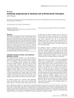
Figure 2
The domain structures of an antibody molecule and its derivatives. (a) An antibody molecule. (b) A fragment antigen-binding (Fab) fragment.
(c) A non-covalently linked VH and VL domains (Fv). (d) A single-chain Fv. (e) A receptor-immunoglobulin fusion protein. CH, heavy chain constant
domain; CL, light chain constant domain; Fc, fragment crystallisable; VH, heavy chain variable domain; VL, light chain variable domain.
optimise the ‘fit’ and ultimate affinity, the chosen human V
gene was generally one that closely resembled that of the
parent mouse mAb. The main theoretical advantage of
humanisation was a further reduction in immunogenicity,
although the selected V-region backbone was not always one
that was used commonly by the natural human antibody
repertoire [17]. In a small study, however, the first humanised
therapeutic mAb, CAMPATH-1H (alemtuzumab), was shown
to be minimally immunogenic in patients with rheumatoid
arthritis (RA) [18]. This drug is highly effective at killing
lymphocytes and is now licensed for the treatment of chronic
lymphocytic leukaemia whilst continuing to be developed for
a number of autoimmune indications. Tocilizumab, a
humanised mAb against the interleukin-6 receptor that is
currently in phase III development for RA, was also developed
by CDR grafting, as were ocrelizumab, an anti-CD20 mAb
that is currently in phase III trials for RA, and epratuzumab, an
anti-CD22 mAb currently being evaluated in systemic lupus
erythematosus (SLE) and Sjögren syndrome (Table 1).
A number of techniques have subsequently evolved for
generating humanised and ‘human’ mAbs. Because of their
murine CDRs, humanised mAbs theoretically retain a degree
of immunogenicity (human anti-human, or HAHA, responses)
although trials show this to be relatively low. For a number of
reasons, the ‘obvious’ solution, to generate human hybridomas,
was not feasible: it was not appropriate to immunise a human
expressly for the generation of a mAb, attempts to make
mAbs from venous blood (as opposed to spleen) were
unsuccessful or provided low-affinity IgM mAbs in small
quantities from unstable cell lines, and immunological
tolerance provided a significant barrier to raising human
mAbs against human targets.
Human antibodies
In 1989, Orlandi and colleagues [19] showed that it was
possible to use the polymerase chain reaction (PCR) to clone
immunoglobulin V domains. Subsequently, ‘libraries’ of immuno-
globulin VH and VL sequences were created within plasmid
and phagemid vectors, allowing the expression of a huge
diversity of antibodies [20]. Sequence conservation meant
that a relatively small number of ‘forward’ (3′) and ‘backward’
(5′) primers could be used to amplify a large proportion of the
V-domain repertoire from an appropriate source, including
peripheral blood. The incorporation of restriction endo-
nuclease recognition sites into primers facilitated the sub-
sequent in-frame cloning of amplified V-domain sequences.
An extension of the technology allowed the mutation of a
cloned V domain using a number of methods. For example, in
‘spiked PCR’, the forward primer is synthesised under
conditions that introduce low-frequency random mutations,
providing a mixed population of many subtly different primers.
Because the forward primer encodes CDR3, the resulting
PCR product encodes a V-domain mixture with subtly
variable CDR3s and hence fine specificities. In contrast,
Arthritis Research & Therapy Vol 11 No 3 Isaacs
Page 4 of 11
(page number not for citation purposes)
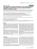
Figure 3
Antibody heavy-chain gene rearrangement, transcription, and translation. In step 1, any V segment (in this case, V2) rearranges to any D segment
(in this case, D1). In step 2, the VD segment rearranges to one of the six J segments (in this case, J5). Primary RNA transcripts extend from the
rearranged VDJ segments through to the Cδ gene (step 3). Finally, RNA processing results in the incorporation of either Cμ or Cδ by the
transcripts, encoding for an IgM or IgD antibody, respectively.
‘error-prone’ PCR (using non-stringent amplification condi-
tions or non-proofreading polymerases) results in sequence
variability throughout the amplified V domains. These and
similar techniques, when applied to a cloned V domain,
generate variants of altered affinity in a manner analogous to
affinity maturation. Other techniques include ‘chain shuffling’,
in which a ‘fixed’ VH or VL domain is allowed to pair with a
library of partner domains, biasing the resulting Fvs toward a
desired specificity [21]. Guided selection enabled the deriva-
tion of a human mAb starting from a murine sequence [22].
This technology had several advantages. The ability to rapidly
capture and clone a significant proportion of the V-domain
repertoire from a biological sample was a major advance.
Critically, the new technology bypassed the need to use
animals for mAb generation – libraries could be created from
human blood samples. Furthermore, because the VH- and VL-
domain libraries could be randomly combined and mutated, it
became possible to generate specificities absent from the
natural repertoire of the source tissue, bypassing immune
tolerance mechanisms.
To fully exploit these advances, novel techniques were
needed to screen the massive V-domain libraries for desired
specificities. Thus, through the use of peripheral blood B cells
from a non-immunised individual, PCR amplification might
result in 10
7
VH sequences and a similar number of VL
sequences. Random pairing of these would result in a ‘library’
of 10
14
different combinations, each cloned into a plasmid.
Transformation of a bacterial culture with this library could
result in 10
9
distinct Fv specificities (limited largely by
transformation efficiency). Phage display technology provided
a method for screening such libraries. Filamentous bacterio-
phages are simple viruses that infect bacteria. They comprise
a nucleic acid core and a surrounding protein capsid. By
cloning V domains in-frame with specific capsid proteins, the
encoded Fv could be expressed at the phage surface. In
particular, functional scFvs (Figure 2d) could be expressed.
These molecules comprise a VH and a VL joined by a short,
flexible, peptide linker. In this way, libraries of VH and VL
domains could be converted into an antibody fragment phage
library, each phage displaying a distinct specificity on its
surface [23,24].
Each phage is effectively a ‘recombinant genetic display
package’ expressing an Fv on its surface and containing the
encoding DNA within. This physical linking of specificity and
DNA provided a major advance. To select phage expressing
Fv of desired specificity, it was necessary simply to incubate
supernatant from a phage-infected bacterial culture with a
solid support (for example, test tube or Petri dish) to which
the target antigen was attached, a process termed ‘panning’.
Available online />Page 5 of 11
(page number not for citation purposes)
Table 1
Antibody classification according to structure, with examples of products that are licensed or under development
mAb category Suffix Examples Specificity Reference
Chimeric -ximab Infliximab (Remicade
®
) TNF-α [59]
Rituximab (Rituxan
®
, Mabthera
®
) CD20 [60]
Humanised -zumab Alemtuzumab (MabCampath
®
) CD52 [18]
Tocilizumab (RoActemra
®
) IL-6R [61]
Ocrelizumab CD20 [62]
Epratuzumab CD22 [63]
Certolizumab pegol (PEGylated Fab fragment) (Cimzia
®
) TNF-α [64]
Otelixizumab (Aglycosyl) CD3 [42]
Teplizumab (Fc-mutated) CD3 [65]
Visilizumab (Fc-mutated) CD3 [44]
‘Fully human’ -mumab Adalimumab (Humira
®
) TNF-α [66]
Ofatumumab (Humax-CD20
®
) CD20 [67]
Belimumab (LymphoStat-B
®
) BLyS [68]
Golimumab TNF-α [69]
Fusion proteins -cept Etanercept (Enbrel
®
) TNF-α [70]
Abatacept (Orencia
®
) CD80/CD86 [71]
Atacicept BLyS/BAFF [72]
BAFF, B-cell activating factor; BLyS, B-lymphocyte stimulator; Fab, fragment antigen-binding; Fc, fragment crystallisable; mAb, monoclonal
antibody; TNF-α, tumour necrosis factor-alpha.
Unbound phage could be washed away, leaving bound
phage, a proportion of which was specific for the target
antigen. Bound phage then could be eluted and further
enriched by infecting a second bacterial culture and repeat-
ing the panning process a number of times (Figure 4a). Once
an Fv of appropriate specificity and affinity was identified, it
could be recloned into a vector containing appropriate C
domains for further drug development. The complex structure
of a full mAb required a mammalian cell for its assembly,
glycosylation, and secretion, whereas functional fragments
such as Fabs could be produced in bacteria.
The ability to produce a ‘fully human’ mAb of any desired
specificity was a major advance over earlier technologies.
Adalimumab, a ‘fully human’ anti-TNF mAb, was developed in
this way and is licensed for use both in RA and severe Crohn
disease. Belimumab is a mAb against B-lymphocyte stimu-
lator (BLyS) which was developed using this technology and
is in the early phase of development for a number of rheumatic
indications (Table 1). Despite the theoretical advantage of
fully human mAbs in terms of immunogenicity, however,
CDR3 is not germline-encoded by definition. Therefore, this
portion of any immunoglobulin molecule is not subject to
conventional immune tolerance mechanisms and may remain
immunogenic, particularly on repeated administration.
Human immunoglobulin transgenic mice
A further technique that has significantly contributed to the
development of ‘fully human’ antibodies is the development of
mice that are transgenic for the human immunoglobulin locus.
These mice have been manipulated such that their
endogenous immunoglobulin genes are disrupted and are
replaced by their human counterparts [25,26]. In some cases,
all human immunoglobulin genes have been inserted,
including all heavy-chain classes [27]. When these mice are
immunised, they produce ‘human’ antibodies via physiological
processes that include affinity maturation. mAbs then can be
developed using conventional fusion technology or even
phage display technology. Ofatumumab and golimumab, fully
human antibodies against CD20 and TNF-α, respectively,
both currently in phase III development for RA, were derived
using this approach (Table 1).
Although a number of ‘fully human’ therapeutic mAbs have
been developed by both phage display and transgenic mouse
technology, it is too early to say whether one approach has
specific advantages over the other. As highlighted in a recent
review [28], phage display may provide a more limited
potential repertoire than transgenic mice due to restrictions
on antibody expression in bacteria. Furthermore, a higher
proportion of mAbs derived from phage display require ‘lead
optimisation’ to improve their affinity, presumably due to the
lack of in vivo affinity maturation. However, both types of mAb
have proven clinical efficacy, suggesting that these are
complementary technologies with important roles in future
mAb development.
Fusion proteins and non-monoclonal antibody entities
A number of biologics used to treat rheumatological disease
are fusion proteins, in which the extracellular domain of a cell
surface receptor is fused to part of an immunoglobulin C
region, generally human IgG1, to create a soluble form of the
receptor (Figure 2e and Table 1). Etanercept is the best-
recognised example in rheumatological practice, representing
a soluble form of the p75 TNF receptor that inhibits TNF-α
activity. The IgG1 C region increases the size and hence the
half-life of fusion proteins but potentially also imparts other
functions such as complement activation and FcγR binding
[29]. Abatacept, a fusion protein of CTLA4 and human IgG1,
competes with CD28 for binding to CD80 and CD86,
thereby interfering with T-cell activation. In this example, the
C region has been mutated to reduce complement activation
(see below). Atacicept (TACI-Ig) is a soluble form of the
transmembrane activator and calcium modulator and cyclo-
philin ligand interactor (TACI). TACI is a ligand for both BLyS
and BAFF (B-cell activating factor) and atacicept therefore
neutralises both of these B-cell growth factors, distinguishing
it from both belimumab and the BLyS receptor fusion protein,
BR3-Fc, which neutralise BLyS only [30]. Thus, fusion
proteins are generally simple to design and, as with
abatacept and atacicept, can exploit the ligand redundancy of
certain receptors, providing a broader specificity than anti-
ligand or anti-receptor mAbs.
The modular design of mAbs provides the template to create
completely bespoke therapeutic entities, a concept exploited
by Trubion Pharmaceuticals Inc. (Seattle, WA, USA) in the
creation of small modular immunopharmaceuticals (SMIPs™).
These are single-chain polypeptides that are engineered for
full ligand binding and effector function but that are one third
to one half the size of a conventional mAb [31]. TRU-015,
directed against CD20, comprises an anti-CD20 Fv attached
via a linker to an Fc that has been modified to reduce
complement activation but to maintain FcγR binding. It is
currently undergoing early-phase studies in RA and SLE. The
SMIP™ technology equally permits the incorporation of
receptor fragments in place of an Fv and, for example, toxins
in place of an Fc.
Whereas smaller biological entities may require more
frequent dosing, potential advantages include improved
tissue penetration that, in RA, might provide greater access
to inflamed synovium. The smallest antibody fragment drugs
currently under development are single VH or VL domains
(nanobodies
®
and domain antibodies or dAbs™) [32-34].
Aside from their small size, potential advantages include ease
of production and greatly enhanced stability, potentially
allowing oral administration. If required, the half-life of such
antibody fragments can be extended using PEGylation or via
fusion to an Fc region. Such an approach was taken for the
development of an anti-TNF dAb that is currently being tested
in phase II trials in psoriasis [35]. Dual-specificity agents that
neutralise two distinct cytokines simultaneously or bring a
Arthritis Research & Therapy Vol 11 No 3 Isaacs
Page 6 of 11
(page number not for citation purposes)
Available online />Page 7 of 11
(page number not for citation purposes)
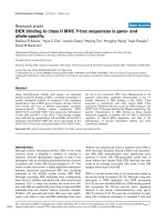
Figure 4
Developing a fully human monoclonal antibody (mAb) using (a) phage display technology and (b) transgenic mouse technology. (a) Step 1: A
suitable source of starting material (for example, human blood) is subjected to polymerase chain reaction using appropriate primers, providing
‘libraries’ of heavy chain V domain (VH) and light chain V domain (VL) sequences. Step 2: Randomly combined VH and VL sequences, connected
via a short linker, are incorporated into the genome of a bacteriophage such that they will be expressed at the phage surface. The combination
marked with an asterisk encodes the desired specificity. Step 3: The phage library is used to infect a bacterial culture, and the resulting
supernatant, containing single-chain Fv-expressing phage particles, is incubated with an appropriate source of target antigen (panning). This can
be on a column, Petri dish, and so on. Phage with appropriate specificity adheres to the antigen source. Step 4: Adherent phage is eluted and
enriched for the appropriate specificity by further rounds of panning. Step 5: After several rounds of panning, adherent phage is sequenced. A
successful procedure should lead to the presence of just one or a few Fv specificities, which can be individually cloned and their specificity
checked. At this stage, in vitro affinity maturation procedures can be performed if required (see ‘Human antibodies’ section for details). Ultimately,
the desired specificity is recloned into an appropriate vector containing full-length mAb sequence for expression in a mammalian cell line. (b) Step
1: A transgenic mouse that produces human antibodies is created by targeted disruption of the endogenous murine immunoglobulin heavy- and
light-chain genetic loci and their replacement by the equivalent human sequences. Step 2: The mouse, now containing human immunoglobulin
genes, is immunised in a conventional manner using the target antigen. Step 3: Splenocytes from the immunised mouse are used to generate
hybridomas via conventional fusion technology. Step 4: Resulting hybridomas are screened, leading to isolation and cloning of a hybridoma-
secreting high-affinity mAb against the target antigen. Note: In theory, phage display rather than fusion technology can be applied from stage 3
onwards.
target and effector cell into apposition can also be created.
The latter approach was pioneered many years ago in the
form of bispecific antibodies [36].
Fc modifications
For several years, the main focus of biotech activity has been,
quite reasonably, the mAb V region – developing mAbs with
novel specificities or improved affinities. However, the
‘downstream’ effects of mAbs and fusion proteins, following
ligand binding, rely on the C region/Fc – and not all sequelae
are desirable. For example, most CD4 mAbs studied in RA
trials were profoundly depleting, whereas non-depleting mAbs
were more potent tolerogens in animal models. Similarly, it is
thought that complement activation is responsible for some of
the infusion-associated adverse effects of mAbs. A profound
example of the consequences of FcγR binding was witnessed
following the administration of TGN1412 to six healthy
volunteers in a phase I clinical trial in 2006 [37]. Massive
cytokine release was triggered when the Fc of the ‘agonistic’
CD28 mAb bound to human FcγR. The isotype of TGN1412
was human IgG4, which has a lower affinity than IgG1 for
FcγR and does not activate complement. The lack of
interaction between human IgG4 and monkey FcγR probably
explains why the mAb appeared safe in primate studies.
Engineering of mAb Fcs is now relatively common, following
the identification of key residues that underlie both
complement and FcγR binding [2-5]. In general, modification
is performed to reduce effector function, although it may also
be enhanced [38]. For example, the CTLA4-Ig Fc is mutated
to reduce complement activation, which may reduce the
incidence of infusion reactions. Certolizumab pegol has a
unique structure among mAb therapeutics. It comprises the
Fab fragment of a humanised TNF-α mAb conjugated to
polyethylene glycol. By definition, this molecule has no Fc-
related functions, acting as a pure TNF-α antagonist.
PEGylation increases the half-life of the molecule, which
remains smaller than a conventional mAb [39]. It is effica-
cious in RA and Crohn disease, which attests to the impor-
tance of TNF-α neutralisation in their treatment, without an
absolute requirement for Fc-mediated effector mechanisms.
Several engineered CD3 mAbs are currently in development
for indications that include psoriatic arthritis and RA. These
have been modified to reduce FcγR binding to harness the
efficacy of CD3 blockade with reduced side effects. The
original murine CD3 mAb, OKT3, potently reversed allograft
rejection but caused a profound cytokine release syndrome
on initial dosing, mediated via FcγR binding [40].
Otelixizumab is a humanised rat mAb in which asparagine has
been replaced by alanine at residue 297 of the human IgG1
Fc. This is the o-linked glycosylation site, where carbohydrate
is incorporated into the mAb structure. The mutation
therefore creates an aglycosyl mAb that in vitro and pre-
clinical data suggest has significantly reduced effector
function [5], and this has been confirmed by clinical studies in
allograft recipients and type-1 diabetics [41,42]. Teplizumab
is a humanised Fc-mutated version of OKT3. It has been
rendered ‘non-mitogenic’ by the mutation of two key FcγR-
binding residues and has demonstrated efficacy in psoriatic
arthritis [43]. A third CD3 mAb with similar properties is
visilizumab, although in this case inflammatory bowel disease
trials have demonstrated that its efficacy is accompanied by
significant first dose-associated cytokine release [44].
Advances in glycobiology have led to an explosion of
knowledge around carbohydrate structure-function relation-
ships, which is now being exploited in glyco-engineering.
Sugar contributes between 3% and 12% of the mass of an
immunoglobulin molecule, the precise Fc sugar content and
structure influencing effector function [45,46]. This can be
modified either chemically or by producing mAbs in cell lines
expressing particular sugar-modifying enzymes. For example,
a glyco-engineered form of rituximab that has enhanced
ADCC (antibody-dependent cellular cytotoxicity) activity has
been created [47].
Notwithstanding the above discussion, it is important to
recognise the importance of target antigen with respect to
mAb effector function. Even a mAb that potently activates
complement and strongly binds FcγR will not necessarily lyse
cells expressing its target antigen. Conversely, some targets
are particularly attractive for cell lysis. CD52 is one such
target and even a human IgG4 CD52 mAb (IgG4-CAMPATH
or IgG4-alemtuzumab) induced profound lymphopenia despite
absent complement activation and weak FcγR binding [48].
Similarly, mAbs against distinct epitopes of the same antigen
can have widely differing cytotoxic characteristics [49]. The
critical features of the target antigen have not been fully
defined, but close apposition between mAb and target cell
membrane is a key parameter, as is the case with alemtuzu-
mab and CD52 [50]. Interestingly, alemtuzumab has a
relatively low affinity for CD52, demonstrating that high
affinity is not required for potent cytotoxicity.
Outstanding issues
Understanding monoclonal antibody pharmacology
The uniqueness of mAbs underpins a sometimes enigmatic
aspect of their biology. As highlighted in a recent review [15],
the ‘obvious’ mode of action for a mAb is sometimes difficult
to substantiate in the clinic. This has been the case
particularly for TNF-α mAbs in RA, in which simple neutralisa-
tion of soluble TNF-α cannot always explain the observed
benefits of therapy. The situation can be even more complex
for mAbs with a cell surface target, such as anti-T cell mAbs.
A lack of target identity means that the therapeutic mAb
cannot usually be tested for biological activity in animal
models. In such cases, it may be necessary to develop a
surrogate mAb against the mouse or rat homologue to test
biological activity in animal models. However, under these
circumstances, it may not be possible to extrapolate precisely
the expected clinical effects, and consequently, potential
Arthritis Research & Therapy Vol 11 No 3 Isaacs
Page 8 of 11
(page number not for citation purposes)
beneficial and adverse effects cannot necessarily be
predicted. Furthermore, the complexities of the immune
system render most in vitro models of limited use in terms of
predicting effector function; therefore, in vivo biological
activity can only be conjectured and, as with anti-CD4 mAbs,
often erroneously [15]. Notably, even when the in vivo
consequences of TGN1412 administration were apparent, it
remained difficult to conceive an in vitro model that predicted
the cytokine storm that underpinned its toxicity [51]. There is
no simple answer to this issue of predictability, apart from
continued careful observation of patients in the clinic
alongside experimental medicine studies on their blood and
tissues, measuring pharmacokinetics and testing
pharmacodynamic hypotheses.
Immunogenicity
Even fully humanised mAbs retain immunogenicity in some
patients. In addition to CDR immunogenicity referred to
earlier, inter-individual genetic variation results in immuno-
globulin allotypes [52]. These V- and C-region allotypic
sequences theoretically can invoke anti-globulin responses in
individuals of alternate allotypes [18]. The only human C
region that is not allotypic is IgG4 [53]. Therapeutic mAbs
are produced from non-human cell lines, and consequently,
their carbohydrates also differ from endogenous immuno-
globulins. In general, this has not been shown to adversely
affect immunogenicity. A recent report, however, demon-
strated hypersensitivity to the galactose-α-1,3-galactose
moiety on cetuximab, a chimeric mAb against the epidermal
growth factor receptor produced in the SP2/0 mouse cell line
[54]. Pre-existing IgE antibodies against this oligosaccharide,
which is closely related to substances in the ABO blood
group, predisposed to anaphylactic reactions.
Biosimilars
Equivalent issues are relevant to the concept of ‘generic’
mAbs or biosimilars. Unlike with small-molecule drugs, it may
not be possible to create an identical version of a therapeutic
mAb. Even different clones of a particular cell line may impart
subtle changes on a mAb molecule, and only the original
mAb-encoding DNA clone and master cell bank can be
guaranteed to generate a consistent product, provided
culture conditions are carefully maintained. Even then, subtle
modifications to downstream manufacturing processes can
result in significant changes to properties such as
immunogenicity or even effector function [55,56]. Legislation
and regulations concerning the development of ‘biosimilar’
mAbs remain to be fully defined, but as current patents start
to expire, this situation must soon change [57].
Economics
It is important to recognise that the identification of a
potential mAb specificity is only the start of a long and
expensive process that may or may not culminate in a
marketable and profitable product. Even after mAb-encoding
DNA is cloned and characterised and the protein product
demonstrates appropriate bioactivity, significant work follows
in order to optimise and standardise the manufacturing
process. For example, considerable effort is required to
define the optimal production cell line and growth conditions
for high yields, and downstream purification and formulation
processes may also be complex and require precise
standardisation. This is reflected in the high cost of most
licensed biologic drugs [58].
In contrast to mammalian cell lines, bacteria provide a highly
efficient means of mAb production, a fact exploited by
certolizumab pegol which is produced in Escherichia coli.
This is possible because Fab fragments do not require as
much processing by the producer cells as do full-length
mAbs: bacterial cells cannot glycosylate nor can they
assemble complex multichain macromolecules. A disadvan-
tage of bacterial production is that the downstream process
must ensure complete freedom of the final product from
bacterial molecules such as endotoxin. Yields are significantly
higher, however, and it seems likely that bacterial production
processes will be further exploited in the future, particularly in
relation to some of the novel mAb fragments referred to earlier.
Conclusions
The original mAb revolution, precipitated by the discovery of
fusion technology, has been superseded by an even more
profound transformation catalysed by antibody engineering.
Indeed, all of the currently licensed biologics used in
rheumatological practice, as well as those in development,
have been engineered in one way or another. Future
advances are likely to involve glyco-engineering and small
mAb fragments, whilst bacterial production processes and
biosimilars may provide cheaper therapeutics. This is critical
because the current high cost of biologics means that many
patients still cannot access these highly effective drugs. From
an academic viewpoint, it remains paramount that we
continue to study these drugs from an experimental medicine
perspective to ensure that we fully understand their
capabilities and the potential consequences of their adminis-
tration to our patients.
Available online />Page 9 of 11
(page number not for citation purposes)
This article is part of a special collection of reviews, The
Scientific Basis of Rheumatology: A Decade of
Progress, published to mark Arthritis Research &
Therapy’s 10th anniversary.
Other articles in this series can be found at:
/>The Scientific Basis
of Rheumatology:
A Decade of Progress
Competing interests
JDI has consulted for and/or has served on advisory boards
of Bristol-Myers Squibb Company (Princeton, NJ, USA),
MedImmune Limited (formerly known as Cambridge Antibody
Technology, Gaithersburg, MD, USA), GlaxoSmithKline
(Uxbridge, Middlesex, UK), Genzyme (Cambridge, MA, USA),
Roche (Basel, Switzerland), and TolerRx (Cambridge, MA,
USA). He is named as co-inventor on a European patent
relating to the use of non-mitogenic anti-CD3 mAb in
inflammatory arthritis.
References
1. Junghans RP, Anderson CL: The protection receptor for IgG
catabolism is the beta2-microglobulin-containing neonatal
intestinal transport receptor. Proc Natl Acad Sci U S A 1996,
93:5512-5516.
2. Duncan A, Winter G: The binding site for C1q on IgG. Nature
1988, 332:738-740.
3. Duncan A, Woof J, Partridge L, Burton D, Winter G: Localization
of the binding site for the human high-affinity Fc receptor on
IgG. Nature 1988, 332:563-564.
4. Lund J, Winter G, Jones PT, Pound JD, Tanaka T, Walker MR,
Artymiuk PJ, Arata Y, Burton DR, Jefferis R, Woof JM: Human Fc
gamma RI and Fc gamma RII interact with distinct but over-
lapping sites on human IgG. J Immunol 1991, 147:2657-2662.
5. Isaacs J, Greenwood J, Waldmann H: Therapy with monoclonal
antibodies II. The contribution of Fcg receptor binding and the
influence of Ch1 and Ch3 domains on in vivo effector function.
J Immunol 1998, 161:3863-3869.
6. Lefranc MP, Giudicelli V, Ginestoux C, Bodmer J, Muller W,
Bontrop R, Lemaitre M, Malik A, Barbie V, Chaume D: IMGT, the
international ImMunoGeneTics database. Nucleic Acids Res
1999, 27:209-212.
7. Kohler G, Milstein C: Continuous cultures of fused cells secreting
antibody of predefined specificity. Nature 1975, 256:495-497.
8. Herzog C, Walker C, Pichler W, Aeschlimann A, Wassmer P,
Stockinger H, Knapp W, Rieber P, Muller W: Monoclonal anti-
CD4 in arthritis. Lancet 1987, 8573:1461-1462.
9. Schroeder TJ, First MR, Mansour ME, Hurtubise PE, Hariharan S,
Ryckman FC, Munda R, Melvin DB, Penn I, Ballistreri WF, Alexan-
der JW: Antimurine antibody formation following OKT3
therapy. Transplantation 1990, 49:48-51.
10. Neuberger MS, Williams GT, Mitchell EB, Jouhal SS, Flanagan
JG, Rabbitts TH: A hapten-specific chimaeric IgE antibody with
human physiological effector function. Nature 1985, 314:268-
270.
11. Bruggemann M, Williams G, Bindon C, Clark M, Walker M, Jef-
feris R, Waldmann H, Neuberger M: Comparison of the effector
functions of human immunoglobulins using a matched set of
chimeric antibodies. J Exp Med 1987, 166:1351-1361.
12. Isaacs J, Clark M, Greenwood J, Waldmann H: Therapy with
monoclonal antibodies. An in vivo model for the assessment
of therapeutic potential. J Immunol 1992, 148:3062-3071.
13. Wolbink GJ, Vis M, Lems W, Voskuyl AE, de Groot E, Nurmo-
hamed MT, Stapel S, Tak PP, Aarden L, Dijkmans B: Develop-
ment of antiinfliximab antibodies and relationship to clinical
response in patients with rheumatoid arthritis. Arthritis Rheum
2006,
54:711-715.
14. Baert F, Noman M, Vermeire S, Van Assche G, Haens G, Car-
bonez A, Rutgeerts P: Influence of immunogenicity on the
long-term efficacy of infliximab in Crohn’s disease. N Engl J
Med 2003, 348:601-608.
15. Strand V, Kimberly R, Isaacs JD: Biologic therapies in rheuma-
tology: lessons learned, future directions. Nat Rev Drug Discov
2007, 6:75-92.
16. Jones PT, Dear PH, Foote J, Neuberger MS, Winter G: Replacing
the complementarity-determining regions in a human anti-
body with those from a mouse. Nature 1986, 321:522-525.
17. Clark M: Antibody humanization: a case of the ‘Emperor’s new
clothes’? Immunol Today 2000, 21:397-402.
18. Isaacs J, Watts R, Hazleman B, Hale G, Keogan M, Cobbold S,
Waldmann H: Humanised monoclonal antibody therapy for
rheumatoid arthritis. Lancet 1992, 340:748-752.
19. Orlandi R, Gussow DH, Jones PT, Winter G: Cloning
immunoglobulin variable domains for expression by the poly-
merase chain reaction. Proc Natl Acad Sci U S A 1989, 86:
3833-3837.
20. Persson MA, Caothien RH, Burton DR: Generation of diverse
high-affinity human monoclonal antibodies by repertoire
cloning. Proc Natl Acad Sci U S A 1991, 88:2432-2436.
21. Marks JD, Griffiths AD, Malmqvist M, Clackson TP, Bye JM,
Winter G: By-passing immunization: building high affinity
human antibodies by chain shuffling. Biotechnology (N Y)
1992, 10:779-783.
22. Jespers LS, Roberts A, Mahler SM, Winter G, Hoogenboom HR:
Guiding the selection of human antibodies from phage
display repertoires to a single epitope of an antigen. Biotech-
nology (N Y) 1994, 12:899-903.
23. Griffiths AD, Williams SC, Hartley O, Tomlinson IM, Waterhouse
P, Crosby WL, Kontermann RE, Jones PT, Low NM, Allison TJ,
Prospero TD, Hoogenboom HR, Nissim A, Cox JPL, Harrison JL,
Zaccolo M, Gherardi E, Winter G: Isolation of high affinity
human antibodies directly from large synthetic repertoires.
EMBO J 1994, 13:3245-3260.
24. McCafferty J, Griffiths AD, Winter G, Chiswell DJ: Phage anti-
bodies: filamentous phage displaying antibody variable
domains. Nature 1990, 348:552-554.
25. Bruggemann M, Caskey HM, Teale C, Waldmann H, Williams GT,
Surani MA, Neuberger MS: A repertoire of monoclonal antibod-
ies with human heavy chains from transgenic mice. Proc Natl
Acad Sci U S A 1989, 86:6709-6713.
26. Lonberg N: Human antibodies from transgenic animals. Nat
Biotechnol 2005, 23:1117-1125.
27. Medarex - UltiMAb Human Antibody Development System
®
[ />28. Lonberg N: Fully human antibodies from transgenic mouse
and phage display platforms. Curr Opin Immunol 2008, 20:
450-459.
29. Nesbitt A, Fossati G, Bergin M, Stephens P, Stephens S, Foulkes
R, Brown D, Robinson M, Bourne T: Mechanism of action of cer-
tolizumab pegol (CDP870): in vitro comparison with other
anti-tumor necrosis factor alpha agents. Inflamm Bowel Dis
2007, 13:1323-1332.
30. Dillon SR, Gross JA, Ansell SM, Novak AJ: An APRIL to remem-
ber: novel TNF ligands as therapeutic targets. Nat Rev Drug
Discov 2006, 5:235-246.
31. Trubion Pharmaceuticals, SMIP™ therapeutics [http://www.
trubion.com/products/technology/smip-therapeutics/].
32. Ablynx, Nanobody
®
Platform [ />index.htm].
33. Domantis Limited homepage [].
34. Ward ES, Gussow D, Griffiths AD, Jones PT, Winter G: Binding
activities of a repertoire of single immunoglobulin variable
domains secreted from Escherichia coli. Nature 1989, 341:
544-546.
35. Arana Therapeutics, ART621 - Phase II Trial Underway [http://
www.arana.com/inflammation_franchise_art621.htm].
36. Milstein C, Cuello AC: Hybrid hybridomas and their use in
immunohistochemistry. Nature 1983, 305:537-540.
37. Suntharalingam G, Perry MR, Ward S, Brett SJ, Castello-Cortes
A, Brunner MD, Panoskaltsis N: Cytokine storm in a phase 1
trial of the anti-CD28 monoclonal antibody TGN1412. N Engl J
Med 2006, 355:1018-1028.
38. Natsume A, In M, Takamura H, Nakagawa T, Shimizu Y, Kitajima K,
Wakitani M, Ohta S, Satoh M, Shitara K, Niwa R: Engineered
antibodies of IgG1/IgG3 mixed isotype with enhanced cyto-
toxic activities. Cancer Res 2008, 68:3863-3872.
39. Melmed GY, Targan SR, Yasothan U, Hanicq D, Kirkpatrick P:
Certolizumab pegol. Nat Rev Drug Discov 2008, 7:641-642.
40. Chatenoud L, Ferran C, Legendre C, Franshimont P, Reuter A,
Kreis H, Bach J: Clinical use of OKT3: the role of cytokine
release and xenosensitization. J Autoimmun 1988, 1:631-640.
41. Friend PJ, Hale G, Chatenoud L, Rebello P, Bradley J, Thiru S,
Phillips JM, Waldmann H: Phase I study of an engineered agly-
cosylated humanized CD3 antibody in renal transplant rejec-
tion. Transplantation 1999, 68:1632-1637.
42. Keymeulen B, Vandemeulebroucke E, Ziegler A, Mathieu C,
Kaufman L, Hale G, Gorus F, Goldman M, Walter M, Candon S,
Schandene L, Crenier L, De Block C, Seigneurin JM, De Pauw P,
Pierard D, Weets I, Rebello P, Bird P, Berrie E, Frewin M, Wald-
Arthritis Research & Therapy Vol 11 No 3 Isaacs
Page 10 of 11
(page number not for citation purposes)
mann H, Bach JF, Pipeleers D, Chatenoud L: Insulin needs after
CD3-antibody therapy in new-onset type 1 diabetes. N Engl J
Med 2005, 352:2598-2608.
43. Utset TO, Auger JA, Peace D, Zivin RA, Xu D, Jolliffe L, Alegre ML,
Bluestone JA, Clark MR: Modified anti-CD3 therapy in psoriatic
arthritis: a phase I/II clinical trial. J Rheumatol 2002, 29:1907-
1913.
44. Plevy S, Salzberg B, Van Assche G, Regueiro M, Hommes D,
Sandborn W, Hanauer S, Targan S, Mayer L, Mahadevan U,
Frankel M, Lowder J: A phase I study of visilizumab, a human-
ized anti-CD3 monoclonal antibody, in severe steroid-refrac-
tory ulcerative colitis. Gastroenterology 2007, 133:1414-1422.
45. Ferrara C, Brunker P, Suter T, Moser S, Puntener U, Umana P:
Modulation of therapeutic antibody effector functions by gly-
cosylation engineering: influence of Golgi enzyme localization
domain and co-expression of heterologous beta1, 4-N-acetyl-
glucosaminyltransferase III and Golgi alpha-mannosidase II.
Biotechnol Bioeng 2006, 93:851-861.
46. Kanda Y, Yamada T, Mori K, Okazaki A, Inoue M, Kitajima-Miyama
K, Kuni-Kamochi R, Nakano R, Yano K, Kakita S, Shitara K, Satoh
M: Comparison of biological activity among nonfucosylated
therapeutic IgG1 antibodies with three different N-linked Fc
oligosaccharides: the high-mannose, hybrid, and complex
types. Glycobiology 2007, 17:104-118.
47. Davies J, Jiang L, Pan LZ, LaBarre MJ, Anderson D, Reff M:
Expression of GnTIII in a recombinant anti-CD20 CHO pro-
duction cell line: expression of antibodies with altered glyco-
forms leads to an increase in ADCC through higher affinity for
FC gamma RIII. Biotechnol Bioeng 2001, 74:288-294.
48. Isaacs J, Wing M, Greenwood J, Hazleman B, Hale G, Waldmann
H: A therapeutic human IgG4 monoclonal antibody that
depletes target cells in humans. Clin Exp Immunol 1996, 106:
427-433.
49. Bindon C, Hale G, Waldmann H: Importance of antigen speci-
ficity for complement mediated lysis by monoclonal antibod-
ies. Eur J Immunol 1988, 18:1507-1514.
50. Xia M, Hale G, Waldmann H: Efficient complement-mediated
lysis of cells containing the CAMPATH-1 (CDw52) antigen.
Mol Immunol 1993, 30:1089-1096.
51. Stebbings R, Findlay L, Edwards C, Eastwood D, Bird C, North D,
Mistry Y, Dilger P, Liefooghe E, Cludts I, Fox B, Tarrant G, Robin-
son J, Meager T, Dolman C, Thorpe SJ, Bristow A, Wadhwa M,
Thorpe R, Poole S: “Cytokine storm” in the phase I trial of
monoclonal antibody TGN1412: better understanding the
causes to improve preclinical testing of immunotherapeutics.
J Immunol 2007, 179:3325-3331.
52. Bruggemann M, Winter G, Waldmann H, Neuberger MS: The
immunogenicity of chimeric antibodies. J Exp Med 1989, 170:
2153-2157.
53. Gorman SD, Clark MR: Humanisation of monoclonal antibod-
ies for therapy. Semin Immunol 1990, 2:457-466.
54. Chung CH, Mirakhur B, Chan E, Le QT, Berlin J, Morse M, Murphy
BA, Satinover SM, Hosen J, Mauro D, Slebos RJ, Zhou Q, Gold D,
Hatley T, Hicklin DJ, Platts-Mills TA: Cetuximab-induced ana-
phylaxis and IgE specific for galactose-alpha-1,3-galactose. N
Engl J Med 2008, 358:1109-1117.
55. Eckardt K, Casadevall N: Pure red-cell aplasia due to anti-ery-
thropoietin antibodies. Nephrol Dial Transplant 2003, 18:865-
869.
56. Mason U, Aldrich J, Breedveld F, Davis C, Elliott M, Jackson M,
Jorgensen C, Keystone E, Levy R, Tesser J, Totoritis M, Truneh A,
Weisman M, Wiesenhutter C, Yocum D, Zhu J: CD4 coating, but
not CD4 depletion, is a predictor of efficacy with primatized
monoclonal anti-CD4 treatment of active rheumatoid arthritis.
J Rheumatol 2002, 29:220-229.
57. Genazzani AA, Biggio G, Caputi AP, Del Tacca M, Drago F, Fan-
tozzi R, Canonico PL: Biosimilar drugs: concerns and opportu-
nities. BioDrugs 2007, 21:351-356.
58. Farid SS: Process economics of industrial monoclonal anti-
body manufacture. J Chromatogr A 2007, 848:8-18.
59. Elliott MJ, Maini RN, Feldmann M, Kalden JR, Antoni C, Smolen
JS, Leeb B, Breedveld FC, Macfarlane JD, Bijl H, Woody JN: Ran-
domised double-blind comparison of chimeric monoclonal
antibody to tumour necrosis factor alpha (cA2) versus
placebo in rheumatoid arthritis. Lancet 1994, 344:1105-1110.
60. Edwards J, Szczepanski L, Szechinski J, Filipowicz-Sosnowska A,
Emery P, Close D, Stevens R, Shaw T: Efficacy of B-cell-tar-
geted therapy with rituximab in patients with rheumatoid
arthritis. N Engl J Med 2004, 350:2572-2581.
61. Maini RN, Taylor PC, Szechinski J, Pavelka K, Bröll J, Balint G,
Emery P, Raemen F, Petersen J, Smolen J, Thomson D, Kishimoto
T; CHARISMA Study Group: Double-blind randomized con-
trolled clinical trial of the interleukin-6 receptor antagonist,
tocilizumab, in European patients with rheumatoid arthritis
who had an incomplete response to methotrexate. Arthritis
Rheum 2006, 54:2817-2829.
62. Genovese MC, Kaine JL, Lowenstein MB, Giudice JD, Baldassare
A, Schechtman J, Fudman E, Kohen M, Gujrathi S, Trapp RG,
Sweiss NJ, Spaniolo G, Dummer W; ACTION Study Group:
Ocrelizumab, a humanized anti-CD20 monoclonal antibody, in
the treatment of patients with rheumatoid arthritis: a phase
I/II randomized, blinded, placebo-controlled, dose-ranging
study. Arthritis Rheum 2008, 58:2652-2661.
63. Dorner T, Kaufmann J, Wegener W, Teoh N, Goldenberg D,
Burmester G: Initial clinical trial of epratuzumab (humanized
anti-CD22 antibody) for immunotherapy of systemic lupus
erythematosus. Arthritis Res Ther 2006, 8:R74.
64. Keystone E, Heijde D, Mason D Jr., Landewe R, Vollenhoven RV,
Combe B, Emery P, Strand V, Mease P, Desai C, Pavelka K: Cer-
tolizumab pegol plus methotrexate is significantly more effec-
tive than placebo plus methotrexate in active rheumatoid
arthritis: findings of a fifty-two-week, phase III, multicenter,
randomized, double-blind, placebo-controlled, parallel-group
study. Arthritis Rheum 2008, 58:3319-3329.
65. Herold KC, Hagopian W, Auger JA, Poumian-Ruiz E, Taylor L,
Donaldson D, Gitelman SE, Harlan DM, Xu D, Zivin RA, Bluestone
JA: Anti-CD3 monoclonal antibody in new-onset type 1 dia-
betes mellitus. N Engl J Med 2002, 346:1692-1698.
66. Weinblatt ME, Keystone EC, Furst DE, Moreland LW, Weisman
MH, Birbara CA, Teoh LA, Fischkoff SA, Chartash EK: Adali-
mumab, a fully human anti-tumor necrosis factor alpha mono-
clonal antibody, for the treatment of rheumatoid arthritis in
patients taking concomitant methotrexate: the ARMADA trial.
Arthritis Rheum 2003, 48:35-45.
67. Dorner T, Burmester GR: New approaches of B-cell-directed
therapy: beyond rituximab. Curr Opin Rheumatol 2008, 20:263-
268.
68. Furie R, Stohl W, Ginzler EM, Becker M, Mishra N, Chatham W,
Merrill JT, Weinstein A, McCune WJ, Zhong J, Cai W, Freimuth
W; Belimumab Study Group: Biologic activity and safety of beli-
mumab, a neutralizing anti-B-lymphocyte stimulator (BLyS)
monoclonal antibody: a phase I trial in patients with systemic
lupus erythematosus. Arthritis Res Ther 2008, 10:R109.
69. Keystone EC, Genovese MC, Klareskog L, Hsia EC, Hall ST,
Miranda PC, Pazdur J, Bae SC, Palmer W, Zrubek J, Wiekowski
M, Visvanathan S, Wu Z, Rahman MU: Golimumab, a human
antibody to TNF-{alpha} given by monthly subcutaneous
injections, in active rheumatoid arthritis despite methotrexate:
The GO-FORWARD Study. Ann Rheum Dis 2008, Dec 11.
[Epub ahead of print].
70. Weinblatt ME, Kremer JM, Bankhurst AD, Bulpitt KJ, Fleischmann
RM, Fox RI, Jackson CG, Lange M, Burge DJ: A trial of etaner-
cept, a recombinant tumor necrosis factor receptor:Fc fusion
protein, in patients with rheumatoid arthritis receiving
methotrexate. N Engl J Med 1999, 340:253-259.
71. Genovese MC, Becker JC, Schiff M, Luggen M, Sherrer Y, Kremer
J, Birbara C, Box J, Natarajan K, Nuamah I, Li T, Aranda R, Hagerty
DT, Dougados M: Abatacept for rheumatoid arthritis refractory
to tumor necrosis factor alpha inhibition. N Engl J Med 2005,
353:1114-1123.
72. Tak PP, Thurlings RM, Rossier C, Nestorov I, Dimic A, Mircetic V,
Rischmueller M, Nasonov E, Shmidt E, Emery P, Munafo A: Ataci-
cept in patients with rheumatoid arthritis: results of a multi-
center, phase ib, double-blind, placebo-controlled,
dose-escalating, single- and repeated-dose study. Arthritis
Rheum 2008, 58:61-72.
Available online />Page 11 of 11
(page number not for citation purposes)