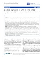
Báo cáo y học: "Elevated expression of caspase-3 inhibitors, survivin and xIAP correlates with low levels of apoptosis in active rheumatoid synovium" potx
Bạn đang xem bản rút gọn của tài liệu. Xem và tải ngay bản đầy đủ của tài liệu tại đây (4.54 MB, 11 trang )
Open Access
Available online />Page 1 of 11
(page number not for citation purposes)
Vol 11 No 1
Research article
Elevated expression of caspase-3 inhibitors, survivin and xIAP
correlates with low levels of apoptosis in active rheumatoid
synovium
Anak ASSK Dharmapatni
1
, Malcolm D Smith
2
, David M Findlay
3
, Christopher A Holding
1
,
Andreas Evdokiou
3
, Michael J Ahern
2
, Helen Weedon
2
, Paul Chen
4
, Gavin Screaton
5
, Xiao N Xu
4
and David R Haynes
1
1
Discipline of Pathology, School of Medical Sciences, Faculty of Health Sciences, University of Adelaide, North Terrace, Adelaide, 5005 South
Australia, Australia
2
Rheumatology Research Unit, Repatriation General Hospital, Daws Road, Adelaide, 5041 South Australia, Australia
3
Discipline of Orthopaedics and Trauma, School of Medicine, Faculty of Health Sciences, University of Adelaide and Hanson Institute, Frome Road,
Adelaide, 5005 South Australia, Australia
4
MRC Human Immunology Unit, Weatherall Institute of Molecular Medicine, John Radcliffe Hospital, Oxford OX3, UK
5
Hammersmith Hospital, Du Cane Road, London W12 0NN, UK
Corresponding author: David R Haynes,
Received: 20 Jun 2008 Revisions requested: 23 Jul 2008 Revisions received: 2 Dec 2008 Accepted: 27 Jan 2009 Published: 27 Jan 2009
Arthritis Research & Therapy 2009, 11:R13 (doi:10.1186/ar2603)
This article is online at: />© 2009 Dharmapatni et al.; licensee BioMed Central Ltd.
This is an open access article distributed under the terms of the Creative Commons Attribution License ( />),
which permits unrestricted use, distribution, and reproduction in any medium, provided the original work is properly cited.
Abstract
Introduction Tumour necrosis factor-related apoptosis-
inducing ligand (TRAIL) is a tumour necrosis factor (TNF) family
member capable of inducing apoptosis in many cell types.
Methods Using immunohistochemistry, terminal
deoxynucleotidyl transferase biotin-dUTP nick end labelling
(TUNEL) and real-time PCR we investigated the expression of
TRAIL, TRAIL receptors and several key molecules of the
intracellular apoptotic pathway in human synovial tissues from
various types of arthritis and normal controls. Synovial tissues
from patients with active rheumatoid arthritis (RA), inactive RA,
osteoarthritis (OA) or spondyloarthritis (SpA) and normal
individuals were studied.
Results Significantly higher levels of TRAIL, TRAIL R1, TRAIL
R2 and TRAIL R4 were observed in synovial tissues from
patients with active RA compared with normal controls (p <
0.05). TRAIL, TRAIL R1 and TRAIL R4 were expressed by many
of the cells expressing CD68 (macrophages). Lower levels of
TUNEL but higher levels of cleaved caspase-3 staining were
detected in tissue from active RA compared with inactive RA
patients (p < 0.05). Higher levels of survivin and x-linked
inhibitor of apoptosis protein (xIAP) were expressed in active RA
synovial tissues compared with inactive RA observed at both the
protein and mRNA levels.
Conclusions This study indicates that the induction of
apoptosis in active RA synovial tissues is inhibited despite
stimulation of the intracellular pathway(s) that lead to apoptosis.
This inhibition of apoptosis was observed downstream of
caspase-3 and may involve the caspase-3 inhibitors, survivin
and xIAP.
Introduction
Decreased apoptosis has been proposed as a possible factor
that contributes to the hyperplasia of the synovial membrane
and accumulation of inflammatory cells observed in the syno-
vitis of patients with active rheumatoid arthritis (RA) [1,2].
Inducing apoptosis in these synovial cells has the potential to
reduce the disease severity and progression similar to that
ACR: American College of Rheumatology; CRP: C-reactive protein; DAS28: disease activity score 28; DMARD: disease modifying antirheumatic
drug; ESR: erythrocyte sedimentation rate; FADD: Fas associated death domain; FLIP: flice inhibitory protein; IAP: inhibitor of apoptosis protein; Ig:
immunoglobulin; OA: osteoarthritis; OPG: osteoprotegerin; PCR: polymerase chain reaction; RA: rheumatoid arthritis; RF: rheumatoid factor; SpA:
spondyloarthropathies; SQA: semiquantitative analysis; TNF: tumour necrosis factor; TRAIL: tumour necrosis factor related apoptosis inducing ligand;
TUNEL: terminal deoxynucleotidyl transferase biotin-dUTP nick end labelling; xIAP: x-linked inhibitor of apoptosis protein.
Arthritis Research & Therapy Vol 11 No 1 Dharmapatni et al.
Page 2 of 11
(page number not for citation purposes)
suggested previously for apoptosis via the FAS-FAS ligand
pathway [3,4].
Tumour necrosis factor-related apoptosis-inducing ligand
(TRAIL) is a member of the tumour necrosis factor (TNF) family
and a type II membrane bound cytokine that is expressed by
many cell types [5,6]. Although TRAIL mainly mediates apop-
tosis, like many other TNF family members, it has many other
roles including regulation of endothelial nitric oxide synthase
and the innate immune system [7,8]. In relation to apoptosis
TRAIL has two types of receptors that differ in their ability to
either initiate or inhibit TRAIL-mediated apoptosis [9]. TRAIL
R1 (death receptor 4) and TRAIL R2 (death receptor 5) induce
apoptotic cell death. The second type of TRAIL receptors act
as decoy receptors and these are TRAIL R3 (DcR1, decoy
receptor 1), TRAIL R4 (DcR2, decoy receptor 2) and osteo-
protegerin (OPG) [10].
TRAIL and TRAIL death receptors form a complex, which
transmits an apoptotic signal via the Fas associated death
domain (FADD). This leads to activation of caspase-8 or other
initiator caspases, which in turn activate downstream cas-
pases (such as caspase-3, 9, 6 and 7) that cause cell death.
Inhibition of apoptosis mediated by TRAIL could occur
upstream or downstream of the pathway. At the upstream lev-
els the inhibition could result from the expression of TRAIL
decoy receptors, while at the intracellular signalling level pro-
teins capable of inhibiting caspase activation, such as FLIP
(flice inhibitory protein) [11], that blocks initiator caspase (cas-
pase-8) and IAP (inhibitor of apoptosis protein) family mem-
bers [12], that block effector caspase (caspase-3) further
downstream, could potentially inhibit apoptosis.
Several studies have reported on the importance of TRAIL and
TRAIL receptor expression in inducing or inhibiting apoptosis
[13-16]. Some studies have shown that TRAIL and its recep-
tor, TRAIL R2, are expressed in the synovial tissues of RA
patients [17,18] and TRAIL R2 is highly expressed in synovial
cells in culture [18-21]. TRAIL gene therapy has been reported
to inhibit development of arthritis in a collagen-induced mouse
model [17,22]. In addition, an agonistic monoclonal antibody
that binds to the TRAIL death receptor, TRAIL R2, has been
reported to induce apoptosis in RA synovial fibroblasts
[18,19]. However, none of the studies comprehensively inves-
tigated TRAIL and all its receptors in the synovial tissue from
patients with various types of arthritis.
In addition to TRAIL and its receptor interaction, recent evi-
dence suggests that intracellular regulators such as FLIP, cas-
pases [23], members of the Bcl2 family [24] and tumour
suppressor proteins such as p53 are often central in determin-
ing whether apoptosis occurs in particular cells [11]. Recently,
survivin, a member of the IAP family, has been reported to be
elevated in serum in RA, with high levels correlating with joint
erosion in active RA [25].
Many of the previous studies [18,19,21,26,27] have focused
on investigating the expression of TRAIL and TRAIL receptors
using synovial fibroblasts in culture. However, this population
of cells may not be representative of the inflammatory cells,
such as lymphocytes and cells of the macrophage/monocyte
lineage. Therefore, the present study investigated the expres-
sion of TRAIL and each of the TRAIL receptors in synovial tis-
sues in situ from a range of arthritic conditions, including RA
and OA. The expression of intracellular pro- and anti-apoptotic
molecules was simultaneously investigated with the extent of
apoptosis in pathogenic and normal human synovial tissues.
Materials and methods
Patients and tissue preparation
Synovial tissue samples were obtained from patients at the
time of knee arthroscopy or total knee replacement (OA
patients) at the Rheumatology unit at the Repatriation General
Hospital, Adelaide, South Australia. Normal synovial tissues
were obtained from patients attending sports medicine clinics
with unexplained knee pain. All patients with active RA or
spondyloarthropathy (SpA) had active joint inflammation.
Patients with inactive RA were in remission after successful
disease modifying antirheumatic drug (DMARD) treatment for
active RA and were undergoing a further knee joint arthros-
copy for routine follow-up.
Informed consent was obtained from the patients before the
procedures and the study was approved by the Repatriation
General Hospital Human Ethics Committee. All patients diag-
nosed with RA fulfilled the 1987 revised criteria of the Ameri-
can College of Rheumatology (ACR) [28]. Osteoarthritis (OA)
patients fulfilled the criteria by Altman and colleages [29] and
SpA patients fulfilled the European Spondyloarthropathy
Study Group criteria [30]. Characteristics of patients are sum-
marised in Table 1.
Clinical and laboratory data were collected from all RA
patients including tender and swollen joint count, patient
assessment of pain and global score of disease activity and
physician score of global disease activity (all on visual ana-
logue scales). In addition, C-reactive protein (CRP), erythro-
cyte sedimentation rate (ESR), rheumatoid factor levels (RF),
disease activity score (DAS28), as well as x-rays of the hands
and feet were assessed for the presence of joint erosions. Fro-
zen sections and formalin-fixed paraffin-embedded tissue
were prepared from the synovial biopsies. When available, an
additional synovial biopsy was placed into RNAlater (Ambion,
Foster City, CA, USA) and stored at 4°C until RNA extraction
was performed.
Available online />Page 3 of 11
(page number not for citation purposes)
Monoclonal antibodies
Recombinant human TRAIL (375-TL/CF) and monoclonal anti-
bodies directed against TRAIL (MAB687, immunoglobulin (Ig)
G1), TRAIL receptor 3/DcR1 (M430, IgG1), rabbit anti-human
survivin (AF886, polyclonal IgG) and anti-human x-linked inhib-
itor of apoptosis protein (xIAP) (MAB8221, IgG1) were pur-
chased from R&D Systems, Inc. (Minneapolis, MN, USA).
Monoclonal antibodies against TRAIL receptor 1/TRAIL R1
(M271, IgG2a), TRAIL receptor2/TRAIL R2 (M413, IgG1) and
TRAIL receptor 4/TRAIL R4 (M444, IgG1) were gifts from
Amgen (Thousand Oaks, CA, USA). Monoclonal antibodies
against TRAIL R2 were used as described previously [31].
Monoclonal antibodies against cleaved caspase-3 (cat no #
9664) were purchased from Cell Signaling Technology Inc.
(Beverley, MA, USA) and monoclonal antibodies against
cleaved caspase-8 (AP1013) were purchased from Calbio-
chem (Darmstad, Germany). Anti-human CD3 monoclonal
antibodies (Clone Sk7) were used to detect T lymphocytes,
anti-CD22 antibodies (clone Mc 64-12) were used to detect
B lymphocytes and anti-CD68 antibodies (clone EBMII) were
used to detect macrophages and were all purchased from
Dako (Botany, NSW, Australia). Anti-human CD55 antibodies
(MCA 1614 GA) were used to detect synovial lining fibrob-
lasts and were obtained from Serotec (Kidlington, Oxford,
UK). Frozen sections were used for immunostaining with all
antibodies except for those studies using TRAIL R2 and TRAIL
R3 in which formalin-fixed paraffin-embedded material was
used.
Immunohistochemical detection
For immunoperoxidase staining a three-step immunohisto-
chemical detection was performed as described previously
[32]. Staining for TRAIL, TRAIL receptors, cleaved caspases,
xIAP and survivin were performed at the same time in all syno-
vial tissues to eliminate day-to-day variability of the staining.
Negative controls consisted of omission of the primary anti-
bodies, and the use of isotype-matched antibody controls
(1B5 for IgG1, 1D4.5 for IgG2a and normal rabbit serum for
polyclonal antibody) [33]. Positive controls were tissues
known to be positive for TRAIL and TRAIL receptors (normal
colon, adenoma and carcinoma of the colon). Specificity of
TRAIL staining was confirmed by antibody absorption, using
recombinant human TRAIL, according to a method published
previously [34].
Double staining immunohistochemistry
Double staining was performed according to a method pub-
lished previously [34] to identify the cell lineages that express
TRAIL, TRAIL R1 and TRAIL R4. Antibodies directed against
CD68 were used to detect macrophage lineage cells, CD55
for synovial fibroblasts in the synovial lining layer, CD22 for B
cells and CD3 for T-lymphocyte lineage cells.
Detection of apoptosis by TUNEL staining
Terminal deoxynucleotidyl transferase biotin-dUTP nick end
labelling (TUNEL) staining was performed on frozen synovial
tissues using a TUNEL detection kit (Roche Diagnostics, Cas-
tle Hill, NSW, Australia). As a positive control, synovial tissue
Table 1
Demographic of patients
Patient's Active RA Inactive RA OA SpA Normal
Number 21 9 7 12 17
Age (years) (mean;SEM) 65.33 (3.32) 72.33 (2.35) 69.7 (2.34) 48.72 (5.66) 33.41 (2.45)
Disease duration (months) (mean;SEM) 3.76 (.50) 21.77 (3.63) NA 72.9 (19.32) NA
Gender (F/M) 11 (10) 3 (6) 1 (6) 5 (7) 7 (10)
DMARDs 17 NSAIDs 2 MTX 6 NSAIDs 8 NSAIDs NA
2 Hydroxy 1 Hydroxy 1 Panadeine-F 2 SSZ
1 Pred 4 Gold 1 SSZ/AZA/pred
1 SSZ 1 SSZ 1 SSZ/NSAIDs
1 gold/MTX
CRP (mg/L) (mean;SEM) 67.67 (13.5) 4.7 (1.03) NA 43.45 (7.65) NA
RF 11 positive 5 positive NA NA NA
Erosions 6 positive 2 positive NA 7 positive NA
DAS 28 (mean;SEM) 5.47 (0.22) 1.08 (0.24) NA NA NA
AZA = azathioprine; CRP = C-reactive protein; DAS28 = disease activity score 28; DMARD = disease modifying antirheumatic drug; F = female;
Gold = intramuscular sodium aurothiomalate injections; Hydroxy = hydroxychloroquine; M = male; MTX = methotrexate; NA = not available;
NSAIDs = nonsteroidal anti-inflammatory drugs; OA = osteoarthritis; Panadeine F = paracetamol/codeine combination; Pred = prednisolone; RA
= rheumatoid arthritis; SEM = standard error of the mean; SpA = spondyloarthropathies; SSZ = sulphasalazine.
Arthritis Research & Therapy Vol 11 No 1 Dharmapatni et al.
Page 4 of 11
(page number not for citation purposes)
was pretreated with 1 μg/mL DNA-ase for 10 minutes at room
temperature before TUNEL detection. The negative control
was synovial tissue incubated with label solution only accord-
ing to the manufacturer's instructions.
Semiquantitative scoring
A semi-quantitative (SQA) scoring system was carried out by
two "blinded" observers (DH and AASSK) to evaluate the per-
centage of cell staining as the results of immunohistochemis-
try and TUNEL staining, using a score of 0 to 4, as described
previously [35]. A score of 0 indicated that there were 0 to
10% positive cells, 1 indicated 11 to 25% positive cells, 2
indicated 26 to 50%, 3 indicated 51 to 75% and 4 indicated
more than 75% positive cells.
RNA extraction and cDNA synthesis
Total RNA was isolated from tissue after homogenisation with
1 mL/100 mg tissue TRIzol (Invitrogen Life Technologies,
Carlsbad, CA, USA), according to the manufacturer's recom-
mendations. One microgram total RNA was reverse tran-
scribed using 250 ng random hexamer (Geneworks, Adelaide,
SA, Australia) and 200 Units Superscript III Reverse Tran-
scriptase as per the manufacturer's recommendations.
Real-time PCR
Real-time PCR was performed using Platinum SYBR Green
qPCR Supermix-UDG (Invitrogen Life Technologies,
Carlsbad, CA, USA) as per the manufacturer's recommenda-
tions. Amplification was carried out in a Rotor-Gene 3000
(Corbett Life Science, Mortlake, NSW, Australia) with SYBR
green detection and melt curve analysis. Oligonucleotide prim-
ers used have been described previously, and are specific for
caspase-3 [36], survivin [37] and xIAP [38]. The endogenous
reference gene hARP [39] was used to normalise C
t
data
obtained from the genes investigated. Reaction mixtures con-
tained 10 ng cDNA, Platinum SYBR Green qPCR Supermix-
UDG, 300 nM each of forward and reverse primer and diethyl
pyrocarbonate treated water to a final volume of 15 μL. All
samples were investigated in triplicate and the melting curves
obtained after each PCR amplification confirmed the specifi-
city of the SYBR green assays. Relative expression of the tar-
get genes in the studied samples was obtained using the
difference in the comparative threshold (ΔΔC
t
) method [40].
Statistical analysis
Statistical analysis was performed using SPSS version 11.5
(Chicago, IL, USA). The Mann Whitney-U test was used to
compare mean rank of the SQAs between two groups and
Kendall's tau_b test was used for the correlation between two
parameters examined with p < 0.05 accepted as indicating
statistical significance.
Results
Expression of TRAIL and TRAIL receptors
TRAIL expression was significantly higher in the synovial tis-
sues from active RA and SpA (p < 0.05) compared with nor-
mal synovial tissues (Table 2; Figure 1). In addition, a marked
increase in the expression of TRAIL R1 was observed in syno-
vial tissues from patients with active RA, OA or SpA when
compared with synovial tissues from either normal subjects or
RA patients with inactive disease (p < 0.05; Table 2). TRAIL
R1 was expressed mostly in the cytoplasm of cells in the syn-
ovial sublining and was virtually absent in synovial tissues from
normal subjects (Figure 1). Expression of TRAIL R2, another
TRAIL death receptor, was also significantly higher in the SpA
group and active RA tissues compared with normal synovial
tissues (p < 0.05; Table 2) with most staining associated with
the nucleus (Figure 1). Although a similar trend was observed
with the decoy receptor TRAIL R3 the differences were not as
marked (Table 2; Figure 1). The other decoy receptor, TRAIL
R4, was expressed in synovial tissues from all forms of arthritis
and, to a lesser extent, in normal synovial tissues (Figure 1).
TRAIL R4 was expressed predominantly in the synovial lining
and appeared to be associated with the nuclear and perinu-
clear regions of cells, consistent with a previous report [41].
Statistical analysis for SQAs of all immunohistochemical label-
ling and TUNEL results is presented in Table 2.
Apoptosis detection
The TUNEL method identified many apoptotic cells in inactive
RA synovium (Figure 2) but very few apoptotic cells were seen
in active RA and normal synovia. This difference was statisti-
cally significant (p < 0.05; Table 2). Conversely, fewer cells
expressing cleaved caspase-3 were observed in inactive RA
but there were many cells expressing cleaved caspase-3 in
active RA and SpA synovia (Figure 2). The difference between
active and inactive RA was statistically significant (p < 0.05;
Table 2). Cleaved caspase-8 was only weakly detected in the
synovial tissues both from active RA and inactive RA patients
(data not shown).
Survivin and xIAP expression
Both xIAP and survivin were highly expressed in the cytoplasm
of cells in synovial tissue from active RA (Figure 2). xIAP was
expressed at significantly higher levels in active RA compared
with normal synovial tissue (p < 0.05; Table 2). In addition, sur-
vivin was expressed at significantly higher levels in synovial tis-
sue from patients with active RA compared with other types of
synovial tissue tested (p < 0.05; Table 2; Figure 2). Dual label-
ling of this tissue for cleaved caspase-3 and xIAP demon-
strated that many, but not all, cells expressing xIAP also
expressed cleaved caspase-3 (Figure 3g,h).
Cells expressing TRAIL and TRAIL receptors
Double labelling was carried out to determine the phenotype
of cells expressing TRAIL and TRAIL receptors R1 and R4
(Figure 3). CD68 positive cells present in the synovial lining
Available online />Page 5 of 11
(page number not for citation purposes)
and sublining strongly expressed TRAIL (Figure 3a). Many
CD68 negative cells expressed TRAIL R1 both in the synovial
lining and sublining (Figure 3b). Like TRAIL, the decoy recep-
tor TRAIL R4 was associated with CD68-positive cells mainly
in the synovial lining (Figure 3c). CD3 positive lymphocytes
expressed TRAIL R1 and, to a lesser extent, TRAIL (Figure
3e,d). TRAIL R4 was poorly expressed by CD3 positive cells
(Figure 3f). Few cells expressing CD22 (B lymphocytes) or
CD55 (synovial fibroblasts) also expressed TRAIL, TRAIL R1
or TRAIL R4 (data not shown). Double labelling of TRAIL R2
or TRAIL R3 with CD68 showed similar co-expression to that
seen for TRAIL/TRAIL R1and CD68 (data not shown).
Strong correlations were observed between TRAIL and TRAIL
R1 SQAs (r = 0.539, p < 0.0001). In addition, TRAIL R1 SQA
was significantly correlated with CRP (r = 0.342, p < 0.009)
and DAS28 (r = 362, p < 0.05). Also, a significant correlation
was noted between survivin and cleaved caspase-3 SQAs (r
= 0.492, p = 0.031) and between xIAP and survivin SQAs (r
= 0.527, p = 0.005).
Real-time PCR
Overall the relative levels of caspase-3, survivin and xIAP
mRNA present in the different synovial tissues reflected the
levels of protein determined immunohistologically above. The
Figure 1
TRAIL, TRAIL R1, TRAIL R2, TRAIL R3 and TRAIL R4 expression patternTRAIL, TRAIL R1, TRAIL R2, TRAIL R3 and TRAIL R4 expression pattern. Positive cells are shown in red staining. Staining pattern in active RA syn-
ovial tissue demonstrated in the area indicated by arrows. Magnification × 200. Synovial tissues were obtained from normal (1st row), active rheuma-
toid arthritis (RA) (2nd row), inactive RA (3rd row), osteoarthritis (OA) (4th row) and spondyloarthropathy (SpA) patients (5th row). TRAIL = tumour
necrosis factor related apoptosis inducing ligand. Regions of interest in some panels are indicated by the circles and arrows and discussed in the
text.
Arthritis Research & Therapy Vol 11 No 1 Dharmapatni et al.
Page 6 of 11
(page number not for citation purposes)
levels of caspase-3 mRNA expression in normal synovia and
active RA revealed similar levels of expression, while there was
a trend towards reduced caspase-3 expression with inactive
RA (Figure 4), although this difference was not shown to be
significant.
A similar pattern was found with survivin mRNA expression to
that of caspase-3. There was a similar level of expression
between normal synovia and active RA; however, survivin
mRNA was expressed at significantly lower levels in inactive
RA compared with active RA (p < 0.05, Figure 4).
xIAP mRNA expression was found to be highest in normal syn-
ovia compared with inactive and active RA; however, this
expression was found to be quite variable within the samples
investigated (Figure 4). In addition, a trend towards higher
xIAP mRNA expression in active RA compared with inactive
RA was also found (Figure 4), although a significant difference
was not observed.
Discussion
To the authors' knowledge, this is the first comprehensive
study to demonstrate the expression of TRAIL and TRAIL
receptors (both death and decoy receptors) in the synovial tis-
sue from patients with a range of inflammatory and non-inflam-
matory arthritides, as well as in normal synovial tissue.
Although OPG was not studied here, our previous work using
tissue from a similar group of patients show that expression of
this possible decoy TRAIL receptor was significantly reduced
in active RA [32]. However, OPG was only expressed by vas-
cular endothelial cells and some synovial macrophage popula-
tions, whereas the other TRAIL receptors studied here were
widely expressed by infiltrating leucocytes.
This report demonstrates a marked increase in TRAIL expres-
sion in synovial tissue from patients with several types of arthri-
tis that was largely due to the increased numbers of
macrophages expressing TRAIL in the inflamed synovial tis-
sues. Due to the co-expression of both death and decoy
receptors in macrophages as seen in our finding, the net effect
of this TRAIL upregulation on synovial tissue inflammatory cells
is difficult to predict. Several studies have shown that TRAIL-
induced apoptosis is regulated by the balance of the death
and decoy receptors expressed by cells [42-44] and the rela-
tive expression of TRAIL death or decoy receptors may be
important in regulating apoptosis in active arthritides. How-
ever, while other upstream molecules stimulating apoptosis
may be involved, the presence of cleaved caspase-3 in active
RA synovial tissues observed in our study is consistent with
the possibility of TRAIL binding to death receptor(s) resulting
in downstream signalling of apoptosis.
The elevation of TRAIL expression is consistent with a recent
report showing that soluble TRAIL levels are higher in RA syn-
ovial fluid compared with OA synovial fluid [45]. In contrast to
the results reported here, Perlman and colleagues [27] were
unable to detect elevation of TRAIL or TRAIL receptors in syn-
ovial fibroblasts but did observe increased TRAIL R3 in RA
synovial fluid macrophages. The different findings may have
resulted from differences in the source of the samples (cul-
tured cells versus intact synovium), technique (flow cytometry
versus histology) and also the different antibodies used in
each study. A study by Ichikawa and colleagues [18] was per-
formed both on synovial fibroblasts in culture and on the syn-
ovial membrane but was limited to the investigation of only
TRAIL R1 and TRAIL R2. Their study reported that only TRAIL
R2 was expressed in the synovial tissues in situ but not TRAIL
R1.
Table 2
Semiquantitative analysis scoring for TRAIL, TRAIL receptors, TUNEL, cleaved caspase-3, survivin and xIAP
Normal Active RA Inactive RA OA SpA
TRAIL 1.0 (0 to 3) 3.0 (1 to 4) * † ‡ 2.0 (1 to 3) 2.0 (1 to 3) 2.0 (1 to 4) *
TRAIL R1 0 (0 to 2) 3.0 (0 to 4) * † ‡ 1.0 (0 to 1) 2.0 (1 to 2) * † 2.0 (0 to 3) * †
TRAIL R2 0 (0 to 3) 2.0 (0 to 4) * 1.0 (0 to 3) 2.0 (0 to 3) 3.0 (1 to 3) *
TRAIL R3 2.0 (0 to 3) 4.0 (1 to 4) 2.0 (0 to 4) 3.0 (1 to 4) 4.0 (3 to 4)
TRAIL R4 1.0 (0 to 2) 3.0 (1 to 4) * ‡ 3.0 (1 to 4) * 2.0 (1 to 3) 3.0 (2 to 4) *
TUNEL 0 (0 to 2) 0 (0) † 2.5 (0 to 3) 1.0 (0 to 2) 1.5 (0 to 3)
Caspase-3 0 (0 to 1) 3.0 (1 to 3) * † ‡ 1.0 (0 to 1) 1.0 (0 to 1) 2.0 (0 to 2) * †
Survivin 2.0 (1 to 2) 3.0 (2 to 3) * † ‡ 2.0 (1 to 2) 2.0 (1 to 2) 2.5 (0 to 3)
xIAP 0.0 (0 to 1) 2.5 (0 to 3)* 1.0 (0 to 1) 2.0 (0 to 3) 0.5 (0.0 to 2.5)
Results are presented as median scores (range).
* p < 0.05 compared with normal synovial tissue.
† p < 0.05 compared with inactive rheumatoid arthritis (RA) synovial tissue.
‡ p < 0.05 compared with osteoarthritis (OA) synovial tissue.
SpA = spondyloarthropathies; TRAIL = tumour necrosis factor related apoptosis inducing ligand; TUNEL = terminal deoxynucleotidyl transferase
biotin-dUTP nick end labelling; xIAP = x-linked inhibitor of apoptosis protein.
Available online />Page 7 of 11
(page number not for citation purposes)
We noted high expression of TRAIL R1 and R2 in the cyto-
plasm and perinuclear regions of the cell, respectively. In con-
trast the cell surface membrane only weakly stained for the
presence of these receptors. This is consistent with studies of
apoptosis in melanoma cells that have reported nuclear local-
isation of TRAIL R2 and may be one possible mechanism of
escape from apoptosis [46].
The discrepancy between TRAIL and TRAIL receptor expres-
sion on cultured synovial fibroblasts and that seen in the syn-
ovial membrane highlights the importance of studying the
Figure 2
TUNEL, cleaved caspase-3, survivin and xIAP protein expression (red) in five patient groupsTUNEL, cleaved caspase-3, survivin and xIAP protein expression (red) in five patient groups. Magnification ×200, except for x-linked inhibitor of
apoptosis protein (xIAP) × 400. OA = osteoarthritis; RA = rheumatoid arthritis; SpA = spondyloarthropathies; TUNEL = terminal deoxynucleotidyl
transferase biotin-dUTP nick end labelling.
Arthritis Research & Therapy Vol 11 No 1 Dharmapatni et al.
Page 8 of 11
(page number not for citation purposes)
expression of TRAIL and its receptors comprehensively in the
intact synovial membrane, as undertaken in this present study.
Studies with synovial fibroblasts are limited because of the
presence in the inflamed synovial tissues of many lymphocytes
and cells of the macrophage/monocyte lineage. The over-
whelming number of non-fibroblastic inflammatory cells are
important in regulating the progression of RA. Therefore, reg-
ulation of apoptosis in these cells may be a key process that
modulates disease. Remission in RA patients, particularly after
DMARD treatment, has been reported to be associated with a
decrease in macrophage content of the synovium [47].
It was expected that the marked elevation in the levels of
TRAIL and TRAIL death receptors observed in the active RA
synovial tissues would result in an increase in apoptosis. How-
ever, this was not the case, with very few TUNEL positive cells
being observed in active RA synovial tissue despite high levels
of activated caspase-3. Although events upstream of activa-
tion of caspase-3 were not investigated and may play a role,
the expression of high levels of caspase-3 in active RA indi-
cate that inhibition of apoptosis may largely occur downstream
of caspase-3 activation. Furthermore, inhibitors of activated
caspase-3, such as the IAP family members, survivin and xIAP,
are likely to be involved because there was a significant corre-
lation between survivin and cleaved caspase-3 expression.
Additionally, our finding that the mRNAs of caspase-3, xIAP
and survivin were generally higher in active RA compared with
inactive RA synovial tissue is consistent with the protein data.
This further supports the contention that these inhibitors have
a role in maintaining active RA. The increase of both xIAP and
survivin in active RA synovial tissue may be even more signifi-
cant because of the known synergy of survivin and xIAP form-
ing a complex that promotes increased xIAP stability against
ubiquitination/proteasomal destruction, as previously reported
[48]. Other molecules may also be indirectly involved in the
regulation of caspase-3. For example, SMAC/Diablo regulates
the IAP family members and may indirectly regulate caspase-3
through this mechanism [49].
Unlike caspase-3, we observed low levels of caspase-8 in
active RA synovial tissues. This could be either because of
activation of caspase-3 by the intrinsic pathway through cas-
pase-9 (instead of through TRAIL receptor activation and cas-
pase-8) or activation of caspase-8 may be inhibited by the over
Figure 3
Double labelling of active RA tissues for expression of (a) TRAIL (red) and CD68 (blue); (b) TRAIL R1 (red) and CD68 (blue); (c) TRAIL R4 (red) and CD68 (blue); (d) TRAIL (red) and CD3 (blue); (e) TRAIL R1 (red) and CD3 (blue); (f) TRAIL R4 (red) and CD3 (blue)Double labelling of active RA tissues for expression of (a) TRAIL (red) and CD68 (blue); (b) TRAIL R1 (red) and CD68 (blue); (c) TRAIL R4 (red)
and CD68 (blue); (d) TRAIL (red) and CD3 (blue); (e) TRAIL R1 (red) and CD3 (blue); (f) TRAIL R4 (red) and CD3 (blue). An arrow indicates from
where the magnified image at the bottom right corner of panel was obtained in panels a-c. Panel g shows double labelling of cleaved caspase-3 and
xIAP. Panel h is a magnified region of the image that is indicated by the arrow and circle in panel g. Magnification of panels is × 200 and magnified
areas are approximately × 600.
Available online />Page 9 of 11
(page number not for citation purposes)
expression of FLIP, which has been reported in active RA syn-
ovial tissue [50].
Although dual labelling was not technically possible due to the
types of antibodies used, we did carry out staining of sequen-
tial sections of tissues and found that the same populations of
cells expressing both TRAIL death receptors and xIAP/survivin
(data not shown). In addition, our data presented here show
that TRAIL death receptors and xIAP/suvivin are expressed by
large numbers of CD68 positive cells in the synovium.
Although RA was the major focus of this study, OA and SpA
synovial tissues were also investigated. As might be expected,
tissue from the other inflammatory arthritis group, SpA patients
had significantly elevated expression of TRAIL and TRAIL
receptors compared with normal tissues, similar to the active
RA tissues. This indicates that TRAIL R1 may be important in
other inflammatory arthritides and is not specific to RA. The
significant correlation between TRAIL R1, CRP and DAS28
observed in this study supports this hypothesis. However, it
was surprising to observe a significant increase in TRAIL R1
expression in OA synovial tissues as OA is not usually associ-
ated with marked synovial inflammation. Although it is possible
that this could be because of the effects of low-grade inflam-
mation of the synovium that is associated with late stages of
OA [49], it would be of interest to determine whether TRAIL is
related to the OA disease process.
The regulation of apoptosis is complex with ligands, receptors
and intracellular signalling pathways playing important roles in
determining whether apoptosis occurs. Apoptosis in active RA
may be important as a way of limiting inflammation by reducing
the number of inflammatory cells and could potentially be a
therapeutic target.
Conclusion
This study indicates that the induction of apoptosis in active
RA synovial tissues is inhibited despite stimulation of the intra-
cellular pathway(s) that lead to apoptosis, possibly through
TRAIL and TRAIL death receptors. In addition to the inhibition
mediated by TRAIL decoy receptors, inhibition of apoptosis
appeared to be downstream of caspase-3 and may involve the
caspase-3 inhibitors survivin and xIAP. Although further in vitro
studies involving culture of the appropriate inflammatory RA
synovial cell types would be needed to confirm our findings,
this study suggests that there are potential therapeutic targets
downstream of caspase-3 cleavage that could be used to con-
trol active RA.
Competing interests
The authors declare that they have no competing interests.
Authors' contributions
AASSKD and DRH were responsible for design of the study,
interpretation of data and preparation of the manuscript.
Figure 4
Expression levels of caspase-3, survivin and xIAP mRNAs in inactive RA, active RA and OA patientsExpression levels of caspase-3, survivin and xIAP mRNAs in inactive
RA, active RA and OA patients. These levels were determined by real-
time PCR. The results are normalised to levels of hARP and have been
expressed relative to OA control tissue. *p < 0.05, compared with inac-
tive RA. n = 3 (OA), n = 3 (inactive RA), n = 5 (active RA). OA = oste-
oarthritis; PCR = polymerase chain reaction; RA = rheumatoid arthritis;
xIAP = x-linked inhibitor of apoptosis protein.
Arthritis Research & Therapy Vol 11 No 1 Dharmapatni et al.
Page 10 of 11
(page number not for citation purposes)
AASSKD carried out the immunohistology. MDS, DMF and AE
contributed to the manuscript's intellectual content. MJA was
responsible for statistical analysis and interpretation of data.
PC, GS and XNX were responsible for development of some
antibodies used in the study. CAH was responsible for acqui-
sition, analysis and interpretation of RT PCR data. HW and
MDS were responsible for acquisition of tissue and patient
data.
Acknowledgements
The authors wish to thank Mark Coleman, Virginia Papangelis and Dale
Caville for their help with this study and preparing the manuscript.
AASSKD was supported by IPRS International Postgraduate Research
Scholarship and UAS (University of Adelaide Scholarship). The study
was supported in part by The National Health and Medical Research
Council of Australia and The Medical Research Council (UK).
References
1. Hsu HC, Wu Y, Mountz JD: Tumor necrosis factor ligand-recep-
tor superfamily and arthritis. Curr Dir Autoimmun 2006,
9:37-54.
2. Vaishnaw AK, McNally JD, Elkon KB: Apoptosis in the rheumatic
diseases. Arthritis Rheum 1997, 40:1917-1927.
3. Fujisawa K, Asahara H, Okamoto K, Aono H, Hasunuma T, Kobata
T, Iwakura Y, Yonehara S, Sumida T, Nishioka K: Therapeutic
effect of the anti-Fas antibody on arthritis in HTLV-1 tax trans-
genic mice. J Clin Invest 1996, 98:271-278.
4. Okamoto K, Asahara H, Kobayashi T, Matsuno H, Hasunuma T,
Kobata T, Sumida T, Nishioka K: Induction of apoptosis in the
rheumatoid synovium by Fas ligand gene transfer. Gene Ther
1998, 5:331-338.
5. Nesterov A, Ivashchenko Y, Kraft AS: Tumor necrosis factor-
related apoptosis-inducing ligand (TRAIL) triggers apoptosis
in normal prostate epithelial cells. Oncogene 2002,
21:1135-1140.
6. Pettersen I, Figenschau Y, Olsen E, Bakkelund W, Smedsrod B,
Sveinbjornsson B: Tumor necrosis factor-related apoptosis-
inducing ligand induces apoptosis in human articular
chondrocytes in vitro. Biochem Biophys Res Commun 2002,
296:671-676.
7. Di Pietro R, Mariggio MA, Guarnieri S, Sancilio S, Giardinelli A, Di
Silvestre S, Consoli A, Zauli G, Pandolfi A: Tumor necrosis fac-
tor-related apoptosis-inducing ligand (TRAIL) regulates
endothelial nitric oxide synthase (eNOS) activity and its local-
ization within the human vein endothelial cells (HUVEC) in
culture. J Cell Biochem 2006, 97:782-794.
8. Diehl GE, Yue HH, Hsieh K, Kuang AA, Ho M, Morici LA, Lenz LL,
Cado D, Riley LW, Winoto A: TRAIL-R as a negative regulator of
innate immune cell responses. Immunity 2004, 21:877-889.
9. Kimberley FC, Screaton GR: Following a TRAIL: update on a lig-
and and its five receptors. Cell Res 2004, 14:359-372.
10. Emery JG, McDonnell P, Burke MB, Deen KC, Lyn S, Silverman C,
Dul E, Appelbaum ER, Eichman C, DiPrinzio R, Dodds RA, James
IE, Rosenberg M, Lee JC, Young PR: Osteoprotegerin is a recep-
tor for the cytotoxic ligand TRAIL. J Biol Chem 1998,
273:14363-14367.
11. Griffith TS, Chin WA, Jackson GC, Lynch DH, Kubin MZ: Intracel-
lular regulation of TRAIL-induced apoptosis in human
melanoma cells. J Immunol
1998, 161:2833-2840.
12. Deveraux QL, Takahashi R, Salvesen GS, Reed JC: X-linked IAP
is a direct inhibitor of cell-death proteases. Nature 1997,
388:300-304.
13. Evdokiou A, Bouralexis S, Atkins GJ, Chai F, Hay S, Clayer M, Find-
lay DM: Chemotherapeutic agents sensitize osteogenic sar-
coma cells, but not normal human bone cells, to Apo2L/TRAIL-
induced apoptosis. Int J Cancer 2002, 99:491-504.
14. Degli-Esposti MA, Dougall WC, Smolak PJ, Waugh JY, Smith CA,
Goodwin RG: The novel receptor TRAIL-R4 induces NF-kap-
paB and protects against TRAIL-mediated apoptosis, yet
retains an incomplete death domain. Immunity 1997,
7:813-820.
15. Zhang XD, Franco A, Myers K, Gray C, Nguyen T, Hersey P: Rela-
tion of TNF-related apoptosis-inducing ligand (TRAIL) recep-
tor and FLICE-inhibitory protein expression to TRAIL-induced
apoptosis of melanoma. Cancer Res 1999, 59:2747-2753.
16. Koornstra JJ, Kleibeuker JH, van Geelen CM, Rijcken FE, Hollema
H, de Vries EG, de Jong S: Expression of TRAIL (TNF-related
apoptosis-inducing ligand) and its receptors in normal colonic
mucosa, adenomas, and carcinomas. J Pathol 2003,
200:327-335.
17. Song K, Chen Y, Goke R, Wilmen A, Seidel C, Goke A, Hilliard B,
Chen Y: Tumor necrosis factor-related apoptosis-inducing lig-
and (TRAIL) is an inhibitor of autoimmune inflammation and
cell cycle progression. J Exp Med 2000, 191:1095-1104.
18. Ichikawa K, Liu W, Fleck M, Zhang H, Zhao L, Ohtsuka T, Wang Z,
Liu D, Mountz JD, Ohtsuki M, Koopman WJ, Kimberly R, Zhou T:
TRAIL-R2 (DR5) mediates apoptosis of synovial fibroblasts in
rheumatoid arthritis. J Immunol 2003, 171:1061-1069.
19. Miranda-Carus ME, Balsa A, Benito-Miguel M, De Ayala CP, Mar-
tin-Mola E: Rheumatoid arthritis synovial fluid fibroblasts
express TRAIL-R2 (DR5) that is functionally active. Arthritis
Rheum 2004, 50:2786-2793.
20. Miyashita T, Kawakami A, Nakashima T, Yamasaki S, Tamai M, Tan-
aka F, Kamachi M, Ida H, Migita K, Origuchi T, Nakao K, Eguchi K:
Osteoprotegerin (OPG) acts as an endogenous decoy recep-
tor in tumour necrosis factor-related apoptosis-inducing lig-
and (TRAIL)-mediated apoptosis of fibroblast-like synovial
cells. Clin Exp Immunol 2004, 137:430-436.
21. Morel J, Audo R, Hahne M, Combe B: Tumor necrosis factor-
related apoptosis-inducing ligand (TRAIL) induces rheuma-
toid arthritis synovial fibroblast proliferation through mitogen-
activated protein kinases and phosphatidylinositol 3-kinase/
Akt. J Biol Chem
2005, 280:15709-15718.
22. Liu Z, Xu X, Hsu HC, Tousson A, Yang PA, Wu Q, Liu C, Yu S,
Zhang HG, Mountz JD: CII-DC-AdTRAIL cell gene therapy inhib-
its infiltration of CII-reactive T cells and CII-induced arthritis. J
Clin Invest 2003, 112:1332-1341.
23. Nicholson DW, Ali A, Thornberry NA, Vaillancourt JP, Ding CK,
Gallant M, Gareau Y, Griffin PR, Labelle M, Lazebnik YA, Munday
NA, Raju SM, Smulson ME, Yamin T, Yu VL, Miller DK: Identifica-
tion and inhibition of the ICE/CED-3 protease necessary for
mammalian apoptosis. Nature 1995, 376:37-43.
24. Schirmer M, Vallejo AN, Weyand CM, Goronzy JJ: Resistance to
apoptosis and elevated expression of Bcl-2 in clonally
expanded CD4+CD28- T cells from rheumatoid arthritis
patients. J Immunol 1998, 161:1018-1025.
25. Bokarewa M, Lindblad S, Bokarew D, Tarkowski A: Balance
between survivin, a key member of the apoptosis inhibitor
family, and its specific antibodies determines erosivity in rheu-
matoid arthritis. Arthritis Res Ther 2005, 7:R349-358.
26. Miyashita T, Kawakami A, Tamai M, Izumi Y, Mingguo H, Tanaka F,
Abiru S, Nakashima K, Iwanaga N, Aratake K, Kamachi M, Arima K,
Ida H, Migita K, Origuchi T, Tagashira S, Nishikaku F, Eguchi K: Akt
is an endogenous inhibitor toward tumor necrosis factor-
related apoptosis inducing ligand-mediated apoptosis in
rheumatoid synovial cells. Biochem Biophys Res Commun
2003, 312:397-404.
27. Perlman H, Nguyen N, Liu H, Eslick J, Esser S, Walsh K, Moore TL,
Pope RM: Rheumatoid arthritis synovial fluid macrophages
express decreased tumor necrosis factor-related apoptosis-
inducing ligand R2 and increased decoy receptor tumor necro-
sis factor-related apoptosis-inducing ligand R3. Arthritis
Rheum 2003, 48:3096-3101.
28. Arnett FC, Edworthy SM, Bloch DA, McShane DJ, Fries JF, Cooper
NS, LA H, Kaplan SR, Liang MH, Luthra HS, Medsger TA, Mitchell
DM, Neustadt DH, Pinals RS, Schaller JG, Sharp JT, Wilder RL,
Hunder GG: The American rheumatism association 1987
revised criteria for the classification of rheumatoid arthritis.
Arthritis Rheum 1988, 31:315-324.
29. Altman R, Bloch DA, Bole G, Borenstain D, Brant K, Christy W,
Cooke TD, Greenwald R, Hochberg M, Howell D, Kaplan D, Koop-
man W, Longley S III, Mankin H, McShane DJ, Medsger T Jr,
Meenan R, Mikkelsen W, Moskowitz R, Murphy W, Rothschild B,
Segal M, Sokoloff L, Wolfe F: Development of criteria for the
classification and reporting of osteoarthritis: classification of
osteoarthritis of the knee. Arthritis Rheum 1986,
29:1039-1049.
Available online />Page 11 of 11
(page number not for citation purposes)
30. Dougados M, Linden S van der, Juhlin R, Huitfeldt B, Amor B, Calin
A, Cats A, Dijkmans B, Olivieri I, Pasero G, et al.: The European
Spondylarthropathy Study Group preliminary criteria for the
classification of spondylarthropathy. Arthritis Rheum 1991,
34:1218-1227.
31. Ch'en PF, Xu XG, Liu XS, Liu Y, Song CJ, Screaton GR, Jin BQ,
Xu XN: Characterisation of monoclonal antibodies to the TNF
and TNF receptor families. Cell Immunol 2005, 236:78-85.
32. Haynes DR, Barg E, Crotti TN, Holding C, Weedon H, Atkins GJ,
Zannetino A, Ahern MJ, Coleman M, Roberts-Thomson PJ, Kraan
M, Tak PP, Smith MD: Osteoprotegerin expression in synovial
tissue from patients with rheumatoid arthritis,
spondyloarthropathies and osteoarthritis and normal controls.
Rheumatology (Oxford) 2003, 42:123-134.
33. Smith MD, O'Donnell J, Highton J, Palmer DG, Rozenbilds M, Rob-
erts-Thomson PJ: Immunohistochemical analysis of synovial
membranes from inflammatory and non-inflammatory
arthritides: scarcity of CD5 positive B cells and IL2 receptor
bearing T cells. Pathology 1992, 24:19-26.
34. Crotti TN, Smith MD, Weedon H, Ahern MJ, Findlay DM, Kraan M,
Tak PP, Haynes DR: Receptor activator NF-kappaB ligand
(RANKL) expression in synovial tissue from patients with
rheumatoid arthritis, spondyloarthropathy, osteoarthritis, and
from normal patients: semiquantitative and quantitative
analysis. Ann Rheum Dis 2002, 61:1047-1054.
35. Tak PP, Lubbe PA van der, Cauli A, Daha MR, Smeets TJ, Kluin PM,
Meinders AE, Yanni G, Panayi GS, Breedveld FC: Reduction of
synovial inflammation after anti-CD4 monoclonal antibody
treatment in early rheumatoid arthritis. Arthritis Rheum 1995,
38:1457-1465.
36. Krepela E, Prochazka J, Fiala P, Zatloukal P, Selinger P: Expres-
sion of apoptosome pathway-related transcripts in non-small
cell lung cancer. J Cancer Res Clin Oncol 2006, 132:57-68.
37. Schultz IJ, Kiemeney LA, Witjes JA, Schalken JA, Willems JL, Swin-
kels DW, de Kok JB: Survivin mRNA expression is elevated in
malignant urothelial cell carcinomas and predicts time to
recurrence. Anticancer Res 2003, 23:3327-3331.
38. Lopes RB, Gangeswaran R, McNeish IA, Wang Y, Lemoine NR:
Expression of the IAP protein family is dysregulated in pancre-
atic cancer cells and is important for resistance to
chemotherapy. Int J Cancer 2007, 120:2344-2352.
39. Franssen ME, Zeeuwen PL, Vierwinden G, Kerkhof PC van de,
Schalkwijk J, van Erp PE: Phenotypical and functional differ-
ences in germinative subpopulations derived from normal and
psoriatic epidermis. J Invest Dermatol 2005, 124:373-383.
40. Livak KJ, Schmittgen TD: Analysis of relative gene expression
data using real-time quantitative PCR and the 2(-Delta Delta
C(T)) Method. Methods 2001, 25:402-408.
41. Zhang L, Fang B: Mechanisms of resistance to TRAIL-induced
apoptosis in cancer. Cancer Gene Ther 2005, 12:228-237.
42. Sheridan JP, Marsters SA, Pitti RM, Gurney A, Skubatch M, Bald-
win D, Ramakrishnan L, Gray CL, Baker K, Wood WI, Goddard AD,
Godowski P, Ashkenazi A: Control of TRAIL-induced apoptosis
by a family of signaling and decoy receptors. Science 1997,
277:818-821.
43. Kim K, Fisher MJ, Xu SQ, el-Deiry WS: Molecular determinants
of response to TRAIL in killing of normal and cancer cells. Clin
Cancer Res 2000, 6:335-346.
44. Marsters SA, Sheridan JP, Pitti RM, Huang A, Skubatch M, Bald-
win D, Yuan J, Gurney A, Goddard AD, Godowski P, Ashkenazi A:
A novel receptor for Apo2L/TRAIL contains a truncated death
domain. Curr Biol 1997, 7:1003-1006.
45. Jungel A, Baresova V, Ospelt C, Simmen BR, Michel BA, Gay RE,
Gay S, Seemayer CA, Neidhart M: Trichostatin A sensitises
rheumatoid arthritis synovial fibroblasts for TRAIL-induced
apoptosis. Ann Rheum Dis 2006, 65:910-912.
46. Zhang XD, Franco AV, Nguyen T, Gray CP, Hersey P: Differential
localization and regulation of death and decoy receptors for
TNF-related apoptosis-inducing ligand (TRAIL) in human
melanoma cells. J Immunol 2000, 164:3961-3970.
47. Smith MD, Kraan MC, Slavotinek J, Au V, Weedon H, Parker A,
Coleman M, Roberts-Thomson PJ, Ahern MJ: Treatment-induced
remission in rheumatoid arthritis patients is characterized by
a reduction in macrophage content of synovial biopsies. Rheu-
matology (Oxford) 2001, 40:367-374.
48. Dohi T, Okada K, Xia F, Wilford CE, Samuel T, Welsh K, Marusawa
H, Zou H, Armstrong R, Matsuzawa S, Salvesen GS, Reed JC,
Altieri DC: An IAP-IAP complex inhibits apoptosis. J Biol Chem
2004, 279:34087-34090.
49. Wu G, Chai J, Suber TL, Wu JW, Du C, Wang X, Shi Y: Structural
basis of IAP recognition by Smac/DIABLO.
Nature 2000,
408:1008-1012.
50. Catrina AI, Ulfgren AK, Lindblad S, Grondal L, Klareskog L: Low
levels of apoptosis and high FLIP expression in early rheuma-
toid arthritis synovium. Ann Rheum Dis 2002, 61:934-936.