Báo cáo y học: " From antibody insult to fibrosis in neonatal lupus – the heart of the matter" pps
Bạn đang xem bản rút gọn của tài liệu. Xem và tải ngay bản đầy đủ của tài liệu tại đây (514.86 KB, 5 trang )
266
AV = atrioventricular; CHB = congenital heart block; Fc = crystallizable fragment; FITC = fluorescein isothiocyanate; TUNEL = terminal deoxy-
nucleotidyl transferase-mediated 2′-deoxyuridine 5′-triphosphate (dUTP) nick end-labeling.
Arthritis Research & Therapy Vol 5 No 6 Buyon and Clancy
Introduction
Following the seminal observation in the early 1980s that
sera from nearly all mothers of children with isolated
congenital heart block (CHB) contain specific autoantibod-
ies [1,2], this disease, previously of interest only to the disci-
ples of cardiology, became an important model of passively
acquired autoimmunity. The identification of CHB in a fetus,
particularly in the late second trimester and in the absence of
structural abnormalities, currently predicts with at least 85%
certainty that the mother will have autoantibodies to SSA/Ro
alone or in conjunction with antibodies to SSB/La ribonucle-
oproteins [3]. Disease in the fetus is entirely independent of
whether the mother has systemic lupus erythematosus or
Sjögren’s syndrome, or is totally asymptomatic [3,4].
One of the most intriguing aspects of CHB is that it is an
injury unique to some phase(s) of development, since it
has never been reported in the maternal heart despite the
presence of identical antibodies in the maternal circula-
tion. CHB carries a substantial mortality (approaching
20%) and morbidity, with over 60% of affected children
requiring lifelong pacemakers [5]. To date, third-degree
atrioventricular (AV) block is irreversible. With advances in
fetal echocardiography, first-degree and second-degree
blocks have also been detected in utero, an observation
that suggests a window of opportunity with regard to
treatment. Based on a review of records from the
Research Registry for Neonatal Lupus [6], however, we
have recently learned that incomplete AV block can
progress postnatally despite the clearance of the candi-
date maternal antibodies from the neonatal circulation [7].
The present model of passively acquired autoimmunity
offers an exceptional opportunity to examine the effector
Commentary
From antibody insult to fibrosis in neonatal lupus –
the heart of the matter
Jill P Buyon and Robert M Clancy
Department of Rheumatology, Hospital for Joint Diseases, New York University School of Medicine, New York, USA
Corresponding author: Robert M Clancy (e-mail: )
Received: 14 Mar 2003 Accepted: 26 Mar 2003 Published: 25 Sep 2003
Arthritis Res Ther 2003, 5:266-270 (DOI 10.1186/ar763)
© 2003 BioMed Central Ltd (Print ISSN 1478-6354; Online ISSN 1478-6362)
Abstract
Few diseases exemplify the integration of research from bench to bedside as well as neonatal lupus,
often referred to as a model of passively acquired autoimmunity. In essence, this disease encompasses
two patients, both the mother and her child. The signature histologic lesion of autoimmune-associated
congenital heart block is fibrosis of the conducting tissue, and in some cases the surrounding
myocardium. It is astounding how rapid and, in most cases, irreversible is the fibrotic response to injury.
The mechanism by which maternal anti-SSA/Ro–SSB/La antibodies initiate and perpetuate
inflammation, and eventuate in scarring of the atrioventricular node, is not yet defined. In vitro and
in vivo studies suggest that one pathologic cascade leading to scarring may be initiated via apoptosis,
resulting in translocation of SSA/Ro–SSB/La antigens and subsequent surface binding by maternal
autoantibodies. These opsonized cardiocytes are phagocytosed by macrophages, which secrete
factors that transdifferentiate fibroblasts into myofibroblasts, a scarring phenotype. Dissecting the
individual components in this fibrotic pathway should provide insights into the rarity of irreversible injury
and should form the basis of rational approaches to prevention and treatment.
Keywords: anti-SSA/Ro and SSB/La antibodies, congenital heart block, myofibroblasts, neonatal lupus
267
Available online />arm of immunity and to define the pathogenicity of an
autoantibody in mediating tissue injury. The study of CHB
exemplifies not only translational research, which inher-
ently draws upon clinical observations and explores them
in the laboratory, but also ‘integrational’ research, which
attempts to fit together critical clinical and basic observa-
tions, even those seemingly at odds. The pathologic
cascade that eventuates in irreversible fibrosis, character-
istic of this autoimmune-associated cardiac disease, has
been difficult to define at the molecular level. The chal-
lenge rests on integrating the initial antibody insult with the
final cardiac injury, and on reconciling the facts that the
target antigens are intracellular and that most infants born
to mothers with the candidate antibodies do not have clini-
cally detectable AV block [8]. Antibodies are therefore
necessary but insufficient to cause CHB, and the final
pathway leading to fibrosis may be variable, kept totally in
check in most fetuses (normal sinus rhythm), being sub-
clinical in others (first-degree block) and being fully exe-
cuted in very few (advanced block).
Pathologic cascade from antibody insult to
fibrosis
Accessibility of intracellular target antigen to the
extracellular antibodies
In consideration of surface binding, one hypothesis is that
apoptosis might result in translocation of intracellular anti-
gens to the external leaflet of the membrane. Casciola-
Rosen and colleagues first demonstrated, by confocal
microscopy, the presence of SSA/Ro and SSB/La in
surface blebs of apoptotic keratinocytes [9]. Applicability
of apoptosis to the pathogenesis of CHB is supported by
several observations. It is a selective process of physiolog-
ical cell deletion in embryogenesis and normal tissue
turnover, plays an important role in shaping morphological
and functional maturity [10,11], and affects scattered
single cells rather than tracts of contiguous cells [12].
Perhaps a novel view of apoptosis is that it facilitates the
placing of target autoantigens in a position to be recog-
nized by previously generated antibodies. In auto-
immune-associated CHB, the newly accessible antigen is
not ‘inducing’ an immune response (i.e. immunogenic), but
rather becomes a target of cognate maternal autoantibod-
ies already present in the fetal circulation (i.e. antigenic).
Our laboratory initially addressed apoptosis in vitro using
cultured human fetal cardiocytes. Incubation of 4-day cul-
tured human fetal cardiocytes with 0.5 µM staurosporine
or with 0.3 mM 2,3-dimethoxy-1,4-naphthoquinone
induced the characteristic morphologic changes of apop-
tosis, the internucleosomal cleavage of DNA, and the sig-
nature 85 kDa cleavage fragment of poly ADP-ribose
polymerase [13]. Apoptosis could also be induced by cul-
turing the cells on poly(2-)hydroxyethylmethacrylate [14].
The surface expression of 48 kDa SSB/La and of 52 kDa
and 60 kDa SSA/Ro was demonstrated by confocal
microscopy and scanning electron microscopy using
affinity-purified antibodies to each of the respective anti-
gens [13,15].
Tran and colleagues have recently identified physiologic
apoptosis, translocation of SSB/La, and binding of anti-
SSB/La antibodies in the developing murine heart [16,17].
We have extended this in vivo work and examined cardiac
sections from several available autopsies. As assessed by
TUNEL (FITC and immunoperoxidase detection), apoptosis
was increased in available sections including septal tissue
(containing the conduction system), the right ventricle and
left ventricle from two fetuses (20 and 22 weeks gesta-
tional age) dying with CHB, compared to age-matched
normal abortuses from elective termination. Notably, apop-
totic cardiocytes were not present in contiguous tracts, but
rather were diffusely scattered between nonapoptotic cells
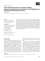
(Fig. 1A). Apoptosis was most pronounced in the septal
regions. Furthermore, human IgG was colocalized to the
apoptotic cells (Fig. 1C). Although apoptosis had not been
previously examined, earlier studies have shown deposition
of IgG in the hearts from two infants dying of CHB and
hydrops (29 and 30 weeks gestation, respectively) in
several areas of the heart, including the conduction system
[18,19]. Lee and colleagues reported that, in some areas,
“IgG appeared to outline cells” [19].
What these experiments do not address is the specificity
of the anti-SSA/Ro–SSB/La antibodies and the unique
vulnerability of the fetal heart. Dieude and colleagues have
recently reported (also confirmed in our laboratory) that
lamin B
1
is redistributed during apoptosis but, unlike
SSA/Ro or SSB/La, is not bound by cognate antibodies
[20]. These findings support discordance in the final cellu-
lar destination of translocated nuclear autoantigens during
the process of apoptosis. In the case of lamin B
1
, physio-
logic noninflammatory clearance of apoptotic cells should
proceed uneventfully even in the presence of circulating
cognate antibodies. In CHB, however, the maternal anti-
SSA/Ro–SSB/La antibodies result in opsonization and
inflammatory/fibrotic sequelae. Even if it is found that
SSA/Ro–SSB/ La are not absolutely unique in this regard,
there may be other factors such as complement binding of
certain antigens or degradation of antigens that facilitate
clearing without further sequelae. Establishing the fact
that at least one other nuclear autoantigen is not surface
bound during apoptosis of human fetal cardiomyocytes is
a step forward.
The inflammatory component
The consequences of antibody-bound (opsonized) apop-
totic cardiocytes were initially explored in vitro using a
coculturing system [15]. Macrophages coincubated with
these opsonized cells secreted increased levels of tumor
necrosis factor alpha over basal conditions or coculture
with apoptotic cardiocytes incubated with IgG from a
268
Arthritis Research & Therapy Vol 5 No 6 Buyon and Clancy
healthy control. Other investigators have also demon-
strated that phagocytosis of opsonized apoptotic cells is
proinflammatory [21,22]; for example, the observation that
ingestion of apoptotic cells bound by anticardiolipin anti-
bodies results in the release of tumor necrosis factor alpha
from cocultured macrophages [22]. Histologic studies
confirmed the in vitro coculturing model. Giant cells and
macrophages (frequently seen proximal to IgG) were
present in septal regions as well as in areas of thickened
fibrous subendocardium, most apparent in the two fetuses
dying before 23 weeks. These studies extend previous
reports of a mononuclear cell infiltration in the myocardium
of a fetus dying in utero at 18 weeks of gestation [23] and
the demonstration of patchy lymphoid aggregates through-
out the myocardium of an infant delivered at 30 weeks and
dying in the immediate postnatal period [19].
Macrophages potentially contribute to several aspects of
the pathologic process mediated by maternal autoantibod-
ies. Although the pathways of clearance and cytokine
secretion may vary, these scavengers phagocytose both
nonopsonized and opsonized apoptotic cells. Concomi-
tantly or alternatively, macrophages may present antigen
to lymphocytes (perhaps those of either maternal or fetal
origin), further contributing to an inflammatory process.
Moreover, macrophages may provide a critical link
between inflammation and ultimate scarring by secretion
of alkaline phosphatases, resulting in increased calcifica-
tion [24]. In fact, macrophages could be seen contained in
areas of calcification, particularly in the early cases. In a
full-term neonate with CHB who died at birth, however,
macrophages were less abundant and not associated with
calcified areas, suggesting a diminished role in inflamma-
tion as the pathologic process evolves.
The fibrosing end of the line
Perhaps CHB occurs as a consequence of unresolved
scarring of the AV node secondary to the transdifferentia-
tion of cardiac fibroblasts to unchecked proliferating myo-
fibroblasts (scarring phenotype in which smooth muscle
actin is expressed). Histologic support for this hypothesis
was provided by the detection of myofibroblasts in all the
affected CHB fetuses regardless of the timing of death rel-
ative to detection. As expected, myofibroblasts were
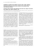
located in areas of fibrosis. In the 20-week CHB fetus,
clusters of macrophages in close proximity to myofibro-
blasts were present in scar tissue near the AV groove as
well as the thickened fibrous subendocardium (Fig. 2). Sec-
tions from the septum of the 22-week CHB fetus showed
myofibroblasts associated with the extensive fibrous matrix,
and showed marked calcification in the inferior portion of
the atrial wall where the AV node is likely to reside. In the
full-term neonate dying at birth of CHB, myofibroblasts
were observed in areas of scar. In contrast, these cells
were not observed in either septal or ventricular tissue from
the control 22-week and 23-week abortuses and from a
term neonate dying of noncardiac causes. For the 20-week
and 22-week CHB hearts, there was a strong positive cor-
relation between the absolute number of macrophages and
the content of myofibroblasts.
The functional implication of the cellular colocalization
demonstrated on the histologic sections was examined by
in vitro studies in which cultured human fetal cardiac
fibroblasts, exposed to supernatants obtained from
macrophages incubated with opsonized apoptotic cardio-
cytes, markedly increased the expression of the myofi-
broblast marker smooth muscle actin (scarring phenotype)
[14]. The addition of neutralizing anti-transforming growth
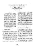
Figure 1
Histological evidence of increased apoptosis and deposition of human IgG in conduction tissue of a 20-week fetus with congenital heart block
(CHB). (A) A longitudinal section through the septal tissue of a 20-week fetus with CHB, counterstained with hematoxylin and eosin. Apoptotic
cells are identified by TUNEL peroxidase and are scattered among healthy cells. (B) The same tissue section stained with alkaline phosphatase
conjugated to anti-human IgG. (C) The same tissue section double-stained with TUNEL peroxidase and alkaline phosphatase conjugated to anti-
human IgG to demonstrate colocalization of apoptosis and of IgG, respectively.
269
Available online />factor beta antibodies to the ‘opsonized’ supernatant
blocked expression of smooth muscle actin, supporting a
potential role of transforming growth factor beta in the final
pathologic cascade to scarring. Of relevance, preliminary
genotyping data suggest that children with CHB have a
higher frequency of the fibrosis-promoting polymorphism
at codon 10 of transforming growth factor beta than do
unaffected siblings [25].
While it is often assumed that fibrosis is simply the end
result of an inflammatory insult, a recently published patho-
logic description of Lyme carditis associated with second-
degree heart block prompts a reappraisal of the elements
of tissue injury, response, and ultimate repair or scar in the
human heart. Right ventricular biopsy revealed mono-
nuclear cells around the myocardial microvasculature and
within the endocardium [26]. Despite prolonged inflamma-
tion (second-degree heart block was present for 8 weeks),
the cascade to fibrosis was not irrevocably programmed
since the block resolved following antibiotic therapy. This
absence of permanent injury stands in strong contrast to
the rapid progression to scarring seen in autoantibody-
associated CHB. The expression of specific combinations
of cytokines may ultimately provide the explanation.
Conclusions
In summary, immunohistological analyses of available
cardiac sections from several cases of CHB/myocarditis
with varying degrees of pathology parallel the results
obtained exploiting in vitro coculturing systems. Physiologic
apoptosis may initiate an inflammatory process via antibody
binding and ingestion by macrophages that not only fuels
continued apoptosis, but contributes to the transdifferentia-
tion of cardiac fibroblasts to a scarring phenotype.
The heart block of neonatal lupus is not only progressive
(second to third degree) but also characteristically irre-
versible, despite brief exposure to autoantibodies and a
limited period of inflammation. This is underscored by the
finding of extensive fibrosis even in the earliest deaths.
Furthermore, persistence of this phenotype even after
birth may be related to the progression of block seen post-
partum in some infants [7]. Moreover, fibrosis of the AV
node contradicts the paradigm that fetal wounds heal
without scarring [27].
Disruption of healing may involve the continued presence
of myofibroblasts, a consequence of protracted stimula-
tion from the macrophages. Irreversible fibrotic replace-
ment of normal tissue may be unique to heart block
acquired in utero following autoantibody-initiated inflam-
mation. Other inflammatory stimuli, as in Lyme disease,
induce transient block [26], arguing against the assump-
tion that fibroblast transdifferentiation is merely a common
final pathway of inflammation.
It seems reasonable to predict that there are both suscep-
tibility and regulatory factors, such as fetal polymorphisms
of Fc receptors and cytokines, each of which could influ-
ence the extent of the proposed pathologic cascade to
result in permanent third-degree heart block. Dissecting
the individual components in this fibrotic pathway should
elucidate the pathogenesis of antibody-associated CHB
and the rarity of irreversible injury, and may provide a ratio-
nale for therapy.
Competing interests
None declared.
Acknowledgements
The original work described herein was supported by US National Insti-
tutes of Health Grant No AR42455 (JPB), Grant No AR48409 (RMC),
and Contract No AR42220 (Research Registry for Neonatal Lupus).
References
1. Scott JS, Maddison PJ, Taylor PV, Esscher E, Scott O, Skinner RP:
Connective-tissue disease, antibodies to ribonucleoprotein,
and congenital heart block. N Engl J Med 1983, 309:209-212.
2. Lee LA, Reed BR, Harmon C: Autoantibodies to SS-A/Ro in
congenital heart block [abstract]. Arthritis Rheum 1983, 20
(suppl):S24.
3. Buyon JP: Neonatal lupus syndromes. In Systemic Lupus Ery-
thematosus, 3rd edition. Edited by Lahita RG. San Diego, CA:
Academic Press; 1999:337-359.
4. Lee LA: Neonatal lupus erythematosus. J Invest Derm 1993,
100:9s-13s.
5. Waltuck J, Buyon JP: Autoantibody-associated congenital
heart block: outcome in mothers and children. Annals Int Med
1994, 120:544-551.
6. Buyon JP, Hiebert R, Copel J, Craft J, Friedman D, Katholi M, Lee
L, Marston K, Provost T, Reichlin M, Rider L, Rupel A, Saleeb S,
Weston W, Skovron ML: Autoimmune-associated congenital
heart block: mortality, morbidity, and recurrence rates
obtained from a national neonatal lupus registry. J Am Coll
Cardiol 1998, 31:1658-1666.
7. Askanase AD, Friedman DM, Dische MR, Dubin A, Starc T, Katholi
MC, Buyon JP: Spectrum and progression of conduction
abnormalities in infants born to mothers with anti-SSA/Ro-
SSB/La antibodies. Lupus 2002, 11:145-151.
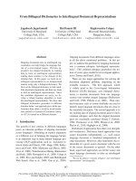
Figure 2
Proximity of macrophages and myofibroblasts in the septum of the
heart from a 20-week fetus with congenital heart block (CHB).
Longitudinal sections through the septum of a 20-week CHB heart.
Tissue was first incubated with (A) anti-CD68 (macrophage) or (B)
anti-smooth muscle actin (myofibroblast and blood vessel smooth
muscle cells), and then stained with alkaline phosphatase.
270
8. Brucato A, Frassi M, Franceschini F, Cimaz R, Faden D, Pisoni
MP, Muscara M, Vignati G, Stramba-Badiale M, Catelli L, Loja-
cono A, Cavazzana I, Ghirardello A, Vescovi F, Gambari PF, Doria
A, Meroni PL, Tincani A: Risk of congenital congenital heart
block in newborns of mothers with anti-Ro/SSA antibodies
detected by counterimmunoelectrophoresis. Arthritis Rheum
2001, 44:1832-1835.
9. Casciola-Rosen LA, Anhalt G, Rosen A: Autoantigens targeted
in systemic lupus erythematosus are clustered in two popula-
tions of surface structures on apoptotic keratinocytes. J Exp
Med 1994, 179:1317-1330.
10. Ucker DS: Death by suicide: one way to go in mammalian cel-
lular development? New Biol 1991, 3:103-109.
11. Hale AJ, Smith CA, Sutherland LC, Stoneman VEA, Longthorne
VL, Culhane AC, Williams GT: Apoptosis: molecular regulation
of cell death. Eur J Biochem 1996, 236:1-26.
12. Takeda K, Yu ZX, Nishikawa T, Tanaka M, Hosoda S, Ferrans VJ,
Kasajima T: Apoptosis and DNA fragmentation in the bulbus
cordis of the developing rat heart. J Mol Cell Cardiol 1996, 28:
209-215.
13. Miranda-Carus ME, Tseng CE, Rashbaum W, Ochs RL, Casiano
CA, DiDonato F, Chan EKL, Buyon JP: Accessibility of SSA/Ro
and SSB/La antigens to maternal autoantibodies in apoptotic
human fetal cardiac myocytes. J Immunol 1998, 161:5061-
5069.
14. Clancy RM, Askanase AD, Kapur RP, Chiopelas E, Azar N,
Miranda-Carus ME, Buyon JP: Transdifferentiation of cardiac
fibroblasts, a fetal factor in anti-SSA/Ro-SSB/La antibody-
mediated congenital heart block. J Immunol 2002, 169:2156-
2163.
15. Miranda-Carús ME, Dinu Askanase A, Clancy RM, Di Donato F,
Chou TM, Libera MR, Chan EKL, Buyon JP: Anti-SSA/Ro and
anti-SSB/La autoantibodies bind the surface of apoptotic
fetal cardiocytes and promote secretion of tumor necrosis
factor
αα
by macrophages. J Immunol 2000, 165:5345-5351.
16. Tran HB, Ohlsson M, Beroukas D, Hiscock J, Bradley J, Buyon JP,
Gordon TP: Subcellular redistribution of La(SS-B) autoantigen
during physiologic apoptosis in the fetal mouse heart and
conduction system: a clue to the pathogenesis of congenital
heart block. Arthritis Rheum 2002, 46:202-208.
17. Tran HB, Macardle PJ, Hiscock J, Cavill D, Bradley J, Buyon JP,
Gordon TP: Anti-La (SS-B) antibodies transported across the
placenta bind apoptotic cells in fetal organs targeted in
neonatal lupus. Arthritis Rheum 2002, 46:1572-1579.
18. Litsey SE, Noonan JA, O’Connor WN, Cottrill CM, Mitchell B:
Maternal connective tissue disease and congenital heart
block. Demonstration of immunoglobulin in cardiac tissue.
N Engl J Med 1985, 312:98-100.
19. Lee LA, Coulter S, Erner S, Chu H: Cardiac immunoglobulin
deposition in congenital heart block associated with maternal
anti-Ro antibody. Am J Med 1987, 83:793-796.
20. Dieude M, Senecal JL, Rauch J, Hanly JG, Fortin P, Brassard N,
Raymond Y: Association of autoantibodies to nuclear lamin B1
with thromboprotection in systemic lupus erythematosus:
lack of evidence for a direct role of lamin B1 in apoptotic
blebs. Arthritis Rheum 2002, 46:2695-2707.
21. Fadok VA, Bratton DA, Konowal A, Freed P, Westcott JY, Henson
P: Macrophages that have ingested apoptotic cells in vitro
inhibit proinflammatory cytokine production through
autocrine/paracrine mechanisms involving TGF-
ββ
, PGE2 and
PAF. J Clin Invest 1998, 101:890-898.
22. Manfredi AA, Rovere P, Galati G, Heltai S, Bozzolo E, Soldini L,
Davoust J, Balestrieri G, Tincani A, Sabbadini MG: Apoptotic cell
clearance in systemic lupus erythematosus. I. Opsonization
by antiphospholipid antibodies. Arthritis Rheum 1998, 41:205-
214.
23. Herreman G, Galezewski N: Maternal connective tissue disease
and congenital heart block [letter]. N Engl J Med 1985, 312:
1329.
24. Tintut Y, Patel J, Territo M, Saini T, Parhami F, Demer LL: Mono-
cyte/macrophage regulation of vascular calcification in vitro.
Circulation 2002, 105:650-655
25. Backer C, Loma-Sanner I, Yin X, Chandrashekhar S, Sullivan K,
Clancy R, Buyon J: Increased frequency of the high produce
genotype TT (codon 10) of TGF beta in congenital heart block
and TNF2 allele in families with neonatal lupus [abstract].
Arthritis Rheum 2002, 46(suppl):S261.
26. Hajjar RJ, Kradin RL: Case records of the Massachusetts
General Hospital: case 17-2002 — a 55-year-old man with
second-degree atrioventricular block and chest pain. N Engl J
Med 2002, 346:1732-1738.
27. Moulin V, Tam BY, Castilloux G, Auger FA, O’Connor-McCourt
MD, Philip A, Germain L: Fetal and adult human skin fibroblasts
display intrinsic differences in contractile capacity. J Cell
Physiol 2001, 188:211-222.
Correspondence
Robert M Clancy, Department of Rheumatology, Room 1608, Hospital for
Joint Diseases, 301 East 17th Street, New York, NY 10003, USA. Tel: +1
212 598 6173; fax: +1 212 598 6168; e-mail:
Arthritis Research & Therapy Vol 5 No 6 Buyon and Clancy