Báo cáo y học: "The synovial proteome: analysis of fibroblast-like synoviocytes" doc
Bạn đang xem bản rút gọn của tài liệu. Xem và tải ngay bản đầy đủ của tài liệu tại đây (1.19 MB, 8 trang )
Introduction
The synovial membrane is a thin lining layer within the joint
cavity that is responsible for maintaining normal joint func-
tion and homeostasis. Within the synovial membrane, the
cells most closely associated with this homeostatic func-
tion are the normally highly synthetic fibroblast-like syn-
ovial (FLS) cells. These are the primary source of articular
hyaluronic acid and other glycoproteins such as lubricin
[1,2]. In chronic inflammatory disorders such as rheuma-
toid arthritis (RA), the synovial membrane becomes the
target of a persistent inflammatory process that leads to
fundamental changes in the phenotype and function of
FLS cells. Although the pathogenesis of this phenotypic
change remains uncertain, available data suggest that this
may involve the acquisition of a combination of increased
proliferative potential and resistance to apoptosis [3]. This
leads to a marked increase in the number of FLS cells in
the synovium. These FLS cells participate in complex
autocrine and paracrine activation networks with
macrophages, lymphocytes, and dendritic cells, which
serve to sustain the synovitis and to enhance its destruc-
tive potential.
Studying the characteristics and behavior of FLS cells in
vitro has generated much of the understanding of the phe-
notypic changes they undergo in RA. The relative ease
with which RA FLS cells proliferate in culture has greatly
facilitated such studies. Indeed, the behavior of these cells
in culture shares many similarities with that of cancer cells,
and the concept of a ‘transformed’ phenotype has been
applied to RA FLS cells [4]. The fact that after multiple cell
passages RA FLS cells appear to adopt a more benign
phenotype that resembles other fibroblasts has led to the
suggestion that the transformed phenotype is induced by
the intense cytokine and growth factor stimulation to
which FLS cells are exposed in the RA synovial microenvi-
2D-PAGE = two-dimensional polyacrylamide gel electrophoresis; DDAH = N
ω
-N
ω
-dimethylarginine dimethylaminohydrolase; DMEM = Dulbecco’s
modified Eagle’s medium; FLS = fibroblast-like synovial; IL = interleukin; MALDI = matrix-assisted laser desorption ionization; ORF = open reading
frame; PBS = phosphate-buffered saline; RA = rheumatoid arthritis.
Available online />Research article
The synovial proteome: analysis of fibroblast-like synoviocytes
Kumar Dasuri
1
, Mihaela Antonovici
2
, Keding Chen
1
, Ken Wong
1
, Kenneth Standing
2,3
,
Werner Ens
2,3
, Hani El-Gabalawy
1
and John A Wilkins
1,2,3
1
Rheumatic Diseases Research Laboratory, University of Manitoba, Winnipeg, Canada
2
Manitoba Centre for Proteomics, Department of Medicine, University of Manitoba, Winnipeg, Canada
3
Time of Flight Laboratory, Department of Physics and Astronomy, University of Manitoba, Winnipeg, Canada
Corresponding author: John A Wilkins, (e-mail: )
Received: 17 Dec 2003 Revisions requested: 13 Jan 2004 Revisions received: 13 Jan 2004 Accepted: 21 Jan 2004 Published: 16 Feb 2004
Arthritis Res Ther 2004, 6:R161-R168 (DOI 10.1186/ar1153)
© 2004 Dasuri et al., licensee BioMed Central Ltd (Print ISSN 1478-6354; Online ISSN 1478-6362). This is an Open Access article: verbatim
copying and redistribution of this article are permitted in all media for any purpose, provided this notice is preserved along with the article's original
URL.
Abstract
The present studies were initiated to determine the protein
expression patterns of fibroblast-like synovial (FLS) cells
derived from the synovia of rheumatoid arthritis patients. The
cellular proteins were separated by two-dimensional
polyacrylamide gel electrophoresis and the in-gel digested
proteins were analyzed by matrix-assisted laser desorption
ionization mass spectrometry. A total of 368 spots were
examined and 254 identifications were made. The studies
identified a number of proteins that have been implicated in the
normal or pathological FLS function (e.g. uridine
diphosphoglucose dehydrogenase, galectin 1 and galectin 3)
or that have been characterized as potential autoantigens in
rheumatoid arthritis (e.g. BiP, colligin, HC gp-39). A novel
uncharacterized protein product of chromosome 19 open
reading frame 10 was also detected as an apparently major
component of FLS cells. These results demonstrate the utility
of high-content proteomic approaches in the analysis of FLS
composition.
Keywords: autoantigens, fibroblast-like synovial cells, galectins, matrix-assisted laser desorption ionization mass spectrometry, proteomics
Open Access
R161
R162
Arthritis Research & Therapy Vol 6 No 2 Dasuri et al.
ronment [5]. The ability to apparently reinduce this pheno-
type with cytokine stimulation supports this contention. It
remains unclear whether RA FLS cells in culture represent
a single population of cells derived from the synovium that
are capable of extensive phenotypic deviation, or whether
RA FLS cells represent heterogeneous populations of
cells, with expansion of specific subpopulations depend-
ing on the microenvironment. It seems that the latter possi-
bility is the more probable [6].
Microarray-based analysis of mRNA representation has
been used extensively to examine cells and tissues from
normal and pathologic sites. The approach is very sensitive
and it is amenable to adaptation for high-throughput analy-
sis [7]. However, several studies have demonstrated a poor
correlation between the levels of mRNA and the actual
expression levels of the corresponding gene products [8].
This was found to be particularly problematic in the cases
of low abundance mRNA species. A comparative analysis
of mRNA and protein levels in synovial tissues derived from
osteoarthritis patients and RA patients recently highlighted
this problem [9]. Protein expression was monitored using a
western blot-based approach in which a commercial array
of 791 antibodies was used to probe SDS-PAGE-sepa-
rated extracts from these tissues. A total of 260 antigens
were detected, of which 29 proteins were upregulated and
42 were downregulated in the RA sample relative to the
osteoarthritis sample. The authors noted that only 28% of
the changes observed in these proteins correlated with
those detected in the mRNA analysis. These results high-
light the importance of confirming gene expression data
with direct quantitation of protein levels.
Proteomic approaches employ mass spectrometry and
bioinformatics to identify proteins [10,11]. In peptide fin-
gerprinting, proteins are digested with enzymes with a
known cleavage pattern. The locations and masses of the
peptides of any protein sequence (real or hypothetical)
can thus be accurately predicted. For example, trypsin
cuts peptides C terminal to an arginine or lysine residue.
The masses of the individual peptides from a digest of an
unknown protein can be determined by mass spectrome-
try. The peak lists are used to search sequence databases
to identify those proteins that match the observed frag-
ment patterns. Using statistical-based bioinformatic
approaches it is possible to use the data to identify pro-
teins with a high level of confidence [12,13].
Proteins can be post-translationally modified in a number
of ways that are not reflected in the mRNA. In a two-
dimensional analysis of the yeast proteome using narrow
isoelectric point range analysis, it was suggested that
there could be as many as 20,000–30,000 proteins [14].
This represents approximately threefold to fivefold more
than the number of open reading frames (ORFs) present
in the yeast genome (6139 ORFs). These observations
highlight the importance of direct protein analysis of
pathological samples. Proteomic analysis undertakes pro-
viding a complete characterization of all of the species of
proteins in the target cell or tissue. This not only provides
a direct indication of what species are expressed, but
there is also the potential to examine post-translationally
modified species.
The present studies were initiated to determine the protein
expression patterns of FLS cells derived from the synovia
of RA patients. The cellular proteins were separated by
two-dimensional polyarylamide gel electrophoresis
(2D-PAGE) and the in-gel digested proteins were ana-
lyzed by mass spectrometry. Several categories of pro-
teins were identified: those proteins involved in FLS
function in health and disease, those proteins that have
been characterized as potential autoantigens in RA, and
novel protein species not previously described in any cell
type.
Materials and methods
Isolation and culture of FLS cells
Synovial tissue was obtained from RA patients undergoing
total knee arthroplasty. All samples were obtained accord-
ing to the guidelines approved by the Ethics Committee of
the University of Manitoba. All patients met American
College of Rheumatology criteria. FLS cells were isolated
as previously described [15]. Briefly, synovial tissue was
dissected from the connective tissue, and digested for
1–2 hours at 37°C with collagenase (1 mg/ml) and
hyaluronidase (0.05 mg/ml) (Sigma Chemicals, Oakville,
Ontario, Canada) in Hank’s buffer (ICN Biomedicals,
Costa Mesa, CA, USA). Cells were washed with modified
DMEM medium (supplemented with 1 mM sodium pyru-
vate and 0.1 mM nonessential amino acids) containing
10% fetal bovine serum and collected by centrifugation at
800 g for 10 min. Cells were cultured overnight, at 37°C
in a humidified 10% CO
2
environment. The nonadherent
cells were discarded and the adherent cells were cultured
in fresh medium. Once the cell layers were confluent, they
were trypsinized and subcultured. Cells were used
between the second and fourth passages.
Sample preparation and 2D-PAGE analysis
Confluent synovial cells were washed once with Hank’s
buffered saline and removed with trypsin. The cells were
collected by centrifugation at 800 g for 10 min and
washed twice with PBS and once with isotonic sucrose
solution (0.35 M) to remove the contaminating salts. The
cell pellet was dissolved in a sample buffer containing 7 M
urea, 2 M thiourea 4% CHAPS, 0.3% (w/v) Bio-lyte
ampholytes (pH 3–10) and 75 mM dithiothreitol along
with complete protease inhibitor cocktail (Roche Molecu-
lar Biochemicals, Laval, Quebec, Canada). Protein levels
were determined using a modified RC DC protein assay
kit (BioRad Laboratories, Mississauga, Ontario, Canada).
R163
Preparative 2D-PAGE was performed on 1 mg total cellu-
lar protein dissolved in sample buffer. Immobilized pH gra-
dient strips (17 cm, pH 3–10, nonlinear) were rehydrated
overnight with sample in an IEF protean cell (BioRad) at
50 V. Electrofocusing was carried at 9000 V as the upper
limit for a total 60 kV hours. Prior to analysis in the second
dimension, separated proteins in the strips were reduced
for 20 min at room temperature with 50 mM Tris (pH 8.8),
6 M urea, 2% sodium dodecyl sulfate, 20% glycerol and
2% (w/v) dithiothreitol, and then alkylated with same
buffer containing 2.5% (w/v) iodoacetamide for 20 min.
Second dimension electrophoresis (SDS-PAGE) was
carried on 12% SDS-PAGE gels (18.5 cm × 20 cm,
1 mm) (25 mA/gel at 20°C) using the PROTEAN II XL
system (BioRad). Gels were fixed and stained using col-
loidal Coomassie blue G250 (Pierce Biotechnology,
Rockford, IL, USA). Gels were scanned and documented
with Phoretix image analysis software (Perkin Elmer Life
Sciences Inc., Boston, MA, USA).
In-gel digestion and mass spectrometry
Spots were manually excised, destained and in-gel digested
with trypsin [16]. Peptides were recovered by extracting the
gel pieces with 25 mM ammonium bicarbonate containing
0.1% trifluoroacetic acid and 40% acetonitrile. The extracts
were lyophilized and dissolved in 10 µl of 0.1% trifluo-
roacetic acid and 10% acetonitrile. Samples were mixed
with an equal volume (0.5 µl) of 16% dihydroxybenzoic acid
in 50% acetonitrile, deposited on a matrix-assisted laser
desorption ionization (MALDI) target and air-dried.
The digests of individual spots were analyzed by an in-
house constructed MALDI quadrupole time of flight mass
spectrometer [17]. Peak lists were generated using
Knexus Automation (Proteometrics Canada, Winnipeg,
Manitoba, Canada) and samples were identified using
ProFound [13] with National Center for Biotechnology
nonredundant human databases. Search parameters
allowed for one missed cleavage site with partial oxidation
of methionine residues. A mass tolerance of 20 parts per
million was routinely used.
Results and discussion
The intent of these studies was to acquire information
regarding the protein expression patterns of typical
RA FLS cells. A total of four samples derived from two
patients were analyzed for these studies. The cells were
cultured for two to four passages and grown to conflu-
ence, at which point they appeared to be exclusively
fibroblast-like based on their morphology and on immuno-
chemical staining for uridine diphosphoglucose dehydro-
genase. The cells were harvested directly without trypsin
by directly solubilizing them in sample buffer.
Total cell lysates were separated on nonlinear immobilized
pH gradient strips (pH 3–10) and fractionated in the
second dimension on large-format SDS-PAGE gels. The
separated proteins were visualized by staining with col-
loidal Coomassie blue. The spots were excised, destained
and digested in gel with trypsin. The peptides were
extracted and analyzed by MALDI mass spectrometry
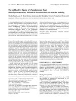
(Fig. 1). In excess of 1500 spots were detected with
Coomassie Blue (Fig. 2). The two-dimensional patterns for
FLS cells presented in the present article are representa-
tive of the results of several samples, and the patterns
were found to be highly reproducible.
Protein identification of the components in a spot was
based on the matching of the observed mass to charge
ratio of the tryptic fragments of the protein with the pre-
dicted values derived from theoretical digests of all pro-
teins in the nonredundant human database. This
fingerprinting approach is dependent on high mass accu-
racy measurements and on relatively simple protein mix-
tures in a given digest. While there may be several
molecular species in a single spot, two-dimensional sepa-
ration markedly reduces the sample complexity in a given
spot making this approach feasible. The instrument
employed in the present study has extremely high mass
accuracy (10 parts per million) and resolution (10,000 full
width at half maximum), making the approach feasible
without liquid chromatography separation of the spot
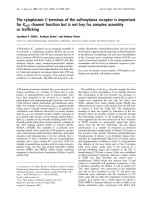
digests. Representative MALDI mass spectrometry
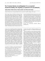
Available online />Figure 1
Schematic of the method used for the separation and identification of
fibroblast-like synovial cellular proteins. MW, molecular weight; pI,
isoelectric point.
spectra are provided in Fig. 3. These properties allowed
us to obtain identifications with a very high level of confi-
dence (expectation values of 10
–3
or less).
A total of 368 spots were selected for mass spectrometric
analysis. The spots were selected based on their intensity
of staining. Approximately 70% of the spots (n = 254)
were identified with 15–90% coverage of the protein
sequence detected. In total, 192 distinct proteins were
identified because of the redundancy of the proteins in the
gel (Additional file 1). This duplication of protein represen-
tation derives from the fact that a single protein can
undergo multiple post-translational modifications with
each species displaying a different electrophoretic mobil-
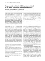
ity. Examples of this are shown in Fig. 4 for lamin
(280 spot series) and for vimentin (215 spot series). In
other cases, isoforms of the same protein display slightly
different mobilities due to amino acid sequence differ-
ences (e.g. 237 spot series of actin).
The theoretical molecular weight and the isoelectric point
can be calculated for a protein based on the amino acid
sequence. This information can then be used to narrow
database search parameters. However, a comparison of
the theoretical and observed values of these parameters
for all of the identified proteins indicates that although
there is generally a strong correlation between values,
there are some clear discrepancies (Fig. 5). These results
highlight the need for caution in using theoretical values of
protein properties as a component of the search parame-
ters for protein identification. These parameters were not
used in our analysis.
The FLS cellular proteins that were identified could be
broadly classified into several functional categories
(Fig. 6). It should, however, be apparent that there can be
significant functional overlap such that a single protein
species may be involved in several different aspects of
cellular function. This type of categorization is thus a very
general guide not an absolute assignment of function.
Accepting these limitations, the predominant functional
groups represented in identified proteins were involved in
aspects of cell structure (cytoskeleton), of signaling, of
metabolism or of transcription translation (Fig. 5).
Analysis of the FLS cellular proteome identified a number
of proteins involved in the normal functions of these cells,
Arthritis Research & Therapy Vol 6 No 2 Dasuri et al.
R164
Figure 2
A representative example of a two-dimensional separation of fibroblast-
like synovial cellular proteins. Spot numbers correspond to identifiers
in Additional file 1.
Figure 3
Representative examples of matrix-assisted laser desorption ionization
quadrupole time of flight mass spectrometer spectra of samples
digested in gel with trypsin. (a) Galectin 3 and (b) DDAH2.
in particular the cytosolic enzyme uridine diphosphoglu-
cose dehydrogenase. This enzyme is critically involved in
the synthesis of hyaluronic acid, which is a major secretory
product of FLS cells needed for the maintenance of joint
fluid viscosity and the health of the articular cartilage. We
have previously [18] identified this enzyme histochemically
in the synovial lining layer, as have Edwards and col-
leagues [19], and have demonstrated that its expression
levels appear to decrease in highly inflamed RA synovium
[20].
HC gp-39 is a major secretory protein of FLS cells,
macrophages and chondrocytes [21]. Although HC gp-39
displays structural homology with members of the Family
18 chitinases it lacks enzymatic activity, raising questions
as to what the mechanism of action might be. Based on
the cellular sources and sites of production of HC gp-39,
it has been suggested that the protein may be involved in
tissue repair and remodeling or possibly in innate host
responses to pathogens containing chitinous elements
[22]. HC gp-39 is of relevance, in the context of RA,
because peptides derived from it are stimulatory for T cells
derived from some patients. The injection of the intact
protein into BALB/c mice was associated with induction
of arthritis [23,24].
BiP is a member of the heat shock protein 70 family of
chaperones [25]. Similar to other members in the family,
BiP plays a central role in the proper folding and assembly
of proteins. Under conditions of misfolding or endoplasmic
reticulum accumulation of proteins, the levels of heat
shock protein can be upregulated to accommodate the
load. Recent studies in RA have demonstrated that BiP
can function as an autoantigen for both antibody and
T-cell responses [26,27]. Immune responses to BiP have
also been observed in experimental models of arthritis.
Furthermore, pretreatment of animals with BiP prior to
induction of adjuvant or collagen-induced arthritis can
reduce the severity of disease in these animal models.
These results suggest that there may be some association
between BiP responses and RA.
Both galectin 1 and galectin 3 were identified in FLS pro-
teome. The galectins are animal-type lectins that share a
common carbohydrate-recognition domain, which recog-
nizes galactose-containing ligands [28]. Several members
of the galectin family have been shown to have profound
effects on cell survival and they have been implicated as
major regulators of inflammatory responses [29]. Recom-
binant galectin 1 inhibits a number of experimental autoim-
mune diseases, including collagen-induced arthritis [30].
In contrast, galectin 3 has antiapoptotic activities and it
can stimulate fibroblast proliferation [31]. Galectin 3 has
also been reported to promote monocyte chemotaxis. It is
clear that the activities of the galectins vary markedly
depending on the responding cell type. Thus it is difficult
Available online />R165
Figure 5
A comparison of the theoretical and observed (a) molecular weights
(MW) and (b) isoelectric point (pI) values for the synovial proteins
identified in these studies. Note the poor correlation between the
expected and the observed values.
Figure 4
Detail of an area of a two-dimensional gel of separated fibroblast-like
synovial cellular proteins. The circled areas include the same species
of proteins with different mobilities reflecting differences in isoelectric
points due post-translational modifications: 215 spot series, vimentin;
237 spot series, beta actin; 278 and 280 spot series, lamin A/C;
279 spot series, caldesmon.
to predict the impact of these molecules alone or in com-
bination on FLS cell functions. Recent reports demon-
strated the presence of galectin 1 and galectin 3 in the
synovial tissues of RA patients [32,33]. Galectin 3 was
widely distributed in the synovium, with clear accumula-
tions at sites of cartilage invasion [33]. In contrast,
galectin 1 appeared to be excluded from the sites of inva-
sion. These results raised the possibility that the galectins
were modifying cellular functions associated with different
processes in the RA synovium.
Protein methylation is thought to represent a mechanism
for regulating protein turnover and function. Some of the
degradation products from these modified proteins,
NMMA and ADMA, are inhibitors of nitric oxide synthase
[34]. The enzyme DDAH2 removes aminomethyl groups
of methylarginines by catabolizing them to citrulline and
methylamines. Previous studies of DDAH2 expression
indicated that the enzyme was widely distributed in
normal adult and fetal tissues [35]. Recent studies
suggest that N
ω
-N
ω
-dimethylarginine dimethylaminohy-
drolase (DDAH) levels are reduced in a hypoxia-induced
rat hypertension model [36]. Overexpression of DDAH in
endothelial cell lines also results in enhanced expression
of vascular endothelial growth factor providing a link for
neovascularization of the synovium [37]. Collectively the
results suggest that DDAH plays a critical role in vascu-
lar function and development. Based on the present
results it appears that DDAH2 is a major product of cul-
tured FLS cells, raising the possibility of a role for this
enzyme in the RA synovium.
The product of chromosome 19 ORF 10 was originally
identified as a product of bone marrow-derived stromal
cells and designated as IL-25 [38]. The published
descriptions of the biological activity of the protein were
subsequently retracted and the official designation of the
protein is now a product of C19 ORF 10 [39]. There are
also suggestions of an IL-27 designation but this is not
consistent with what has been described in the literature
as IL-27 [40]. Although the mRNA of the C19 ORF 10
gene is widely expressed, it does not appear to be
lineage restricted and it remains unclear as to what the
biological activity of this protein is. Based on the staining
intensity the protein is well represented in lysates of syn-
ovial cells, suggesting that it may be a significant
product of FLS cells. The molecule clearly warrants
further investigation.
Conclusions
The present studies provide a preliminary analysis of the
synovial proteome. It should also be appreciated that the
current analysis describes only the major FLS cellular pro-
teins. Future studies will require the development of
enrichment steps for low abundance proteins. Despite
Arthritis Research & Therapy Vol 6 No 2 Dasuri et al.
R166
Figure 7
The in-gel locations of synovial proteins of potential functional or
pathogenic significance in rheumatoid arthritis. UDPGDH, uridine
diphosphoglucose dehydrogenase.
Figure 6
Functional categorization of the fibroblast-like synovial proteome based
on Swissprot and Tremble assigned functions.
Available online />R167
these limitations, the results have identified a number of
novel molecular species that may contribute to inflamma-
tory events in vivo (Fig. 7). The data also suggest that FLS
cells may be reasonable surrogates of their in vivo coun-
terparts for compositional and functional analysis.
Additional files
Competing interests
None declared.
Acknowledgements
The authors thank Ms Sheryl Hagenstein for her assistance in the
preparation of the manuscript. This research was supported by grants
from the Canadian Institutes for Health Research (HEG, JAW), from
the Manitoba Health Research Council (HEG, JAW), and from the
Canadian Arthritis Network.
References
1. Yamanishi Y, Firestein GS: Pathogenesis of rheumatoid arthri-
tis: the role of synoviocytes. Rheum Dis Clin North Am 2001,
27:355-371.
2. Jay GD, Harris DA, Cha CJ: Boundary lubrication by lubricin is
mediated by O-linked beta (1-3) Gal-GalNAc oligosaccha-
rides. Glycoconj J 2001, 18:807-815.
3. Firestein GS: Evolving concepts of rheumatoid arthritis. Nature
2003, 423:356-361.
4. Konttinen YT, Li TF, Hukkanen M, Ma J, Xu JW, Virtanen I: Fibro-
blast biology. Signals targeting the synovial fibroblast in
arthritis. Arthritis Res 2000, 2:348-355.
5. Zimmermann T, Kunisch E, Pfeiffer R, Hirth A, Stahl HD, Sack U,
Laube A, Liesaus E, Roth A, Palombo-Kinne E, Emmrich F, Kinne
RW: Isolation and characterization of rheumatoid arthritis
synovial fibroblasts from primary culture—primary culture
cells markedly differ from fourth-passage cells. Arthritis Res
2001, 3:72-76.
6. Marinova-Mutafchieva L, Taylor P, Funa K, Maini RN, Zvaifler NJ:
Mesenchymal cells expressing bone morphogenetic protein
receptors are present in the rheumatoid arthritis joint. Arthritis
Rheum 2000, 43:2046-2055.
7. Schena M, Heller RA, Theriault TP, Konrad K, Lachenmeier E,
Davis RW: Microarrays: biotechnology’s discovery platform for
functional genomics. Trends Biotechnol 1998, 16:301-306.
8. Greenbaum D, Colangelo C, Williams K, Gerstein M: Comparing
protein abundance and mRNA expression levels on a
genomic scale [letter]. Genome Biol 2003, 4:117.
9. Lorenz P, Ruschpler P, Koczan D, Stiehl P, Thiesen HJ: From
transcriptome to proteome: differentially expressed proteins
identified in synovial tissue of patients suffering from
rheumatoid arthritis and osteoarthritis by an initial screen
with a panel of 791 antibodies. Proteomics 2003, 3:991-1002.
10. Zhu H, Bilgin M, Snyder M: Proteomics. Annu Rev Biochem
2003, 72:783-812.
11. Aebersold R, Mann M: Mass spectrometry-based proteomics.
Nature 2003, 422:198-207.
12. Fenyo D, Beavis RC: Informatics and data management in pro-
teomics. Trends Biotechnol 2002, 20(12 Suppl):S35-S38.
13. Zhang W, Chait BT: ProFound: an expert system for protein
identification using mass spectrometric peptide mapping
information. Anal Chem 2000, 72:2482-2489.
14. Gygi SP, Corthals GL, Zhang Y, Rochon Y, Aebersold R: Evalua-
tion of two-dimensional gel electrophoresis-based proteome
analysis technology. Proc Natl Acad Sci USA 2000, 97:9390-
9395.
15. Hitchon C, Wong K, Ma G, Reed J, Lyttle D, El-Gabalawy H:
Hypoxia-induced production of stromal cell-derived factor 1
(CXCL12) and vascular endothelial growth factor by synovial
fibroblasts. Arthritis Rheum 2002, 46:2587-2597.
16. Krokhin O, Li Y, Andonov A, Feldmann H, Flick R, Jones S, Stroe-
her U, Bastien N, Dasuri KV, Cheng K, Simonsen JN, Perreault H,
Wilkins J, Ens W, Plummer F, Standing KG: Mass spectrometric
characterization of proteins from the SARS virus: a prelimi-
nary report. Mol Cell Proteom 2003, 2:346-356.
17. Shevchenko A, Loboda A, Shevchenko A, Ens W, Standing KG:
MALDI quadrupole time-of-flight mass spectrometry: a pow-
erful tool for proteomic research. Anal Chem 2000, 72:2132-
2141.
18. El-Gabalawy H, King R, Bernstein C, Ma G, Mou Y, Alguacil-
Garcia A, Fritzler M, Wilkins J: Expression of N-acetyl-
D-galac-
tosamine associated epitope in synovium: a potential marker
of glycoprotein production. J Rheumatol 1997, 24:1355-1363.
19. Edwards JC, Wilkinson LS, Pitsillides AA: Palisading cells of
rheumatoid nodules: comparison with synovial intimal cells.
Ann Rheum Dis 1993, 52:801-805.
20. Uzuki H, Watanabe T, Katsura Y, Sawai T: Quantitative histo-
chemical study of hyaluronic acid binding protein and the
activity of uridine diphosphoglucose dehydrogenase in the
synovium of patients with rheumatoid arthritis. Anal Quant
Cytol Histol 1999, 21:75-80.
21. Hakala BE, White C, Recklies AD: Human cartilage gp-39, a
major secretory product of articular chondrocytes and syn-
ovial cells, is a mammalian member of a chitinase protein
family. J Biol Chem 1993, 268:25803-25810.
22. Fusetti F, Pijning T, Kalk KH, Bos E, Dijkstra BW: Crystal struc-
ture and carbohydrate-binding properties of the human carti-
lage glycoprotein-39. J Biol Chem 2003, 278:37753-37760.
23. Verheijden GF, Rijnders AW, Bos E, Coenen-de Roo CJ, van
Staveren CJ, Miltenburg AM, Meijerink JH, Elewaut D, de Keyser
F, Veys E, Boots AM: Human cartilage glycoprotein-39 as a
candidate autoantigen in rheumatoid arthritis. Arthritis Rheum
1997, 40:1115-1125.
24. Tsark EC, Wang W, Teng YC, Arkfeld D, Dodge GR, Kovats S:
Differential MHC class II-mediated presentation of rheuma-
toid arthritis autoantigens by human dendritic cells and
macrophages. J Immunol 2002, 169:6625-6633.
25. Gething MJ: Role and regulation of the ER chaperone BiP.
Semin Cell Dev Biol 1999, 10:465-472.
26. Corrigall VM, Bodman-Smith MD, Fife MS, Canas B, Myers LK,
Wooley PH, Soh C, Staines NA, Pappin DJC, Berlo SE, van Eden
W, van der Zee R, Lanchbury JS, Panayi GS: The human endo-
plasmic reticulum molecular chaperone BiP is an autoantigen
for rheumatoid arthritis and prevents the induction of experi-
mental arthritis. J Immunol 2001, 166:1492-1498.
27. Blass S, Union A, Raymackers J, Schumann F, Ungethum U, Muller-
Steinbach S, De Keyser F, Engel JM, Burmester GR: The stress
protein BiP is overexpressed and is a major B and T cell target
in rheumatoid arthritis. Arthritis Rheum 2001, 44:761-771.
28. Yang RY, Liu FT: Galectins in cell growth and apoptosis. Cell
Mol Life Sci 2003, 60:267-276.
29. Rabinovich GA, Rubinstein N, Toscano MA: Role of galectins in
inflammatory and immunomodulatory processes. Biochim
Biophys Acta 2002, 1572:274-284.
30. Rabinovich GA, Daly G, Dreja H, Tailor H, Riera CM, Hirabayashi
J, Chernajovsky Y: Recombinant galectin-1 and its genetic
delivery suppress collagen-induced arthritis via T cell apopto-
sis. J Exp Med 1999, 190:385-398.
31. Inohara H, Akahani S, Raz A: Galectin-3 stimulates cell prolifer-
ation. Exp Cell Res 1998, 245:294-302.
32. Ohshima S, Kuchen S, Seemayer CA, Kyburz D, Hirt A, Klinzing S,
Michel BA, Gay RE, Liu FT, Gay S, Neidhart M: Galectin 3 and its
binding protein in rheumatoid arthritis. Arthritis Rheum 2003,
48:2788-2795.
The following Additional file is available online:
Additional file 1
An Excel file containing the results of MALDI mass
spectrometry analysis of 2D-PAGE isolated FLS cellular
proteins. The identities of 254 spots are listed.
See />supplementary/ar1153-s1.xls
Arthritis Research & Therapy Vol 6 No 2 Dasuri et al.
R168
33. Neidhart M, Seemayer CA, Hummel KM, Michel BA, Gay RE, Gay
S: Functional characterization of adherent synovial fluid cells
in rheumatoid arthritis: destructive potential in vitro and in
vivo. Arthritis Rheum 2003, 48:1873-1880.
34. Dayoub H, Achan V, Adimoolam S, Jacobi J, Stuehlinger MC,
Wang BY, Tsao PS, Kimoto M, Vallance P, Patterson AJ, Cooke
JP: Dimethylarginine dimethylaminohydrolase regulates nitric
oxide synthesis. Genetic and physiological evidence. Circula-
tion 2003, 108:3042-3047.
35. Leiper JM, Santa Maria J, Chubb A, MacAllister RJ, Charles IG,
Whitley GS, Vallance P: Identification of two human dimethy-
larginine dimethylaminohydrolases with distinct tissue distri-
butions and homology with microbial arginine deiminases.
Biochem J 1999, 343:209-214.
36. Millatt LJ, Whitley GS, Li D, Leiper JM, Siragy HM, Carey RM,
Johns RA: Evidence for dysregulation of dimethylarginine
dimethylaminohydrolase I in chronic hypoxia-induced pul-
monary hypertension. Circulation 2003, 108:1493-1498.
37. Smith CL, Birdsey GM, Anthony S, Arrigoni FI, Leiper JM, Vallance
P: Dimethylarginine dimethylaminohydrolase activity modu-
lates ADMA levels, VEGF expression, and cell phenotype.
Biochem Biophys Res Commun 2003, 308:984-989.
38. Tulin EE, Onoda N, Nakata Y, Maeda M, Hasegawa M, Nomura H,
Kitamura T: SF20/IL-25, a novel bone marrow stroma-derived
growth factor that binds to mouse thymic shared antigen-1
and supports lymphoid cell proliferation. J Immunol 2001, 167:
6338-6347.
39. Tulin EE, Onoda N, Nakata Y, Maeda M, Hasegawa M, Nomura H,
Kitamura T: SF20/IL-25, a novel bone marrow stroma-derived
growth factor that binds to mouse thymic shared antigen-1
and supports lymphoid cell proliferation [letter]. J Immunol
2003, 170:1593.
40. Pflanz S, Timans JC, Cheung J, Rosales R, Kanzler H, Gilbert J,
Hibbert L, Churakova T, Travis M, Vaisberg E, Blumenschein WM,
Mattson JD, Wagner JL, To W, Zurawski S, McClanahan TK,
Gorman DM, Bazan JF, de Waal Malefyt R, Rennick D, Kastelein
RA: IL-27, a heterodimeric cytokine composed of EBI3 and
p28 protein, induces proliferation of naive CD4(+) T cells.
Immunity 2002, 16:779-790.
Correspondence
Dr John A Wilkins, Rheumatic Diseases Research Laboratory, 805
John Buhler Research Centre, 715 McDermot Avenue, Winnipeg,
Manitoba, Canada R3E 3P4. Tel: +1 204 789 3835; fax: +1 204 789
3987; e-mail: