Báo cáo y học: "Towards in vivo application of RNA interference – new toys, old problems" pdf
Bạn đang xem bản rút gọn của tài liệu. Xem và tải ngay bản đầy đủ của tài liệu tại đây (593.21 KB, 8 trang )
78
dsRNA = double-stranded RNA; ES = embryonic stem (cell); nt = nucleotide; RISC = RNA-induced silencing protein complex; RNAi = RNA inter-
ference; shRNA = small hairpin RNA; siRNA = small interfering RNA.
Arthritis Research & Therapy Vol 6 No 2 Rutz and Scheffold
Introduction
In the postgenomic era it has become a major challenge to
develop efficient reverse genetic approaches (i.e. from
genotype to phenotype) to evaluate the function of a vast
number of newly identified genes. Furthermore, specific
silencing of disease-relevant genes (e.g. from tumours,
pathogens, or inflammatory mediators) is an interesting
therapeutic strategy. In this respect RNA interference
(RNAi) technology, which allows targeted ‘knockdown’ of
individual genes by so-called small interfering RNAs
(siRNAs) [1], has already opened up new avenues for
functional analyses in vitro, and holds great promise for
analytical as well as therapeutic applications in vivo.
Although other gene silencing approaches, using anti-
sense oligonucleotides, ribozymes, or DNAzymes, have
been introduced over the past 25 years, their application
has been restricted to certain areas. Only one antisense-
based pharmacological agent has thus far been approved.
In contrast to those technologies, RNAi represents a
physiological process that occurs naturally in many
eukaryotes, where it has evolved probably as a mechanism
to defend against invading nucleic acids such as viruses
and transposons [2,3], and therefore it is easily applicable
to a large variety of organisms, cell types and genes. The
technology has remarkable target specificity and requires
only low amounts of siRNA effector molecules per cell,
which can even be expressed directly in situ, allowing
long-term silencing of target genes. This makes RNAi an
interesting tool for the analysis of loss-of-function pheno-
types in vivo and it may also lead to the development of
new gene therapeutic approaches.
As for all gene silencing approaches, the critical step
toward application of RNAi in mammals is the delivery of
effector molecules into the target cell. What has been
accomplished rather easily in cell lines represents a much
greater challenge in hard-to-transfect primary mammalian
cells, which are of course the ultimate targets.
This review briefly summarizes our current knowledge of
the mechanism of RNAi, the technical basis for its
application to functional gene analysis in mammalian cells
in vitro and in vivo, and potential therapeutic applications.
Technology review
Towards
in vivo
application of RNA interference – new toys, old
problems
Sascha Rutz and Alexander Scheffold
Deutsches Rheuma-Forschungszentrum Berlin, Berlin, Germany
Corresponding author: Alexander Scheffold (e-mail: )
Received: 17 Dec 2003 Revisions requested: 11 Feb 2004 Revisions received: 25 Feb 2004 Accepted: 26 Feb 2004 Published: 10 Mar 2004
Arthritis Res Ther 2004, 6:78-85 (DOI 10.1186/ar1168)
© 2004 BioMed Central Ltd (Print ISSN 1478-6354; Online ISSN 1478-6362)
Abstract
RNA interference (RNAi) is the sequence-specific degradation of mRNA by short double-stranded
RNA molecules. The technology, introduced only 5 years ago, has stimulated many fantasies regarding
the future of functional gene analysis and gene therapy. Given its ease of application, its high efficiency
and remarkable specificity, RNAi holds great promise for broad in vitro and in vivo application in all
areas of biomedicine. Despite its potential, the major obstacle to the use of RNAi (as for all previous
gene silencing approaches) is the need for efficient and sustained delivery of small interfering RNA into
primary mammalian cells, and specific targeting of particular cell types in vivo.
Keywords: functional genomics, gene silencing, primary mammalian cell, small interfering RNA, transfection
79
Available online />Mechanism of RNA interference
The phenomenon of RNAi, originally described in the
nematode worm C. elegans by Fire and colleagues [4] in
1998, has been recognized as a general mechanism in
many organisms (Fig. 1) [1,5]. Basically, RNAi is induced
within the cytoplasm when long, double-stranded RNA
(dsRNA) is recognized by Dicer, a multidomain RNase III
enzyme. Dicer processes dsRNA into short (21–25
nucleotide [nt]) duplexes that are termed siRNAs [6–10].
Like products of other RNase III enzymes, siRNA duplexes
contain 5′ phosphate and 3′ hydroxyl termini, and two
single-stranded nucleotide overhangs on their 3′ ends
[10]. These structural features are important for the entry
of siRNAs into the RNAi pathway because blunt-ended
siRNAs or those that lack a 5′ phosphate group are
ineffective in triggering gene silencing [11,12]. The
generated siRNA associates with a multiprotein complex,
the RNA-induced silencing protein complex (RISC), which
becomes activated on ATP-dependent unwinding of the
siRNA duplex [6,12]. One of the two siRNA strands is
retained within the complex and confers sequence
specificity in targeting of the mRNA by Watson–Crick
base-pairing [6,11,13,14]. A perfectly homologous mRNA
is cleaved at a single site in the centre of the duplex region
formed with the guide siRNA, 10 nt from the 5′ end of the
latter [10,12,13,15]. Finally, RISC is released and the
cleaved mRNA is further degraded by cellular exo-
nucleases [16]. The specific degradation of mRNA in turn
leads to decreased synthesis of the respective protein and
eventually to a loss of protein function.
Concentrations of only a few siRNA molecules per cell
can lead to a pronounced silencing effect, demonstrating
the catalytic action of RISC [1,4]. Generally, although
greatly diminished, residual mRNA levels can be detected.
Hence, the RNAi-mediated silencing of a particular gene is
generally referred to as a ‘knockdown’ rather than a
‘knockout’.
RNA interference in mammalian cells
Originally, the RNAi pathway was thought to be
nonfunctional in mammalian cells, where dsRNA longer
than 30 base pairs induces a nonspecific antiviral
response. This so-called interferon response is character-
ized by the activation of the RNA-dependent protein
kinase [17], leading to phosphorylation of the translation
initiation factor eIF-2α and thereby to a nonspecific arrest
in translation and induction of apoptosis [18]. Moreover,
the synthesis of 2′–5′ polyadenylic acid results in the
activation of the sequence nonspecific RNaseL [19].
The breakthrough for the use of RNAi in mammalian cells
came when Elbashir and coworkers [20] and Caplen and
colleagues [21] showed that siRNA, when directly
introduced into mammalian cells, does not trigger the
RNA-dependent protein kinase response but effectively
elicits RNAi, presumably by directly associating with
RISC. Targeted gene silencing in mammalian cells by the
application of siRNA is well established. The high degree
of sequence specificity inherent to the technology is
emphasized by several reports showing that even a 1–2 nt
mismatch in the siRNA sequence hampers targeted gene
silencing [11,16,20,22,23].
Recently, evaluation of target gene specificity on a
genome-wide level by applying gene expression profiling
led to conflicting results. In two studies [24,25] no effects
on nontarget genes were observed, although high concen-
trations (100 nmol/l) of siRNA were shown to induce
stress-response and apoptosis-related genes. In contrast,
Jackson and coworkers [26] challenged the idea of
perfect sequence specificity of siRNA; they detected
silencing of nontargeted genes with limited sequence
similarity. As few as 11 contiguous nucleotides of identity
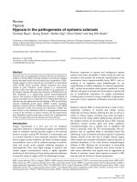
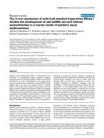
Figure 1
The RNA interference pathway. Long double-stranded RNA (dsRNA)
or small hairpin RNA (shRNA) is processed by Dicer to form a small
interfering RNA (siRNA), which associates with RNA-induced silencing
protein complex (RISC) and mediates target sequence specificity for
subsequent mRNA cleavage. (See text for further details.)
80
Arthritis Research & Therapy Vol 6 No 2 Rutz and Scheffold
to the siRNA were sufficient. Apparently, this off-target
silencing was mediated not only by the antisense but also
the sense strand of the siRNA. These findings highlight
the need for careful selection of the siRNA sequences and
appropriate specificity controls to verify functional effects.
Small interfering RNA selection
A synthetic siRNA consists of a 19 base-pair double-
stranded region that is complementary to the gene of
interest, contains 5′ phosphate and 3′ hydroxyl termini,
and possesses two single-stranded nucleotides on the 3′
ends [20].
Tuschl and coworkers [27] reported a number of
guidelines for the design of siRNA molecules (Table 1).
Several design tools are also available from the internet
(Table 1). Although one can follow these guidelines it is
still necessary to test several siRNAs, targeting distinct
regions within the gene of interest, because there is great
variability in the capacity of an individual siRNA to induce
silencing [16,28]. One may have to test three or four
siRNAs in order to find one that results in more than 90%
reduction in target gene expression (unpublished data).
The reason for this is not entirely understood but it may be
related to one or more of the following factors: incorpor-
ation of siRNA into RISC and stability of RISC; base
pairing with mRNA; cleavage of mRNA and turnover after
mRNA cleavage; secondary and tertiary structures of
mRNA; and binding of mRNA-associated proteins. Accord-
ingly, Vickers and coworkers [28] found a significant
correlation between mRNA sites that are RNase H
sensitive (i.e. accessible) and sites that promote efficient
siRNA-mediated mRNA degradation. Moreover, placing
the recognition site of an efficient siRNA into a highly
structured RNA region abrogated silencing.
Two recent reports [29,30] found that the decision
regarding which of the two strands of a siRNA molecule is
Table 1
Guidelines for siRNA design
General guidelines for siRNA design Select 23-nt long sequences from the mRNA conforming to the consensus 5′-AA[N19]UU-3′ or
5′-NA[N19]NN-3′ (where N is any nucleotide)
Avoid targeting of regions that are likely to bind regulatory proteins, such as 5′-UTR, 3′-UTR and
regions close to the start site
Choose sequences with GC content between 30% and 70%
Avoid highly G-rich sequences
Design sense and antisense N19 sequences, add two 2-deoxythymidine residues to the 3′ ends
Perform BLAST search to exclude potential homology to other genes
Additional considerations for Avoid more than three consecutive As or Ts in the targeting sequence
vector-based siRNA expression
U6 promotor requires a guanine at position +1
H1 promotor prefers adenosine at position +1
Design oligonucleotides containing N19 targeting sequence, a loop forming spacer sequence
(often 5′-TTCAAGAGA-3′), followed by the reverse complementary targeting sequence and five to six
consecutive thymidine residues for termination of transcription
Add respective restriction sites for cloning
siRNA design tools on the internet
/>
/>Rules for the design of synthetic siRNAs according to Tuschl and coworkers [27] and some further considerations for vector-based small hairpin
RNA (shRNA) expression are given. A collection of links to small interfering RNA (siRNA) design tools on the internet is provided. nt, nucleotide;
UTR, untranslated region.
81
incorporated into RISC was crucial in determining the
efficiency of gene silencing. In order to target specifically a
given mRNA for degradation, the antisense strand of the
siRNA duplex, which is complementary to the mRNA, must
be incorporated into the activated RISC. Schwarz and
coworkers [29] and Khvorova and colleagues [30] found
that the absolute and relative stabilities of the base pairs
at the 5′ ends of the two siRNA strands determine the
degree to which each strand participates in the RNAi
pathway. The strand with lower 5′ end stability is
preferred. As a consequence, a highly functional siRNA is
characterized by lower internal stability at the 5′ end of the
antisense strand as compared with less effective
duplexes. A further improved algorithm for the prediction
of siRNA efficiency is highly desirable and will enable us to
to improve quality and efficiency, and reduce the cost of
the technology.
Modes of application and routes into the cell
To induce RNAi in mammalian cells, siRNA can either be
directly transfected or produced endogenously within the
target cell from expression plasmids [22,31–34]. Synthetic
siRNA can be generated by chemical synthesis, by in vitro
transcription using a T7 polymerase [34,35], or by Dicer
digestion of long dsRNA [36]. Synthesized siRNA induces
potent silencing at concentrations of 1–10 nmol/l [13].
siRNA expression vectors utilize mostly U6-snRNA or H1
(RNase P) promoters, both of which are members of the
RNA polymerase III promoter family, which lack down-
stream transcriptional elements and produce a transcript
without a cap or poly-A tail [37]. Transcription is
terminated at a stretch of five to six thymidine residues,
leading to the incorporation of two to three uracil residues
at the 3′ end, which is compatible with the two or three nt
overhangs that are found to be indispensable for silencing
activity in natural siRNAs.
Sense and antisense strands are either produced from
two independent promoters and anneal within the cell
[31], or more commonly the two strands are linked by a
9 base pair spacer leading to the expression of a stem-
loop structure termed short hairpin RNA (shRNA). The
hairpin is subsequently cleaved by Dicer to generate
effective siRNA molecules [22,33,34,38] (Fig. 2). By incor-
porating a drug resistance gene or via episomally replicating
plasmids, a long-lived knockdown effect can be achieved
in cultured cells [31,39]. To facilitate the analysis of genes
that are essential for cell survival and cell cycle regulation,
two groups have generated inducible shRNA expression
systems [40,41]. However, the specificity of gene
knockdown must be tightly controlled, because Bridge
and coworkers [42] recently reported the induction of an
interferon response by a substantial number of shRNA
expression vectors tested, perhaps caused by the accumu-
lation of nonprocessed Pol III transcripts within the cell.
Gene silencing occurs very rapidly after the transfection of
an efficient siRNA. Although the kinetics may vary
depending on the gene of interest, usually target mRNA
levels will be diminished after 48 hours, reaching a
minimum at 72 hours after transfection. A knockdown
efficiency of 90–95% reduction in the amount of target
mRNA can be achieved. However, the major drawback of
the method is its transient gene silencing effect. The
duration of the knockdown using synthetic siRNA is
generally in the range of 3–5 days. Protein levels will
return to normal 5–7 days after transfection [16,27]. The
longevity of silencing depends on factors such as the
abundance of target mRNA and protein, the stability of
target protein, transcriptional feedback loops, and the
number of cell divisions diluting the siRNA, rather than on
the degradation of the siRNA itself.
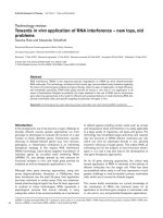
Available online />Figure 2
Approaches to endogenous expression of small interfering RNAs
(siRNAs) in mammalian cells. (a) Sense and antisense strand of the
siRNA duplex are expressed from separate promoters. (b) siRNA
duplex is expressed as a stem-loop structure (small hairpin RNA
[shRNA]) from a single promotor. Sense and antisense strands are
separated by a loop-forming spacer. The construct is further
processed by Dicer within the cell to form a functional siRNA. In both
cases transcription is terminated by six consecutive thymidine
residues.
82
Both chemically synthesized siRNA and shRNA expres-
sion plasmids can be delivered to cells using standard
transfection methods. Thereby, the efficiency mainly
depends on the type of cell that is targeted. Because of
their small size, transfection of synthetic siRNAs is usually
very efficient, even in primary mammalian cells. A number
of cationic lipid-based or liposome-based transfection
reagents optimized for the transfection of oligonucleotides
are commercially available. In cells that are more resistant
to chemical transfection methods (e.g. suspension cells),
electroporation may achieve an efficient induction of RNAi.
Transduction rates with siRNA of up to 80–90% have
been reported for some haematopoietic cell lines and
primary cells [43,44]. Optimized for the transfection of
primary human cells with siRNA, Nucleofection
TM
tech-
nology (Amaxa biosystems, Cologne, Germany) appears
to be a very efficient and convenient approach [45,46].
When using these conventional transfection strategies,
the silencing effect is only transient. Exceptions are
established cell lines that allow selection for integrated
vectors. Viral gene delivery systems are perfectly suited to
overcome these limitations; they are well established tools
for efficient transduction of primary cells and some of
them have the inherent ability to integrate into the host cell
genome, thereby leading to stable transgene expression.
Several adenoviral [47,48], onco-retroviral [49–51] and
lentiviral [52–54] vectors have been utilized for the
efficient delivery of shRNA expression cassettes. Adeno-
viral infection is transient whereas onco-retroviral vectors,
based on the Moloney murine leukaemia virus or the
murine stem cell virus, integrate into the host cell genome,
leading to a prolonged silencing effect. Lentiviral vectors
based on HIV-1 bear the additional advantage of efficiently
transducing both dividing as well as nondividing cells,
such as stem cells and terminally differentiated cells.
Moreover, they are resistant to developmental silencing
after integration of the provirus, and therefore they can be
used to generate transgenic animals. Several groups have
reported the use of lentiviral systems for the silencing of
genes in a variety of cultured as well as primary cells, such
as human and murine T cells [52,53], haematopoietic
stem cells [53] and mouse dendritic cells [53,54].
Although onco-retroviruses and lentiviruses hold great
promise as vehicles for gene therapy, two patients who
were undergoing retroviral based therapy for X-linked
severe combined immunodeficiency developed leukaemia
[55,56]. This indicates that improved safety standards and
ways to control the integration of the provirus are needed
before retroviruses can be used to deliver siRNA for
therapeutic purposes.
Towards
in vivo
application of small
interfering RNA
RNAi has already been proven to be a powerful tool for
dissecting and elucidating gene function, even on a
genome-wide basis. The first example comes from
C. elegans, in which Kamath and coworkers [57] reported
the construction of a library of bacterial clones that
express dsRNA, which corresponds to approximately 86%
of the total gene products made by C. elegans. Also, the
library has been used to screen for genes that are involved
in body fat regulation, longevity and genome stability
[58–60].
Thus far, in vivo gene silencing approaches are very
limited in the mammalian system. Nonetheless, a number
of potential candidate genes, especially in viral infections,
cancers and inherited genetic disorders but also in
chronic inflammatory diseases such as autoimmune
arthritis, has been defined and successfully targeted in
vitro. Consistent with its natural function as an antiviral
defence mechanism, siRNA was found to inhibit in vitro
replication of several viruses effectively, including HIV,
hepatitis C virus and influenza virus, by interfering with
various stages of the virus life cycles [38,52,61–67].
Similarly, several cancer-related genes have been targeted
in proof-of-principle experiments, including cellular onco-
genes and drug resistance genes. In these studies, RNAi
was efficient and highly selective in targeting oncogenes
resulting from chromosomal translocations [43,68] or
carrying single point mutations, without affecting the wild-
type allele [50,69].
Protocols must be established for efficient delivery of
siRNA and selective targeting of specific cell types in order
to allow future therapeutic applications and in vivo verifica-
tion of results obtained from in vitro silencing experiments.
Moreover, it must be determined whether transient gene
silencing, as obtained by introduction of synthetic siRNA
or expression plasmids, is sufficient for treatment, or
whether the target gene must be silenced for an extended
period of time by the use of viral expression systems.
Direct injection of siRNA into the blood would be
ineffective because of rapid degradation of the RNA by
serum ribonucleases. However, it was recently demon-
strated that chemical modification can protect the siRNA
molecule from degradation [70] and might even prolong
the silencing effect due to slower depletion within the cell
[71]. Thus far, synthetic siRNAs have been applied in
animals via hydrodynamic transfection [72] (i.e. the
intravenous injection of a substantial dose of siRNA within
a large volume of liquid), resulting in a knockdown
efficiency up to 70–80%, at least in some organs,
including liver, kidney, spleen, lung and pancreas [73].
Using this method, the silencing of either Fas receptor
[74] or caspase-8 [75] resulted in a clearly measurable
protection from severe Fas-induced liver damage. In vivo
application of siRNA against genes of the hepatitis B virus
also led to an effective inhibition of virus replication [76].
Arthritis Research & Therapy Vol 6 No 2 Rutz and Scheffold
83
This method is of course not applicable to humans. It is
also limited by the fact that siRNA can only be delivered to
a certain set of organs and it is not possible to target
specific organs or cells. Development of cell-specific or
organ-specific delivery systems for siRNA, as is required
for broad in vivo application of this technique, is indeed a
demanding task.
Prolonged gene silencing by stable integration of a siRNA
expression vector is currently only possible in vitro. The
subsequent in vivo adoptive transfer of these in vitro
manipulated cells is an option in situations where a small
number of cells can develop a dominant phenotype in vivo.
This is the case for stem cells (e.g. embryonic stem [ES]
cells) or haematopoietic stem cells, which either give rise to
a complete new animal or at least generate defined organs.
An approach using siRNA-modified stem cells would be
particularly useful for the analysis of gene function in vivo.
So far this has mainly been done in knockout mice, which
carry a nonfunctional mutation of the target gene,
generated by homologous recombination in ES cells. The
technique suffers from a number of limitations that could
be overcome by RNAi technology, such as the need for
cloning of the target gene, the time and effort required for
generating a knockout mouse, and the potential embryonic
lethality. In contrast to the all-or-nothing phenotype
obtained from knockout animals, analysis of gene dosage
effects may be possible by using siRNAs with variable
silencing efficiency. Finally, the combination of multiple
loss-of-function phenotypes in one generation would be
possible. Lentiviral siRNA vectors have been used to
generate stable transgenic ‘knockdown’ animals by
infection of fertilized eggs [77]. In another study, Rubinson
and coworkers [53] used lentiviral vectors expressing
green fluorescent protein as a selection marker and an
siRNA targeting CD8 for embryo infection. Between 25%
and 50% of the resulting mice were transgenic and
expressed both green fluorescent protein and siRNA in all
tissues tested. Transgenic mice exhibited a reduction in
CD8 expression of about 90%; however, the percentage
of cells affected by gene silencing varied among individual
mice and correlated with the number of integrated viruses
per genome. Therefore, different siRNA expression levels
may account for this variance. In an alternative approach,
not involving the use of lentiviral vectors, transgenic
‘knockdown’ mice were generated by transfecting ES
cells with a siRNA expression plasmid containing a drug
resistance gene [78].
The adoptive transfer of in vitro modified cells may also be
applicable to the modulation of an antigen-specific
immune response (e.g. for the treatment of autoimmune
diseases, allergies, or organ rejection). In these situations,
a relatively small population of antigen-specific lympho-
cytes or antigen-presenting cells, previously modified by
siRNA in vitro, may later dominate an antigen-specific
immune response in vivo. This has recently been demon-
strated by transfer of dendritic cells transfected with an
siRNA against the immunomodulatory cytokine interleukin-
12 [79]. However, for therapeutic use in humans, both the
safety of stably transfected cells and the target specificity
of the siRNA must be controlled more closely.
Conclusion
RNAi has rapidly evolved as a potent technology for the
analysis of gene function in many organisms in vitro and in
vivo. In mammals, at present RNAi is mainly restricted to
the analysis of easily transfectable cell lines in vitro, but
here it has already proven its efficiency in targeting a
number of therapeutically relevant genes with high
specificity. Recent work has set the scene for addressing
gene function in primary cells both in vitro and in vivo,
which is more pertinent to the definition of disease-related
pathways and potential therapeutic targets. However, for
therapeutic applications of siRNA in humans, new
strategies must be developed that will allow the efficient
and specific targeting of distinct organs or cell types.
Competing interests
None declared.
Acknowledgements
We were unable to cite all relevant publications because of space con-
straints. Farah Hatam is gratefully acknowledged for critical reading of
the manuscript. SR was supported by a grant from the Boehringer
Ingelheim Fonds.
References
1. Tijsterman M, Ketting RF, Plasterk RH: The genetics of RNA
silencing. Annu Rev Genet 2002, 36:489-519.
2. Vance V, Vaucheret H: RNA silencing in plants: defense and
counterdefense. Science 2001, 292:2277-2280.
3. Plasterk RH: RNA silencing: the genome’s immune system.
Science 2002, 296:1263-1265.
4. Fire A, Xu S, Montgomery MK, Kostas SA, Driver SE, Mello CC:
Potent and specific genetic interference by double-stranded
RNA in Caenorhabditis elegans. Nature 1998, 391:806-811.
5. Hannon GJ: RNA interference. Nature 2002, 418:244-251.
6. Zamore PD, Tuschl T, Sharp PA, Bartel DP: RNAi: double-
stranded RNA directs the ATP-dependent cleavage of mRNA
at 21 to 23 nucleotide intervals. Cell 2000, 101:25-33.
7. Ketting RF, Fischer SE, Bernstein E, Sijen T, Hannon GJ, Plasterk
RH: Dicer functions in RNA interference and in synthesis of
small RNA involved in developmental timing in C. elegans.
Genes Dev 2001, 15:2654-2659.
8. Knight SW, Bass BL: A role for the RNase III enzyme DCR-1 in
RNA interference and germ line development in Caenorhabdi-
tis elegans. Science 2001, 293:2269-2271.
9. Bernstein E, Caudy AA, Hammond SM, Hannon GJ: Role for a
bidentate ribonuclease in the initiation step of RNA interfer-
ence. Nature 2001, 409:363-366.
10. Elbashir SM, Lendeckel W, Tuschl T: RNA interference is medi-
ated by 21- and 22-nucleotide RNAs. Genes Dev 2001, 15:
188-200.
11. Elbashir SM, Martinez J, Patkaniowska A, Lendeckel W, Tuschl T:
Functional anatomy of siRNAs for mediating efficient RNAi in
Drosophila melanogaster embryo lysate. EMBO J 2001, 20:
6877-6888.
12. Nykanen A, Haley B, Zamore PD: ATP requirements and small
interfering RNA structure in the RNA interference pathway.
Cell 2001, 107:309-321.
Available online />84
13. Schwarz DS, Hutvagner G, Haley B, Zamore PD: Evidence that
siRNAs function as guides, not primers, in the Drosophila and
human RNAi pathways. Mol Cell 2002, 10:537-548.
14. Martinez J, Patkaniowska A, Urlaub H, Luhrmann R, Tuschl T:
Single-stranded antisense siRNAs guide target RNA cleavage
in RNAi. Cell 2002, 110:563-574.
15. Hammond SM, Bernstein E, Beach D, Hannon GJ: An RNA-
directed nuclease mediates post-transcriptional gene silenc-
ing in Drosophila cells. Nature 2000, 404:293-296.
16. Holen T, Amarzguioui M, Wiiger MT, Babaie E, Prydz H: Posi-
tional effects of short interfering RNAs targeting the human
coagulation trigger tissue factor. Nucleic Acids Res 2002, 30:
1757-1766.
17. Stark GR, Kerr IM, Williams BR, Silverman RH, Schreiber RD:
How cells respond to interferons. Annu Rev Biochem 1998, 67:
227-264.
18. Gil J, Esteban M: Induction of apoptosis by the dsRNA-depen-
dent protein kinase (PKR): mechanism of action. Apoptosis
2000, 5:107-114.
19. Player MR, Torrence PF: The 2-5A system: modulation of viral
and cellular processes through acceleration of RNA degrada-
tion. Pharmacol Ther 1998, 78:55-113.
20. Elbashir SM, Harborth J, Lendeckel W, Yalcin A, Weber K, Tuschl
T: Duplexes of 21-nucleotide RNAs mediate RNA interference
in cultured mammalian cells. Nature 2001, 411:494-498.
21. Caplen NJ, Parrish S, Imani F, Fire A, Morgan RA: Specific inhibi-
tion of gene expression by small double-stranded RNAs in
invertebrate and vertebrate systems. Proc Natl Acad Sci USA
2001, 98:9742-9747.
22. Brummelkamp TR, Bernards R, Agami R: A system for stable
expression of short interfering RNAs in mammalian cells.
Science 2002, 296:550-553.
23. Harborth J, Elbashir SM, Bechert K, Tuschl T, Weber K: Identifi-
cation of essential genes in cultured mammalian cells using
small interfering RNAs. J Cell Sci 2001, 114:4557-4565.
24. Chi JT, Chang HY, Wang NN, Chang DS, Dunphy N, Brown PO:
Genomewide view of gene silencing by small interfering
RNAs. Proc Natl Acad Sci USA 2003, 100:6343-6346.
25. Semizarov D, Frost L, Sarthy A, Kroeger P, Halbert DN, Fesik SW:
Specificity of short interfering RNA determined through gene
expression signatures. Proc Natl Acad Sci USA 2003, 100:
6347-6352.
26. Jackson AL, Bartz SR, Schelter J, Kobayashi SV, Burchard J, Mao
M, Li B, Cavet G, Linsley PS: Expression profiling reveals off-
target gene regulation by RNAi. Nat Biotechnol 2003, 21:635-
637.
27. Elbashir SM, Harborth J, Weber K, Tuschl T: Analysis of gene
function in somatic mammalian cells using small interfering
RNAs. Methods 2002, 26:199-213.
28. Vickers TA, Koo S, Bennett CF, Crooke ST, Dean NM, Baker BF:
Efficient reduction of target RNAs by small interfering RNA
and RNase H-dependent antisense agents. A comparative
analysis. J Biol Chem 2003, 278:7108-7118.
29. Schwarz DS, Hutvagner G, Du T, Xu Z, Aronin N, Zamore PD:
Asymmetry in the assembly of the RNAi enzyme complex. Cell
2003, 115:199-208.
30. Khvorova A, Reynolds A, Jayasena SD: Functional siRNAs and
miRNAs exhibit strand bias. Cell 2003, 115:209-216.
31. Miyagishi M, Taira K: U6 promoter-driven siRNAs with four
uridine 3’ overhangs efficiently suppress targeted gene
expression in mammalian cells. Nat Biotechnol 2002, 20:497-
500.
32. Paul CP, Good PD, Winer I, Engelke DR: Effective expression of
small interfering RNA in human cells. Nat Biotechnol 2002, 20:
505-508.
33. Paddison PJ, Caudy AA, Bernstein E, Hannon GJ, Conklin DS:
Short hairpin RNAs (shRNAs) induce sequence-specific
silencing in mammalian cells. Genes Dev 2002, 16:948-958.
34. Yu JY, DeRuiter SL, Turner DL: RNA interference by expression
of short-interfering RNAs and hairpin RNAs in mammalian
cells. Proc Natl Acad Sci USA 2002, 99:6047-6052.
35. Donze O, Picard D: RNA interference in mammalian cells using
siRNAs synthesized with T7 RNA polymerase. Nucleic Acids
Res 2002, 30:e46.
36. Myers JW, Jones JT, Meyer T, Ferrell JE Jr: Recombinant Dicer
efficiently converts large dsRNAs into siRNAs suitable for
gene silencing. Nat Biotechnol 2003, 21:324-328.
37. Paule MR, White RJ: Survey and summary: transcription by
RNA polymerases I and III. Nucleic Acids Res 2000, 28:1283-
1298.
38. Sui G, Soohoo C, Affar eB, Gay F, Shi Y, Forrester WC, Shi Y: A
DNA vector-based RNAi technology to suppress gene expres-
sion in mammalian cells. Proc Natl Acad Sci USA 2002, 99:
5515-5520.
39. Wilson JA, Jayasena S, Khvorova A, Sabatinos S, Rodrigue-
Gervais IG, Arya S, Sarangi F, Harris-Brandts M, Beaulieu S,
Richardson CD: RNA interference blocks gene expression and
RNA synthesis from hepatitis C replicons propagated in
human liver cells. Proc Natl Acad Sci USA 2003, 100:2783-
2788.
40. van de WM, Oving I, Muncan V, Pon Fong MT, Brantjes H, van
Leenen D, Holstege FC, Brummelkamp TR, Agami R, Clevers H:
Specific inhibition of gene expression using a stably inte-
grated, inducible small-interfering-RNA vector. EMBO Rep
2003, 4:609-615.
41. Gupta S, Schoer RA, Egan JE, Hannon GJ, Mittal V: From the
cover: inducible, reversible, and stable RNA interference in
mammalian cells. Proc Natl Acad Sci USA 2004, 101:1927-
1932.
42. Bridge AJ, Pebernard S, Ducraux A, Nicoulaz AL, Iggo R: Induc-
tion of an interferon response by RNAi vectors in mammalian
cells. Nat Genet 2003, 34:263-264.
43. Scherr M, Battmer K, Winkler T, Heidenreich O, Ganser A, Eder
M: Specific inhibition of bcr-abl gene expression by small
interfering RNA. Blood 2003, 101:1566-1569.
44. McManus MT, Haines BB, Dillon CP, Whitehurst CE, Van Parijs L,
Chen J, Sharp PA: Small interfering RNA-mediated gene
silencing in T lymphocytes. J Immunol 2002, 169:5754-5760.
45. Bidere N, Lorenzo HK, Carmona S, Laforge M, Harper F, Dumont
C, Senik A: Cathepsin D triggers Bax activation, resulting in
selective apoptosis-inducing factor (AIF) relocation in T lym-
phocytes entering the early commitment phase to apoptosis.
J Biol Chem 2003, 278:31401-31411.
46. Chun HJ, Zheng L, Ahmad M, Wang J, Speirs CK, Siegel RM,
Dale JK, Puck J, Davis J, Hall CG, Skoda-Smith S, Atkinson TP,
Straus SE, Lenardo MJ: Pleiotropic defects in lymphocyte acti-
vation caused by caspase-8 mutations lead to human immun-
odeficiency. Nature 2002, 419:395-399.
47. Xia H, Mao Q, Paulson HL, Davidson BL: siRNA-mediated gene
silencing in vitro and in vivo. Nat Biotechnol 2002, 20:1006-
1010.
48. Shen C, Buck AK, Liu X, Winkler M, Reske SN: Gene silencing
by adenovirus-delivered siRNA. FEBS Lett 2003, 539:111-114.
49. Barton GM, Medzhitov R: Retroviral delivery of small interfering
RNA into primary cells. Proc Natl Acad Sci USA 2002, 99:
14943-14945.
50. Brummelkamp TR, Bernards R, Agami R: Stable suppression of
tumorigenicity by virus-mediated RNA interference. Cancer
Cell 2002, 2:243-247.
51. Devroe E, Silver PA: Retrovirus-delivered siRNA. BMC Biotech-
nol 2002, 2:15.
52. Qin XF, An DS, Chen IS, Baltimore D: Inhibiting HIV-1 infection
in human T cells by lentiviral-mediated delivery of small inter-
fering RNA against CCR5. Proc Natl Acad Sci USA 2003,
100:183-188.
53. Rubinson DA, Dillon CP, Kwiatkowski AV, Sievers C, Yang L,
Kopinja J, Rooney DL, Ihrig MM, McManus MT, Gertler FB, Scott
ML, Van Parijs L: A lentivirus-based system to functionally
silence genes in primary mammalian cells, stem cells and
transgenic mice by RNA interference. Nat Genet 2003, 33:401-
406.
54. Stewart SA, Dykxhoorn DM, Palliser D, Mizuno H, Yu EY, An DS,
Sabatini DM, Chen IS, Hahn WC, Sharp PA, Weinberg RA,
Novina CD: Lentivirus-delivered stable gene silencing by RNAi
in primary cells. RNA 2003, 9:493-501.
55. Hacein-Bey-Abina S, von Kalle C, Schmidt M, Le Deist F, Wulf-
fraat N, McIntyre E, Radford I, Villeval JL, Fraser CC, Cavazzana-
Calvo M, Fischer A: A serious adverse event after successful
gene therapy for X-linked severe combined immunodefi-
ciency. N Engl J Med 2003, 348:255-256.
56. Marshall E: Gene therapy. Second child in French trial is found
to have leukemia. Science 2003, 299:320.
57. Kamath RS, Fraser AG, Dong Y, Poulin G, Durbin R, Gotta M,
Kanapin A, Le Bot N, Moreno S, Sohrmann M, Welchman DP,
Arthritis Research & Therapy Vol 6 No 2 Rutz and Scheffold
85
Zipperlen P, Ahringer J: Systematic functional analysis of the
Caenorhabditis elegans genome using RNAi. Nature 2003,
421:231-237.
58. Ashrafi K, Chang FY, Watts JL, Fraser AG, Kamath RS, Ahringer J,
Ruvkun G: Genome-wide RNAi analysis of Caenorhabditis
elegans fat regulatory genes. Nature 2003, 421:268-272.
59. Lee SS, Lee RY, Fraser AG, Kamath RS, Ahringer J, Ruvkun G: A
systematic RNAi screen identifies a critical role for mitochon-
dria in C. elegans longevity. Nat Genet 2003, 33:40-48.
60. Pothof J, van Haaften G, Thijssen K, Kamath RS, Fraser AG,
Ahringer J, Plasterk RH, Tijsterman M: Identification of genes
that protect the C. elegans genome against mutations by
genome-wide RNAi. Genes Dev 2003, 17:443-448.
61. Randall G, Grakoui A, Rice CM: Clearance of replicating hepati-
tis C virus replicon RNAs in cell culture by small interfering
RNAs. Proc Natl Acad Sci USA 2003, 100:235-240.
62. Kapadia SB, Brideau-Andersen A, Chisari FV: Interference of
hepatitis C virus RNA replication by short interfering RNAs.
Proc Natl Acad Sci USA 2003, 100:2014-2018.
63. Jacque JM, Triques K, Stevenson M: Modulation of HIV-1 repli-
cation by RNA interference. Nature 2002, 418:435-438.
64. Novina CD, Murray MF, Dykxhoorn DM, Beresford PJ, Riess J, Lee
SK, Collman RG, Lieberman J, Shankar P, Sharp PA: siRNA-
directed inhibition of HIV-1 infection. Nat Med 2002, 8:681-
686.
65. Surabhi RM, Gaynor RB: RNA interference directed against
viral and cellular targets inhibits human immunodeficiency
Virus Type 1 replication. J Virol 2002, 76:12963-12973.
66. Song E, Lee SK, Dykxhoorn DM, Novina C, Zhang D, Crawford K,
Cerny J, Sharp PA, Lieberman J, Manjunath N, Shankar P: Sus-
tained small interfering RNA-mediated human immunodefi-
ciency virus type 1 inhibition in primary macrophages. J Virol
2003, 77:7174-7181.
67. Ge Q, McManus MT, Nguyen T, Shen CH, Sharp PA, Eisen HN,
Chen J: RNA interference of influenza virus production by
directly targeting mRNA for degradation and indirectly inhibit-
ing all viral RNA transcription. Proc Natl Acad Sci USA 2003,
100:2718-2723.
68. Wilda M, Fuchs U, Wossmann W, Borkhardt A: Killing of
leukemic cells with a BCR/ABL fusion gene by RNA interfer-
ence (RNAi). Oncogene 2002, 21:5716-5724.
69. Ding H, Schwarz DS, Keene A, Affar eB, Fenton L, Xia X, Shi Y,
Zamore PD, Xu Z: Selective silencing by RNAi of a dominant
allele that causes amyotrophic lateral sclerosis. Aging Cell
2003, 2:209-217.
70. Czauderna F, Fechtner M, Dames S, Aygun H, Klippel A, Pronk
GJ, Giese K, Kaufmann J: Structural variations and stabilising
modifications of synthetic siRNAs in mammalian cells. Nucleic
Acids Res 2003, 31:2705-2716.
71. Amarzguioui M, Holen T, Babaie E, Prydz H: Tolerance for muta-
tions and chemical modifications in a siRNA. Nucleic Acids
Res 2003, 31:589-595.
72. McCaffrey AP, Meuse L, Pham TT, Conklin DS, Hannon GJ, Kay
MA: RNA interference in adult mice. Nature 2002, 418:38-39.
73. Lewis DL, Hagstrom JE, Loomis AG, Wolff JA, Herweijer H: Effi-
cient delivery of siRNA for inhibition of gene expression in
postnatal mice. Nat Genet 2002, 32:107-108.
74. Song E, Lee SK, Wang J, Ince N, Ouyang N, Min J, Chen J,
Shankar P, Lieberman J: RNA interference targeting Fas pro-
tects mice from fulminant hepatitis. Nat Med 2003, 9:347-351.
75. Zender L, Hutker S, Liedtke C, Tillmann HL, Zender S, Mundt B,
Waltemathe M, Gosling T, Flemming P, Malek NP, Trautwein C,
Manns MP, Kuhnel F, Kubicka S: Caspase 8 small interfering
RNA prevents acute liver failure in mice. Proc Natl Acad Sci
USA 2003, 100:7797-7802.
76. McCaffrey AP, Nakai H, Pandey K, Huang Z, Salazar FH, Xu H,
Wieland SF, Marion PL, Kay MA: Inhibition of hepatitis B virus
in mice by RNA interference. Nat Biotechnol 2003, 21:639-644.
77. Tiscornia G, Singer O, Ikawa M, Verma IM: A general method for
gene knockdown in mice by using lentiviral vectors express-
ing small interfering RNA. Proc Natl Acad Sci USA 2003, 100:
1844-1848.
78. Kunath T, Gish G, Lickert H, Jones N, Pawson T, Rossant J:
Transgenic RNA interference in ES cell-derived embryos reca-
pitulates a genetic null phenotype. Nat Biotechnol 2003, 21:
559-561.
79. Hill JA, Ichim TE, Kusznieruk KP, Li M, Huang X, Yan X, Zhong R,
Cairns E, Bell DA, Min WP: Immune modulation by silencing IL-
12 production in dendritic cells using small interfering RNA. J
Immunol 2003, 171:691-696.
Correspondence
Alexander Scheffold, Deutsches Rheuma-Forschungszentrum,
Schumannstr. 21/22, 10117 Berlin, Germany. Tel: +49 30 28460
700; fax: +49 30 28460 603; e-mail:
Available online />