Báo cáo y học: "Patients with systemic lupus erythematosus have abnormally elevated Epstein–Barr virus load in blood" pps
Bạn đang xem bản rút gọn của tài liệu. Xem và tải ngay bản đầy đủ của tài liệu tại đây (231.05 KB, 8 trang )
Open Access
Available online />R295
Vol 6 No 4
Research article
Patients with systemic lupus erythematosus have abnormally
elevated Epstein–Barr virus load in blood
Uk Yeol Moon
1
*, Su Jin Park
1
*, Sang Taek Oh
1
, Wan-Uk Kim
2
, Sung-Hwan Park
2
, Sang-
Heon Lee
2
, Chul-Soo Cho
2
, Ho-Youn Kim
2
, Won-Keun Lee
3
and Suk Kyeong Lee
1
1
Research Institute of Immunobiology, Catholic Research Institutes of Medical Science, Catholic University of Korea, Seoul, Korea
2
Department of Medicine, The Center for Rheumatic Diseases, Kangnam St. Mary's Hospital, Seoul, Korea
3
Department of Biological Sciences, Myongji University, Yongin, Kyunggi-do, Korea
*Contributed equally
Corresponding author: Suk Kyeong Lee,
Received: 4 Nov 2003 Revisions requested: 5 Dec 2003 Revisions received: 22 Mar 2004 Accepted: 1 Apr 2004 Published: 7 May 2004
Arthritis Res Ther 2004, 6:R295-R302 (DOI 10.1186/ar1181)
http://arthr itis-research.com/conte nt/6/4/R295
© 2004 Moon et al.; licensee BioMed Central Ltd. This is an Open Access article: verbatim copying and redistribution of this article are permitted in
all media for any purpose, provided this notice is preserved along with the article's original URL.
Abstract
Various genetic and environmental factors appear to be involved
in systemic lupus erythematosus (SLE). Epstein–Barr virus
(EBV) is among the environmental factors that are suspected of
predisposing to SLE, based on the characteristics of EBV itself
and on sequence homologies between autoantigens and EBV
antigens. In addition, higher titers of anti-EBV antibodies and
increased EBV seroconversion rates have been observed in
SLE patients as compared with healthy control individuals.
Serologic responses do not directly reflect EBV status within
the body. Clarification of the precise status of EBV infection in
SLE patients would help to improve our understanding of the
role played by EBV in this disease. In the present study we
determined EBV types in SLE patients (n = 66) and normal
control individual (n = 63) by direct PCR analysis of mouthwash
samples. We also compared EBV load in blood between SLE
patients (n = 24) and healthy control individuals (n = 29) using
semiquantitative PCR assay. The number of infections and EBV
type distribution were similar between adult SLE patients and
healthy control individuals (98.5% versus 94%). Interestingly,
the EBV burden in peripheral blood mononuclear cells (PBMCs)
was over 15-fold greater in SLE patients than in healthy control
individuals (mean ± standard deviation: 463 ± 570 EBV
genome copies/3 µg PBMC DNA versus 30 ± 29 EBV genome
copies/3 µg PBMC DNA; P = 0.001), suggesting that EBV
infection is abnormally regulated in SLE. The abnormally
increased proportion of EBV-infected B cells in the SLE patients
may contribute to enhanced autoantibody production in this
disease.
Keywords: Epstein–Barr virus, Epstein–Barr virus type, systemic lupus erythematosus, virus burden
Introduction
Systemic lupus erythematosus (SLE) is an idiopathic dis-
ease characterized by variable inflammatory destruction. A
variety of autoantibodies are found in the serum of SLE
patients, indicating that SLE is an autoimmune disease [1].
However, the mechanisms that lead to the aberrant autoim-
mune responses are not clearly understood, and various
genetic and environmental factors are thought to be
involved [2]. Epstein–Barr virus (EBV) is suspected to play
a role in predisposing to SLE for several reasons. First, EBV
promotes proliferation of B cells after infection, and thus it
poses a prolonged antigenic challenge. This prolonged
EBV antigen expression may trigger SLE in genetically
prone individuals. Second, EBV-infected B cells can
become a continuous source of autoantibodies. Third,
sequence homologies exist between SLE autoantigens and
some EBV proteins, such as EBV nuclear antigen (EBNA)-
1 and EBNA-2. The antibodies elicited by these viral anti-
gens may cross-react with autoantigens and trigger SLE
[3-5].
If EBV is indeed involved in the pathogenesis of SLE, then
there must be some association between EBV infection
and SLE [6-9]. Elevated titers of anti-EBV antibodies have
been detected in SLE patients compared with control indi-
viduals [10-12]. It is difficult to prove that there is any asso-
ciation between EBV and SLE by comparing
seroconversion rates between patients and healthy control
bp = base pair; EBNA = Epstein–Barr virus nuclear antigen; EBV = Epstein–Barr virus; PBMC = peripheral blood mononuclear cell; PCR = polymer-
ase chain reaction; SLE = systemic lupus erythematosus.
Arthritis Research & Therapy Vol 6 No 4 Moon et al.
R296
individuals because the majority of adults are seropositive
for EBV [13]. Recently, James and coworkers [14,15]
examined more than 100 SLE patients and found that the
EBV seroconversion rate was significantly greater in SLE
patients than in normal control individuals, both in young
and adult populations. However, these studies do not
prove the existence of a temporal relationship between
EBV infection and development of SLE. In addition, meas-
uring antibodies to EBV antigen does not directly indicate
the status of EBV within the body. This is because the sero-
logic response can be affected not only by the nature of an
antigen but also by immune dysregulation induced by a
patient's underlying disease or treatment. Recent reports
[16,17] indicated that some individuals developed SLE
immediately after an EBV-induced infectious mononucleo-
sis, which supports the hypothesis that EBV infection could
trigger at least some SLE cases. Hence, clarifying the pre-
cise status of an EBV infection in patients would be valua-
ble in improving our understanding of the role played by
EBV in the pathogenesis of SLE.
There have been few reports of EBV loads or EBV types in
SLE patients. Individual EBV isolates are classified into
type 1 and type 2, based on polymorphisms in their EBNA-
2, EBNA-3A, EBNA-3B, and EBNA-3C genes [18]. All
virus isolates can be typed at the DNA level by PCR ampli-
fication across these polymorphic regions [18]. Different
types of EBV produce antigens with different immuno-
genicity [19], and T-cell immunity may be affected by EBV
type. Because an EBV-specific cytotoxic T-cell function
appears to be impaired in SLE patients [20], it is possible
that SLE patients are infected with a specific type of EBV.
In the present study we determined EBV types in SLE
patients and normal control individuals by direct PCR anal-
ysis of mouthwash samples. We also compared EBV loads
in blood between SLE patients and healthy control individ-
uals using a semiquantitative PCR assay.
Materials and methods
Patients and samples
Sixty-six Korean patients with SLE treated at the Depart-
ment of Internal Medicine (Kangnam St. Mary's Hospital,
Seoul, Korea) participated in the study. Diagnosis of SLE
required fulfillment of at least four of the American College
of Rheumatology criteria [1]. Sixty-three healthy volunteers
were also recruited for comparison (control group). The
age (mean ± standard deviation) was 45.7 ± 15.6 years for
the normal control individuals and 38.5 ± 10.8 years for the
SLE patients.
In order to characterize EBV infection, mouthwash samples
were collected from the participants after 45 s of gargling
with 13 ml sterile phosphate-buffered saline. To measure
EBV burden, peripheral blood samples were collected from
some of the participants (24/66 SLE patients and 29/63
healthy volunteers). Informed consent was obtained from all
participants recruited into the study.
Cell culture
BJAB is an EBV-negative Burkitt's lymphoma cell line. ES-
1, B95-8, LCL2, M.2, SNU-99, AG876, and Namalwa are
EBV-transformed cell lines. All cells were grown in RPMI-
1640 medium supplemented with 10% fetal bovine serum
(Gibco BRL, San Diego, CA, USA), 100 U/ml penicillin,
and 100 µg/ml streptomycin at 37°C in 5% carbon dioxide.
DNA purification
Mouthwash samples were centrifuged at 3000 rpm for 10
min to remove cell debris, and the supernatant was centri-
fuged again at 15,000 rpm for 40 min. EBV DNA was
obtained from the pellet by lysing it in 250 µl lysis buffer (10
mmol/l Tris-HCl, 1 mmol/l EDTA, 2% SDS, 1 mg/ml protei-
nase K) overnight at 55°C. The samples were then
extracted with phenol/chloroform and DNA was precipi-
tated with ethanol. DNA from a mouthwash sample was
dissolved in 40 µl TE buffer, and 2 µl was used for each
PCR reaction. Peripheral blood mononuclear cells
(PBMCs) were obtained from blood samples by centrifuga-
tion over a cushion of Ficoll-Hypaque (Amersham Pharma-
cia Biotech, Uppsala, Sweden), as described previously
[21]. Genomic DNA was prepared from cultured cell lines
or PBMC samples by boiling in 0.2× phosphate-buffered
saline and digesting with proteinase K (1 mg/ml) overnight
at 55°C. The samples were then extracted with phenol/
chloroform and DNA was precipitated with ethanol. The
extracted DNA was quantified on a spectrophotometer and
3 µg DNA was used for each PCR reaction.
Analysis of Epstein–Barr virus infection by PCR/
Southern blot
The type of EBV was determined by PCR amplification
across the polymorphic regions of EBNAs (EBNA-2,
EBNA-3B, and EBNA-3C), as previously reported [18]. The
sequences of the primers and the expected PCR product
sizes are listed in Table 1. For every PCR reaction, a 20th
of the purified DNA from a mouthwash sample was used.
PCR was performed in a total volume of 10 µl, which con-
tained 2 µl extracted DNA sample, 1 µl 10× PCR buffer
(with 100 mmol/l Tris-HCl, 500 mmol/l KCl, and 15 mmol/
l MgCl
2
), 2 µl primer pair mix, and 1 U Taq polymerase
(Takara, Tokyo, Japan). The remaining volume was filled
with distilled water. The final concentration of each primer
was 0.25 µmol/l.
Amplification was performed using a thermocycler (model
9600; Perkin-Elmer Corporation, Foster City, CA, USA)
under the conditions shown in Table 1. DNA extracted from
Namalwa (type 1) and AG876 (type 2) cell lines were used
as type-specific EBV-positive controls. DNA purified from
BJAB was used as a negative control. PCR products were
Available online />R297
subjected to electrophoresis on a 2% agarose gel. South-
ern transfer onto a Hybond-N
+
nylon membrane (Amersham
Pharmacia Biotech) was performed to increase the
sensitivity of detection and to authenticate the PCR-ampli-
fied product. The blot was UV cross-linked (Spectronics
Corporation, Westbury, NY, USA) and processed to detect
PCR products using an EBNA-3C-specific probe (Table 1)
and an ECL 3'-oligolabelling/detection system (Amersham
Pharmacia Biotech).
Semiquantitative analysis of Epstein–Barr virus burden
in the blood of SLE patients
EBV burden in the blood of SLE patients was assessed by
EBNA-3C-specific PCR/Southern blot using the DNA puri-
fied from PBMCs. DNA from Namalwa cells, which con-
tains two EBV genome copies per cell [22,23], was used
to prepare a standard curve and to determine the sensitivity
of the assay. Serial 10-fold dilutions of Namalwa cells (cor-
responding to 1 to 1 × 10
7
cells) were mixed with BJAB
cells to yield a total cell number of 1 × 10
7
. DNA was iso-
lated from these cell mixtures by phenol/chloroform extrac-
tion followed by ethanol precipitation. To control for
variation in PCR efficiency, PCR was performed for serially
diluted Namalwa DNA in parallel with sample DNA. PCR
products were analyzed by 2% agarose gel electrophoresis
and were Southern blotted onto a Hybond-N
+
nylon mem-
brane (Amersham Pharmacia Biotech). After blotting, DNA
was UV cross-linked. Probe labeling and hybridization were
carried out using an ECL 3'-oligolabelling and detection
system (Amersham Pharmacia Biotech). For objective eval-
uation, Southern blot results were analyzed on an image
analysis system (Amersham Pharmacia Biotech). Results
obtained from serially diluted Namalwa cells were used to
prepare a standard curve. The density of each sample was
measured and the EBV copies were deduced by interpolat-
ing on the standard curve.
Statistical analysis
Fisher's exact test was used to compare the EBV infection
rates between SLE patients and healthy control individuals.
P < 0.05 was considered statistically significant.
The Mann–Whitney U rank sum test was used to compare
EBV loads between patients and healthy control individu-
als. Spearman correlation analysis was performed to deter-
mine bivariate correlations.
Results
Epstein–Barr virus detection and Epstein–Barr virus
typing in mouthwash samples
To detect EBV infection and to determine the type of infect-
ing EBV, DNA from the mouthwash samples were sub-
jected to PCR/Southern blot across the polymorphic
region of the EBNA-3C gene. Before testing the samples,
the specificity of this method was examined using a panel
of six different EBV-infected cell lines of known EBV type.
As expected, the EBNA-3C-specific PCR yielded products
with different sizes depending on EBV type: a 153 bp prod-
uct for type 1 EBV and a 246 bp product for type 2 EBV
(Fig. 1a).
The mouthwash samples from 63 control individuals and
66 SLE patients were evaluated for EBV infection. Repre-
sentative results are illustrated in Fig. 1b,1c. Some individ-
uals were singly infected with either type 1 or type 2 EBV,
whereas some were co-infected with both types of EBV.
Collectively, among the 63 healthy volunteers, 22 were
infected with type 1 EBV, four were infected with type 2
Table 1
PCR primers and Southern blot probes
Gene Primers and probes Sequence (5'-3') Expected product size PCR conditions
EBNA-3C Forward primer AGAAGGGGAGCGTGTGTTGT Type 1: 153 bp
Type 2: 246 bp
94°, 30 s
61°, 60 s
72°, 60 s
Reverse primer GGCTCGTTTTTGACGTCGGC
Probe TCATAGAGGTGATTGATGTT
EBNA-2 Forward primer AGGCTGCCCACCCTGAGGAT Type 1: 168 bp
Type 2: 184 bp
94°, 30 s
64°, 45 s
72°, 30 s
Reverse primer GCCACCTGGCAGCCCTAAAG
EBNA-3B Forward primer CCCTTGCGGATGCAGCCAAT Type 1: 125 bp
Type 2: 149 bp
94°, 30 s
62°, 60 s
72°, 60 s
Reverse primer GGCTGATATGGAATGTGCCC
EBNA, Epstein–Barr virus nuclear antigen.
Arthritis Research & Therapy Vol 6 No 4 Moon et al.
R298
EBV, 33 were infected with both types of EBV, and four
were negative for EBV infection (Table 2). For the 66 SLE
patients, 26 carried type 1 EBV, three carried type 2 EBV,
36 had dual carriage, and one was negative for both types
of EBV (Table 2).
To reconfirm the EBV types detected by EBNA-3C PCR,
PCR amplification across polymorphic regions of EBNA-2
and EBNA-3B genes was carried out using the type-spe-
cific primers listed in Table 1. Representative results for
EBV DNA detection using the mouthwash samples from
healthy individuals are shown in Fig. 2. Identical EBV type
was detected for each individual by EBNA-2, EBNA-3B,
and EBNA-3C-specific PCR, showing that the results
obtained by EBNA-3C PCR are credible.
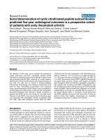
Semiquantitative analysis of Epstein–Barr virus burden
in blood of SLE patients
DNA purified from PBMCs was used to determine the EBV
burden by EBNA-3C-specific PCR/Southern blot. Serial
dilutions of Namalwa DNA were used to establish the sen-
sitivity of the assay system (Fig. 3a). The expected 153 bp
signal was detected even on the lane loaded with DNA
from a single Namalwa cell. The results show that this
method is highly sensitive and capable of detecting as few
as two copies of EBV genome in a background of 10
5
cells
(Fig. 3a).
DNA from PBMCs of 24 SLE patients and 29 healthy indi-
viduals was analyzed to quantify EBV loads. To obtain more
accurate data using a semiquantitative PCR method, the
PCR reaction was stopped before it reached a plateau
state. In addition, serially diluted Namalwa DNA solutions
were included for every set of PCR experiments to control
for variation in PCR efficiency. Duplicate PCR/Southern
reactions were performed for each sample, and the aver-
age values are expressed as EBV genome copies/3 µg
PBMC DNA (Fig. 3b).
In the healthy individuals, the mean EBV load was 30 cop-
ies/3 µg PBMC DNA (range 0–141 copies/3 µg PBMC
DNA). By contrast, in the SLE patients the mean EBV bur-
den was 463 copies/3 µg PBMC DNA (range 0–2440
copies/3 µg PBMC DNA). The difference in EBV burden
between SLE patients and healthy volunteers was statisti-
cally significant (P = 0.001). The median EBV levels for
healthy individuals and SLE patients were 19 and 322 EBV
genome copies/3 µg PBMC DNA, respectively.
To test whether the increased EBV load in SLE patients
was the consequence of an immune suppressive drug
treatment, we divided SLE patients into two groups: those
under immunosuppressive therapy, including high-dose
steroid hormone treatment (n = 8); and those receiving
low-dose steroid hormone and/or hydroxychloroquin (n =
16). EBV loads were similar for these two groups (mean ±
standard deviation: 258 ± 190 EBV genome copies/3 µg
PBMC DNA versus 461 ± 610 EBV genome copies/3 µg
PBMC DNA; P = 0.327, by Spearman's test). In addition,
there was no significant correlation between SLE disease
activity index loads (data not shown). Also, there was no dif-
ference in EBV load between patients with and without
nephritis (data not shown). For each individual from whom
we could collect both samples, the EBV type detected in
the blood sample was identical to that in the mouthwash
sample (data not shown).
Discussion
The present study was undertaken to examine the types of
EBV infecting SLE patients and their viral loads. Different
EBV types were easily recognized from mouthwash sam-
ples by PCR. In healthy control individuals the numbers of
single infections with type 1 or type 2 EBV, as well as num-
bers of co-infection with both types of EBV, were similar to
those described previously [24-26]. Interestingly, there
was no significant difference in EBV type distribution in
Figure 1
Epstein–Barr virus (EBV) typing of normal individuals and patients with systemic lupus erythematosus (SLE) in mouthwash samplesEpstein–Barr virus (EBV) typing of normal individuals and patients with
systemic lupus erythematosus (SLE) in mouthwash samples. (a) PCR/
Southern blot of the EBV nuclear antigen (EBNA)-3C encoding region
for the cell lines carrying type 1 (ES-1, B95-8, LCL2, and Namalwa)
and type 2 (SNU-99 and AG876) EBV. DNA extracted from each EBV
infected cell line (5 ng) was subjected to EBNA-3C-specific PCR/
Southern blot. PCR amplified products were transferred to a membrane
and hybridized with an EBNA-3C probe common to both type 1 and
type 2 EBV. The expected PCR product sizes were 153 bp for type 1
EBV and 246 bp for type 2 EBV. The EBV negative cell line BJAB and
distilled water served as negative controls. (b,c) PCR/Southern blot of
the EBNA-3C encoding region for the DNA from mouthwash samples.
One 20th of the DNA isolated from mouthwash samples was used for
each PCR reaction. Representative results obtained from normal con-
trols (panel b) and SLE patients (panel c) are shown. Namalwa and
AG876 were used as positive controls for type 1 and type 2 EBV,
respectively. Distilled water (dH
2
0) and DNA isolated from BJAB were
used as negative controls.
N 1
N 2
N 3
N 4
N 5
N 6
N 7
N 8
N 9
N 10
N 11
N 12
N 13
dH
2
0
Namalwa
AG876
BJAB
Type 2
Type 1
SLE 1
SLE 2
SLE 3
SLE 4
SLE 5
SLE 6
SLE 7
SLE 8
SLE 9
SLE 10
SLE 11
SLE 12
SLE 13
dH
2
0
Namalwa
AG876
BJAB
Type 2
Type
1
(a)
Type 1
Type
2
BJAB
ES-1
B95-8
LCL2
Namalwa
SNU-99
AG876
dH
2
O
BJAB
(b)
(c)
Available online />R299
SLE patients and normal control individuals. Thus, a spe-
cific type of EBV in SLE patients does not appear to be
responsible for the abnormal T-cell reaction to EBV [20].
We used a semiquantitative PCR assay to evaluate the
level of EBV genome in the peripheral blood of SLE
patients. We could detect and quantify EBV DNA in almost
all of the patients with SLE and the control individuals. The
SLE patients had EBV loads in PBMCs that were more
than 15-fold those in normal control individuals. The EBV
loads we observed in healthy volunteers are comparable to
those reported by others using a real-time PCR method
[27]. The reason for the elevated EBV burden in SLE
patients observed in the present study is not clear. We did
not test whether T-cell function was impaired in the SLE
patients, as has previously been reported [20]. Instead, we
compared EBV loads between patients with and without
strong immunosuppressive therapies, including high-dose
steroids. No difference was observed between the two
groups of SLE patients in terms of EBV load, suggesting no
direct effect of immune function on EBV load. The
increased EBV burden may cause SLE by stimulating
autoantibody production because of the sequence hom-
ology between autoantigens and EBV proteins [3-5]. The
Table 2
Detection of Epstein–Barr virus in mouthwash samples by PCR/Southern blot
Status Healthy volunteers (n [%]) SLE patients (n [%])
EBV-positive 59 (94.0) 65 (98.5)
Type 1 22 (35.0) 26 (39.5)
Type 2 4 (6.0) 3 (4.5)
Types 1 and 2 33 (53.0) 36 (54.5)
EBV-negative 4 (6.0) 1 (1.5)
Total 63 (100) 66 (100)
EBV, Epstein–Barr virus.
Figure 2
Reconfirmation of the Epstein–Barr virus (EBV) typing resultsReconfirmation of the Epstein–Barr virus (EBV) typing results. The mouthwash samples were analyzed by PCR/Southern blot for EBV nuclear anti-
gen (EBNA)-2 and EBNA-3B in addition to EBNA-3C sequences. Namalwa and AG876 were used as positive controls for type 1 and type 2 EBV,
respectively. Distilled water (dH
2
0) was used as a negative control.
Marker
dH
2
O
AG876
Namalwa
1
2
3
4
5
6
7
8
9
10
11
EBNA-2
EBNA-3B
EBNA-3C
300bp
200bp
300bp
200bp
100bp
300bp
200b
p
Type 2 (186bp)
Type 1 (168bp)
Type 2 (149bp)
Type 1 (125bp)
Type 2 (246bp)
Type 1 (153bp)
Arthritis Research & Therapy Vol 6 No 4 Moon et al.
R300
increased EBV loads in SLE appear to be consistent with
the finding that SLE patients often have what appears to be
a primary or reactivated EBV serologic response [28-30].
Approximately 1 in 10
5
–10
6
B cells are latently infected
with EBV in healthy carriers, and one EBV-infected cell usu-
ally contains about 30 EBV episomes [31,32]. Because
one human genome contains approximately 6 pg DNA, the
3 µg PBMC DNA used in our PCR reaction corresponds to
5 × 10
5
blood cells. Thus, it is not surprising that EBV
genome was detected in almost all PBMC samples, bear-
ing in mind that the sensitivity of our PCR assay was two
copies of EBV genome (Fig. 3a). Furthermore, only one out
of 63 SLE patients (1.5%) was EBV-negative, whereas four
out of 66 normal control individuals (6.0%) were EBV-neg-
ative when DNA from the mouthwash sample was tested.
Even though there was a tendency toward increased EBV
infection rate among SLE patients, this difference did not
reach statistical significance.
Our findings are different from those of one study [33] in
which 13 SLE patients were tested by PCR; that study
found no detectable EBV genomes in PBMC DNA or con-
centrated saliva, even though all of the patients exhibited
EBV seroconversion. Another group of researchers also
reported very low rates of EBV positivity for SLE patients
(2/20) and normal control individuals (0/20) using PCR/
Southern methods [13]. The discrepancy between
reported data and our findings may be due to the sensitivity
of the PCR assays used. The sensitivities of the PCR
assays used to detect EBV-infected cells was 80 copies in
one case [33] and 1 pg B95-8 DNA in the other [13].
When James and coworkers [14] evaluated EBV infection
in PBMCs from young SLE patients by PCR analysis,
100% of the SLE patients were EBV-positive whereas only
72% of the matched control individuals were EBV-positive
(P < 0.002). Those investigators needed to recruit young
SLE patients (average age 15.8 ± 2.2 years) in order to
achieve sufficient statistical power in their study, because
about 95% of adults are presumed to carry EBV [34]. How-
ever, the patients who participated in the present study
were considerably older (average age 38.5 ± 10.8 years),
and statistically significant differences in EBV infection
rates between SLE patients and normal control individuals
might not have been detected because of the relatively old
age and small numbers of patients recruited into our study.
EBV has been suspected of being an etiologic agent not
only for SLE but also for other autoimmune diseases. Sera
from patients with rheumatoid arthritis contain more anti-
bodies to EBV than do sera from healthy control individuals
[35]. Furthermore, patients with rheumatoid arthritis have a
decreased T-cell response to EBV gp110 [36,37]. We
[38] and others [39] found that patients with rheumatoid
arthritis have elevated EBV loads in their peripheral blood.
EBV is also frequently detected in salivary glands from
patients with Sjögren's syndrome [40]. In addition, sponta-
neously transformed B-cell lines producing a large amount
of transforming EBV were preferentially established in Sjö-
gren's syndrome patients, probably because of impaired
EBV-specific regulatory mechanisms in this disease [41].
After we had submitted our manuscript, Kang and cowork-
ers [42] reported that EBV titer in SLE was increased by
about 40-fold that in normal control samples. They also
showed that the EBV loads were unaffected by immuno-
suppressive therapies, as we observed. Because they used
real-time PCR to detect EBV loads in PBMC DNA, the
small difference between their data and ours may be due to
the semiquantitative nature of the PCR assay we used.
Figure 3
Epstein–Barr virus (EBV) loads in peripheral blood mononuclear cells (PBMCs) from 29 normal individuals and 24 patients with systemic lupus erythematosus (SLE)Epstein–Barr virus (EBV) loads in peripheral blood mononuclear cells
(PBMCs) from 29 normal individuals and 24 patients with systemic
lupus erythematosus (SLE). (a) Sensitivity of PCR/Southern blot for the
EBV nuclear antigen (EBNA)-3C sequence. DNA was purified from
serial 10-fold dilutions of Namalwa cells (corresponding to 1 to 1 × 10
7
cells) were mixed with BJAB cells to yield a total cell number of 1 ×
10
7
. PCR was performed using a 100th of the purified DNA (corre-
sponding to DNA of 10
5
cells). The PCR products were separated in an
agarose gel, transferred to a membrane, and probed with an EBNA-3C-
specific oligonucleotide. (b) EBV loads of normal individuals and SLE
patients. The mean EBV load of each group is presented as a heavy
horizontal line.
Number of Namalwa cells
0
1
10
100
1,000
10,000
(a)
(b)
EBV copies/3µg PBMC DNA
10,000
1,000
100
10
1
Normal (n = 29) SLE (n = 24)
463
P =
0.001
30
Available online />R301
Conclusion
The type of EBV infecting adult SLE patients is not different
from that in healthy control individuals. However, many
patients with SLE have elevated EBV load in their blood,
suggesting that EBV infection is abnormally regulated in
SLE. The increased numbers of EBV-infected B cells in
SLE patients may contribute to an enhanced autoantibody
production in this disease.
Competing interests
None declared.
Acknowledgements
This work was supported by a grant (R11-2002-098-04006-0) from the
Korea Science & Engineering Foundation through the RRC (Rheuma-
tism Research Center) at the Catholic University. We are grateful to
Young Shik Shim and Sun-A Lee for their valuable technical support.
References
1. Tan EM, Cohen AS, Fries JF, Masi AT, McShane DJ, Rothfield NF,
Schaller JG, Talal N, Winchester RJ: The 1982 revised criteria for
the classification of systemic lupus erythematosus. Arthritis
Rheum 1982, 25:1271-1277.
2. Mok CC, Lau CS: Pathogenesis of systemic lupus
erythematosus. J Clin Pathol 2003, 56:481-490.
3. Sabbatini A, Bombardieri S, Migliorini P: Autoantibodies from
patients with systemic lupus erythematosus bind a shared
sequence of SmD and Epstein–Barr virus-encoded nuclear
antigen EBNA 1. Eur J Immunol 1993, 23:1146-1152.
4. James JA, Scofield RH, Harley JB: Lupus autoimmunity after
short peptide immunization. Ann N Y Acad Sci 1997,
815:124-127.
5. Incaprera M, Rindi L, Bazzichi A, Garzelli C: Potential role of the
Epstein–Barr virus in systemic lupus erythematosus
autoimmunity. Clin Exp Rheumatol 1998, 16:289-294.
6. Ngou J, Segondy M, Seigneurin JM, Graafland H: Antibody
responses against polypeptide components of Epstein–Barr
virus induced early diffuse antigen in patients with connective
tissue diseases. J Med Virol 1990, 32:39-46.
7. Dror Y, Blachar Y, Cohen P, Livni N, Rosenmann E, Ashkenazi A:
Systemic lupus erythematosus associated with acute
Epstein–Barr virus infection. Am J Kidney Dis 1998,
32:825-828.
8. Vaughan JH: The Epstein–Barr virus in autoimmunity. Springer
Semin Immunopathol 1995, 17:203-230.
9. Petersen J, Rhodes G, Roudier J, Vaughan JH: Altered immune
response to glycine-rich sequences of Epstein–Barr nuclear
antigen-1 in patients with rheumatoid arthritis and systemic
lupus erythematosus. Arthritis Rheum 1990, 33:993-1000.
10. Yokochi T, Yanagawa A, Kimura Y, Mizushima Y: High titer of anti-
body to the Epstein–Barr virus membrane antigen in sera from
patients with rheumatoid arthritis and systemic lupus
erythematosus. J Rheumatol 1989, 16:1029-1032.
11. Kitagawa H, Iho S, Yokochi T, Hoshino T: Detection of antibodies
to the Epstein–Barr virus nuclear antigens in the sera from
patients with systemic lupus erythematosus. Immunol Lett
1988, 17:249-252.
12. Verdolini R, Bugatti L, Giangiacomi M, Nicolini M, Filosa G, Cerio
R: Systemic lupus erythematosus induced by Epstein–Barr
virus infection. Br J Dermatol 2002, 146:877-881.
13. Tsai YT, Chiang BL, Kao YF, Hsieh KH: Detection of Epstein–
Barr virus and cytomegalovirus genome in white blood cells
from patients with juvenile rheumatoid arthritis and childhood
systemic lupus erythematosus. Int Arch Allergy Immunol 1995,
106:235-240.
14. James JA, Kaufman KM, Farris AD, Taylor-Albert E, Lehman TJ, Har-
ley JB: An increased prevalence of Epstein–Barr virus infection
in young patients suggests a possible etiology for systemic
lupus erythematosus. J Clin Invest 1997, 100:3019-3026.
15. James JA, Neas BR, Moser KL, Hall T, Bruner GR, Sestak AL, Har-
ley JB: Systemic lupus erythematosus in adults is associated
with previous Epstein–Barr virus exposure. Arthritis Rheum
2001, 44:1122-1126.
16. Verdolini R, Bugatti L, Giangiacomi M, Nicolini M, Filosa G, Cerio
R: Systemic lupus erythematosus induced by Epstein–Barr
virus infection. Br J Dermatol 2002, 146:877-881.
17. Dror Y, Blachar Y, Cohen P, Livni N, Rosenmann E, Ashkenazi A:
Systemic lupus erythematosus associated with acute
Epstein–Barr virus infection. Am J Kidney Dis 1998,
32:825-828.
18. Sample J, Young L, Martin B, Chatman T, Kieff E, Rickinson A, Kieff
E: Epstein–Barr virus types 1 (EBV-1) and 2 (EBV-2) differ in
their EBNA-3A, EBNA-3B, and EBNA-3C genes. J Virol 1990,
64:4084-4092.
19. Moss DJ, Misko IS, Burrows SR, Burman K, McCarthy R, Sculley
TB: Cytotoxic T-cell clones discriminate between A- and B-
type Epstein–Barr virus transformants. Nature 1988,
331:719-721.
20. Tsokos GC, Magrath IT, Balow JE: Epstein–Barr virus induces
normal B cell responses but defective suppressor T cell
responses in patients with systemic lupus erythematosus. J
Immunol 1983, 131:1797-1801.
21. Lee SK, Compton T, Longnecker R: Failure to complement infec-
tivity of EBV and HSV-1 glycoprotein B (gB) deletion mutants
with gBs from different human herpesvirus subfamilies. Virol-
ogy 1997, 237:170-181.
22. Henderson A, Ripley S, Heller M, Kieff E: Chromosome site for
Epstein–Barr virus DNA in a Burkitt tumor cell line and in lym-
phocytes growth-transformed in vitro. Proc Natl Acad Sci USA
1983, 80:1987-1991.
23. Lawrence JB, Villnave CA, Singer RH: Sensitive, high-resolution
chromatin and chromosome mapping in situ: presence and
orientation of two closely integrated copies of EBV in a lym-
phoma line. Cell 1988, 52:51-61.
24. Srivastava G, Wong KY, Chiang AK, Lam KY, Tao Q: Coinfection
of multiple strains of Epstein–Barr virus in immunocompetent
normal individuals: reassessment of the viral carrier state.
Blood 2000, 95:2443-2445.
25. Walling DM, Brown AL, Etienne W, Keitel WA, Ling PD: Multiple
Epstein–Barr virus infections in healthy individuals. J Virol
2003, 77:6546-6550.
26. Srivastava G, Wong KY, Chiang AK, Lam KY, Tao Q: Coinfection
of multiple strains of Epstein–Barr virus in immunocompetent
normal individuals: reassessment of the viral carrier state.
Blood 2000, 95:2443-2245.
27. Balandraud N, Meynard JB, Auger I, Sovran H, Mugnier B, Reviron
D, Roudier J, Roudier C: Epstein–Barr virus load in the periph-
eral blood of patients with rheumatoid arthritis: accurate quan-
tification using real-time polymerase chain reaction. Arthritis
Rheum 2003, 48:1223-1228.
28. Evan AS, Rothfield NF, Niederman JC: Raised antibody titers to
EB virus in systemic lupus erythematosus. Lancet 1971,
1:167-168.
29. Rothfield NF, Evans AS, Niederman JC: Clinical and laboratory
aspects of raised virus antibody titers in systemic lupus
erythematosus. Ann Rheum Dis 1973, 32:38-46.
30. Stancek D, Robensky J: Enhancement of Epstein–Barr virus
antibody production in systemic lupus erythematosus
patients. Acta Virol 1979, 23:168-169.
31. Rocchi G, Felici A, Ragona G, Heinz A: Quantitative evaluation
of Epstein–Barr-virus-infected mononuclear peripheral blood
leukocytes in infectious mononucleosis. N Engl J Med 1977,
296:132-134.
32. Kieff E: Epstein–Barr virus and its replication. In Fields Virology
3rd edition. Edited by: Fields BN, Knipe DM, Howley PM, Chanock
RM, Melnick JL, Monath TP, Roizman B, Straus SE. Philadelphia:
Lippincott-Raven; 1996:2343-2396.
33. Katz BZ, Salimi B, Kim S, Nsiah-Kumi P, Wagner-Weiner L:
Epstein–Barr virus burden in adolescents with systemic lupus
erythematosus. Pediatr Infect Dis J 2001, 20:148-153.
34. Evans AS, Niederman JC: Epstein–Barr virus. In Viral Infections
of Humans, Epidemiology and Control. Edited by: Evans AS. New
York: Plenum Publishing Corporation; 1989:265-292.
35. Yokochi T, Yanagawa A, Kimura Y, Mizushima Y: High titer of anti-
body to the Epstein–Barr virus membrane antigen in sera from
Arthritis Research & Therapy Vol 6 No 4 Moon et al.
R302
patients with rheumatoid arthritis and systemic lupus
erythematosus. J Rheumatol 1989, 16:1029-1032.
36. Toussirot E, Wendling D, Tiberghien P, Luka J, Roudier J:
Decreased T cell precursor frequencies to Epstein–Barr virus
glycoprotein Gp110 in peripheral blood correlate with disease
activity and severity in patients with rheumatoid arthritis. Ann
Rheum Dis 2000, 59:533-538.
37. Depper JM, Bluestein HG, Zvaifler NJ: Impaired regulation of
Epstein–Barr virus-induced lymphocyte proliferation in rheu-
matoid arthritis is due to a T cell defect. J Immunol 1981,
127:1899-1902.
38. Suk Kyeong Lee: Epstein–Barr virus (EBV) and rheumatoid
arthritis. In Proceedings of the Fourth Korea-Japan Combined
Meeting of Rheumatology: 24–25 March 2001; Tokyo.
39. Balandraud N, Meynard JB, Auger I, Sovran H, Mugnier B, Reviron
D, Roudier J, Roudier C: Epstein–Barr virus load in the periph-
eral blood of patients with rheumatoid arthritis: accurate
quantification using real-time polymerase chain reaction.
Arthritis Rheum 2003, 48:1223-1228.
40. Miyasaka N, Yamaoka K, Tateishi M, Nishioka K, Yamamoto K:
Possible involvement of Epstein–Barr virus (EBV) in polyclonal
B-cell activation in Sjogren's syndrome. J Autoimmun 1989,
2:427-432.
41. Wen S, Shimizu N, Yoshiyama H, Mizugaki Y, Shinozaki F, Takada
K: Association of Epstein–Barr virus (EBV) with Sjögren's syn-
drome: differential EBV expression between epithelial cells
and lymphocytes in salivary glands. Am J Pathol 1996,
149:1511-1517.
42. Kang I, Quan T, Nolasco H, Park SH, Hong MS, Crouch J, Pamer
EG, Howe JG, Craft J: Defective control of latent Epstein–Barr
virus infection in systemic lupus erythematosus. J Immunol
2004, 172:1287-1294.