Báo cáo y học: "Mast cells in inflammatory arthritis" pdf
Bạn đang xem bản rút gọn của tài liệu. Xem và tải ngay bản đầy đủ của tài liệu tại đây (720.98 KB, 11 trang )
1
GPI = glucose-6-phosphate isomerase; IL = interleukin; ITAM = immunoreceptor tyrosine-based activation motif; MHC = major histocompatibility
complex; MIP = macrophage inflammatory protein; OA = osteoarthritis; RA = rheumatoid arthritis; SCF = stem cell factor; TGF-β = transforming
growth factor-β; TLR = Toll-like receptor; TNF = tumor necrosis factor.
Available online />Introduction
The mast cell has long been known to mediate important
manifestations of allergic disease. Crosslinking of surface-
bound IgE results in the immediate release of granule
contents, including histamine, and the more gradual
elaboration of other proinflammatory mediators. Clinical
manifestations can range from seasonal allergic rhinitis to
life-threatening anaphylaxis.
However, research over the past two decades has
revealed that the role of mast cells is not limited to IgE-
mediated immune responses. Mast cells express surface
receptors for IgG, complement, and specific pathogen-
associated molecular patterns. Mast cells are capable of
phagocytosis, intracellular killing, and antigen presentation.
Correspondingly, mice deficient in mast cells have been
found to exhibit striking susceptibility to death from certain
types of bacterial infection. Beyond the acute phase of the
immune response, mast cells may participate in the
response of tissue to injury by means of mediators that
promote angiogenesis and fibrosis.
Recently, several laboratories have established that mast
cells have a critical role in the pathogenesis of synovitis in
a murine system with considerable similarity to rheumatoid
arthritis (RA) [1,2]. This finding has renewed interest in
older histological data documenting prominent mast cell
infiltrates in the rheumatoid synovium. We review here the
functions of mast cells as a prelude to the discussion of
the current state of knowledge about the role of mast cells
in murine and human inflammatory arthritis.
Basic biology of mast cells
Mast cells are found principally in mucosae and in
connective tissue, generally clustered at epithelial
surfaces and around nerves and blood vessels [3]. They
originate in bone marrow and circulate as CD34
+
committed progenitor cells, differentiating into mature
mast cells only after entry into the tissue [4,5]. These
mature cells may divide further. Tissue mast cells are
highly heterogeneous, with great variability in size, granule
contents, cytokine production and receptor expression;
both in vitro experience and in vivo data suggest that this
Review
Mast cells in inflammatory arthritis
Peter A Nigrovic
1,2
and David M Lee
1
1
Division of Rheumatology, Immunology and Allergy, Brigham and Women’s Hospital, Boston, Massachusetts, USA
2
Division of Immunology, Children’s Hospital of Boston, Boston, Massachusetts, USA
Corresponding author: David M Lee,
Published: 2 November 2004
Arthritis Res Ther 2005, 7:1-11 (DOI 10.1186/ar1446)
© 2004 BioMed Central Ltd
Abstract
Mast cells are present in limited numbers in normal human synovium, but in rheumatoid arthritis and
other inflammatory joint diseases this population can expand to constitute 5% or more of all synovial
cells. Recent investigations in a murine model have demonstrated that mast cells can have a critical
role in the generation of inflammation within the joint. This finding highlights the results of more than 20
years of research indicating that mast cells are frequent participants in non-allergic immune responses
as well as in allergy. Equipped with a diversity of surface receptors and effector capabilities, mast cells
are sentinels of the immune system, detecting and delivering a first response to invading bacteria and
other insults. Accumulating within inflamed tissues, mast cells produce cytokines and other mediators
that may contribute vitally to ongoing inflammation. Here we review some of the non-allergic functions
of mast cells and focus on the potential role of these cells in murine and human inflammatory arthritis.
Keywords: inflammation, mast cells, rheumatoid arthritis, synovitis, synovium
2
Arthritis Research & Therapy Vol 7 No 1 Nigrovic and Lee
heterogeneity represents an exquisite developmental
sensitivity to local signals [3]. Similarly, the maintenance of
mast cells within tissues is controlled by the local
environment, in particular the production of stem cell
factor (SCF, c-kit ligand) by stromal cells [6]. Mature mast
cells are also capable of trafficking, as shown by their
recruitment to chemotactic stimuli such as RANTES and
their efflux from tissue through lymphatic channels and
possibly blood vessels [7–9].
Functions of mast cells
IgE-mediated activation
Mast cells express the high-affinity IgE receptor FcεR1, a
tetrameric complex of an α chain (to which IgE binds), a β
chain and a dimer of γ chains [10]. The γ chain is shared
with other stimulatory receptors, including the high-affinity
IgG receptor FcγR1 and the low-affinity immune complex
receptor FcγR3a. On crosslinking of the IgE receptor by
multivalent antigen, the immunoreceptor tyrosine-based
activation motifs (ITAMs) on the β and γ chains become
phosphorylated and initiate a signaling cascade, resulting
in three distinct pathways of mediator production: explosive
release of preformed mediators, elaboration of eicosanoids,
and de novo synthesis of cytokines and chemokines.
Explosive release of preformed mediators
Within seconds to minutes of IgE crosslinking, granules in
the cytoplasm of the mast cell fuse with each other and
with the cell surface membrane, ejecting their contents
into the extracellular milieu. The contents of the granules
depend on the conditions under which the mast cell has
matured, but include histamine, proteoglycans (for
example heparin), and a series of neutral proteases
broadly grouped into tryptases, chymases, and carboxy-
peptidases. Histamine promotes vascular permeability;
proteoglycans provide a scaffold within the granule that
allows the packaging of proteases; and the neutral
proteases cleave proteins from matrix and plasma in
addition to activating propeptides such as the precursors
for interleukin-1β (IL-1β) and angiotensin II. The tryptase
mMCP6 (murine mast cell protease 6) also contributes
potently to neutrophil chemotaxis [11]. Certain subsets of
mast cells store tumor necrosis factor (TNF) within the
granules as well, representing the body’s only source of
TNF available for immediate release [12].
Elaboration of eicosanoids
Within minutes of IgE-mediated activation, mast cells
begin to generate eicosanoids derived from cleavage of
arachidonic acid from membrane phospholipids [13].
Important arachidonic acid metabolites include the
leukotrienes (leukotriene B
4
and the cysteinyl leuko-
trienes), which increase vascular permeability, induce
vasoconstriction and recruit leukocytes, and prosta-
glandins including the neutrophil chemoattractant and
vasoactive mediator prostaglandin D
2
.
De novo synthesis of cytokines and chemokines
Within hours, a later phase of mast cell activation through
IgE becomes evident with the induction of new gene
transcription and translation, generating a host of cyto-
kines and chemokines (Table 1). The mix of cytokines
generated by a particular mast cell depends on its
individual state of differentiation.
The importance of IgE-mediated mast cell activation to the
health of the organism is still incompletely defined. The
preservation of this system under evolutionary pressure,
despite allergic diseases and anaphylaxis, is strong
suggestive evidence that there is benefit to the host. One
likely candidate function is resistance to parasitic disease,
because mice deficient in IgE exhibit impaired defense
against the helminths Schistosoma mansoni and
Trichinella spiralis [14,15].
IgE-independent functions of mast cells
Mast cells cluster at sites of contact with the external
world, such as mucosal and epithelial surfaces. Similarly,
they are found near blood vessels and in the linings of
potential spaces such as the peritoneum, pleural space,
and synovial cavity. This localization suggests a role in
surveillance, and indeed mast cells are capable of
detecting pathogens and initiating an inflammatory
response, earning this cell the appellation of immune
sentinel [16]. Further, mast cells accumulate in chronically
inflamed tissue, suggesting that their role might not be
limited to the initiation phase of the immune response.
Mast cells in bacterial infection
The physiological importance of mast cells in defense
against bacteria has been clearly demonstrated. Mast-cell-
deficient W/W
v
mice have impaired clearance of bacterial
infection in the peritoneum [17,18] and lung [18],
accompanied by markedly higher mortality after
experimental infection. This vulnerability was found to be
associated with decreased infiltration of neutrophils to the
site of infection and could be corrected by reconstitution
with wild-type mast cells. Within an hour of peritoneal
infection, lavage fluid shows a striking increase in TNF
levels in the presence of mast cells. Anti-TNF treatment
largely abrogates the effect of mast cell reconstitution,
whereas injection of TNF concurrent with infection
substantially mimics the benefits of reconstitution in mast-
cell-deficient mice. Although mast cells can phagocytose
and kill bacteria [19], the results imply that the critical role
of mast cells in these models is not direct anti-bacterial
action but the generation of TNF and other mediators
(such as leukotrienes [20]) that recruit neutrophils and
possibly other cells to contain the infection.
Mast cells possess multiple mechanisms to detect
bacterial invasion. These include Toll-like receptors (TLRs)
1, 2, 4, and 6, CD48 (a receptor for a Gram-negative
3
fimbrial protein), and receptors for anaphylatoxins C3a
and C5a and the complement opsonin iC3b [21–25].
Interestingly, mast cells triggered by means of these
mechanisms seem capable of responses that are
substantially more differentiated than those unleashed
through IgE/FcεR1. In contrast to the wholesale
‘anaphylactic’ degranulation that characterizes maximal
IgE-mediated stimulation, bacteria can trigger a gradual
and partial (so-called ‘piecemeal’) degranulation proportional
to the stimulus [19,26]. The production of lipid mediators
and cytokines/chemokines seems also to be tailored to
the event, and can even be entirely decoupled from the
release of granule contents (reviewed in [27]).
An important consequence of mast cell activation may be
the mobilization of adaptive immunity. Mast cell leukotriene
B4 recruits memory CD4
+
and CD8
+
T cells, which can
then be activated locally by mast cells presenting
Available online />Table 1
Selected mast cell mediators and their potential roles in arthritis
Mediator Some relevant functions
Granules
Histamine Vascular permeability, leukocyte recruitment, fibroblast/chondrocyte activation
Heparin Angiogenesis, osteoclast differentiation and activation
Neutral proteases Matrix degradation, leukocyte recruitment, fibroblast activation
TNF Leukocyte recruitment, fibroblast/chondrocyte activation
Eicosanoid mediators
PGD
2
Vascular permeability, neutrophil recruitment
LTB
4
Vascular permeability, leukocyte recruitment and activation
Cysteinyl leukotrienes Vascular permeability, immunomodulatory (LTC
4
)
Cytokines/chemokines
IL-1 Leukocyte recruitment, fibroblast/chondrocyte activation, angiogenesis
IL-2 Lymphocyte stimulation
IL-3 Leukocyte growth factor
IL-4 Immunomodulatory, profibrotic
IL-6 Activation of leukocytes and fibroblasts
IL-8 Neutrophil recruitment
IL-10 Immunomodulatory
IL-13 Immunomodulatory, B cell stimulation
IL-18 Angiogenesis, lymphocyte stimulation
TNF Leukocyte recruitment, fibroblast/chondrocyte activation, angiogenesis
IFN-γ Activation of synovial macrophages
TGF-β Immunomodulatory, fibroblast mitogen, angiogenesis
PDGF Fibroblast mitogen
VEGF Fibroblast mitogen, angiogenesis
bFGF Fibroblast mitogen
NGF Fibroblast mitogen
MCP-1 Leukocyte recruitment
MIP-1α, MIP-1β Leukocyte recruitment, osteoclast differentiation
RANTES Leukocyte recruitment
bFGF, basic fibroblast growth factor; IFN, interferon; IL, interleukin; LTB
4
, leukotriene B
4
; LTC
4
, leukotriene C
4
; MCP-1, monocyte chemoattractant
protein-1; MIP, macrophage inflammatory protein; NGF, nerve growth factor; PDGF, platelet-derived growth factor; PGD
2
, prostaglandin D
2
;
RANTES, regulated upon activation, normal T-cell expressed and secreted; TGF-β, transforming growth factor-β; TNF, tumor necrosis factor;
VEGF, vascular endothelial growth factor. See text for references.
4
phagocytosed peptides via both MHC class II and MHC
class I molecules [28–31]. Mast cells might also
potentiate de novo antigen-specific responses by
promoting the migration of dendritic cells to lymph nodes
and recruiting circulating naive T cells to these nodes by
means of TNF and macrophage inflammatory protein-1β
(MIP-1β) [8,32,33]. Although the ultimate physiological
importance of each of these defensive capabilities remains
to be established, it seems probable that antimicrobial
efficacy accounts at least in part for the remarkable
evolutionary conservation of the mast cell.
Mast cells in antibody-mediated disease
As noted, mast cells express receptors for IgG as well as
IgE. These include FcγR2b and FcγR3a, low-affinity IgG
receptors involved principally in the response to immune
complexes and other constellations of colocalized IgG
molecules. Under certain conditions, mast cells can also
express the high-affinity receptor FcγR1 [34]. These
receptors permit mast cells to participate in humoral
defense, but they also enable a role for mast cells in
antibody-induced pathology. Thus, in a mouse model of
peritonitis induced by intraperitoneal injection of antibody
against an antigen injected intravenously (the reverse
passive Arthus reaction), peritoneal mast cells exposed to
immune complexes release a burst of preformed TNF and
recruit neutrophils [35]. Similarly, in an analogous skin
model, mast cells have been shown to potentiate the
response to antibody administered subcutaneously
against an antigen delivered systemically [36]. Optimal
mast cell participation in this reaction requires a functional
complement system, suggesting that complement fixation
by immune complexes provides an important auxiliary
signal to mast cells, in particular via C5a [37]. A related
phenomenon is observed in a model of bullous
pemphigoid: subcutaneous administration of an antibody
against the hemidesmosomal antigen BP180 induces
inflammatory attack, resulting in lysis of the dermal–
epidermal junction. In the absence of mast cells or
complement, inflammation is markedly attenuated [38,39].
As in bacterial peritonitis, the key function of mast cells in
these models of antibody-mediated pathology seems to
be the mobilization of neutrophils, because the wild-type
phenotype can largely be rescued in mast-cell-deficient
animals with injection of neutrophils or neutrophil
chemotactic factors.
Mast cells: a role in chronic inflammation?
In the models discussed so far, the principal function of
mast cells seems to be to ‘jump start’ the immune
response, in particular to initiate the rapid recruitment of
inflammatory cells. Structurally, the mast cell is uniquely
equipped for this task, with its capacity for the immediate
release of preformed mediators and the rapid elaboration
of lipid mediators. However, the mast cell’s activity does
not end with this initial response. Mast cells continue to
elaborate cytokines for hours after a single stimulus, and a
degranulated mast cell can recharge and fire again
[40,41]. Some mast cell mediators have effects such as
the promotion of angiogenesis, whose relevance is more
evident after the acute inflammatory response [42].
Further, mast cells accumulate at sites of chronic
inflammation, prima facie evidence that their role is not
restricted to the initiation of immune responses; examples
include the gut in inflammatory bowel disease or
helminthic infection, the asthmatic airway,
sclerodermatous skin, and lung in interstitial pulmonary
fibrosis [43–46]. Though no pathogenic role has yet been
definitively assigned to the mast cell in these conditions,
potential functions include ongoing recruitment of
inflammatory cells, stimulatory effects on stromal cells
resulting in fibrosis, and the development of new blood
vessels. It is also conceivable that mast cells might in
some cases limit or otherwise modulate local
inflammation, although no data to this effect are available.
Particular proinflammatory mechanisms are discussed
below in detail as they pertain to the potential role of the
synovial mast cell in arthritis.
Mast cells in inflammatory arthritis
Mast cells in normal and inflamed human synovium
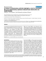
The synovium of patients with RA is an archetypal example
of a chronically inflamed tissue characterized by an
expanded population of mast cells (Fig. 1). In the normal
joint, the synovium consists of a thin lining layer of
macrophages (macrophage-like synoviocytes, ‘Type A’
cells) and fibroblasts (fibroblast-like synoviocytes, ‘Type B’
cells) embedded in a connective tissue matrix and resting
on a sublining of highly vascular loose connective tissue
and adipose tissue. In the absence of inflammation,
scattered mast cells are seen in the sublining, clustered
around vessels and nerves and forming up to 3% of all
cells within the synovium [47]. The role of mast cells in the
normal synovium remains to be defined, although the
importance of mouse peritoneal mast cells for defense
against bacterial peritonitis suggests that one important
function of synovial mast cells might be to monitor the
vulnerable acellular joint cavity for early evidence of
infection.
In RA, the synovial lining thickens from 1–3 cells to
10 cells or more, and the sublining becomes infiltrated
with T cells, B cells, macrophages, and occasional neutro-
phils. Mast cells are commonly markedly increased in
number and can make up 5% or more of the expanded
population of total synovial cells. The number of accumu-
lated mast cells differs substantially from patient to patient,
in general varying directly with the intensity of joint inflam-
mation [17,24,48–55]. Mast cells are present throughout
the synovial sublining, with occasional microanatomic
clustering in the pannus near sites of cartilage and bone
erosion [53,54]. A relative mastocytosis may also be
Arthritis Research & Therapy Vol 7 No 1 Nigrovic and Lee
5
observed in other arthritides, including juvenile rheumatoid
arthritis, systemic lupus erythematosus, psoriatic arthritis,
and some cases of osteoarthritis (OA) [49].
Accompanying the increased numbers of mast cells, mast
cell mediators are also present at higher concentrations in
the synovial fluid of inflamed human joints. These
mediators include histamine and tryptase, both considered
to be specific for mast cells [56–60]. Again, patient-to-
patient variability is considerable. Although mast cells from
RA and OA do not appear distinct histologically, and
express a generally similar panel of surface receptors, RA
but not OA mast cells have been noted to express the
receptor for the anaphylatoxin complement fragment C5a
[24]. Interestingly, whereas normal human synovium
contains mainly mast cells of the so-called ‘connective
tissue’ phenotype, expressing both tryptase and chymase
in their granules (MC
TC
), inflamed synovium also features
mast cells that express only tryptase (MC
T
), a phenotype
more commonly associated with mast cells maturing
under the influence of T cell cytokines at mucosal sites
[24,55,61]. Although the significance of these
subpopulations is uncertain, mast cells with similar
phenotypes isolated from skin and lung exhibit divergent
patterns of cytokine secretion, with IL-4 produced
predominantly by MC
TC
cells whereas MC
T
cells elaborate
IL-5 and IL-6 [62]. If this is true in the synovium, then these
two types of mast cell might have different
pathophysiological roles in inflammatory arthritis, because
IL-4 has profibrotic effects whereas IL-6 may be
stimulatory for T and B lymphocytes (reviewed in [63]).
Correspondingly, MC
TC
cells tend to be found in ‘deeper,’
more fibrotic areas of the inflamed synovium, whereas
MC
T
cells tend to be found more superficially and in
association with lymphoid aggregates [24,61].
Mast cells in arthritis: insights from the K/BxN arthritis
model
Synovial mast cell degranulation was previously noted in
association with arthritis in several animal models, but a
critical functional role in pathogenesis has recently been
firmly established with the K/BxN mouse model
[1,2,64,65]. This arthritis model, mediated by auto-
antibodies against the ubiquitous enzyme glucose-6-
phosphate isomerase (GPI), demonstrates important
similarities to human RA including symmetric joint
involvement, chronicity, a distal-to-proximal gradient of
joint involvement, and histological features including
synovial infiltrates, pannus, and erosions of cartilage and
bone [66].
A key feature of this model is the ability to transfer the
pathogenic autoantibodies passively to induce arthritis in
recipient mice [67]. This passive transfer arthritis
mechanistically ‘disconnects’ the afferent pathogenic
events involving the adaptive immune response and
affords an analytic focus on the efferent pathogenic
mechanisms of synovial inflammation. Given the large and
ever-increasing number of targeted genetic deletions in
mice, it has been possible to apply the power of this
genetic technique to dissect the molecular requirements
for induction of arthritis. Transfer of serum into mice
deficient in various participants in the inflammatory
response has identified a critical role for cytokines (IL-1,
TNF), IgG Fc receptors (especially FcγR3), complement
(C3, C5) and the C5a complement receptor in arthritis
pathogenesis [2,68,69]. Immune complexes are
implicated in the pathogenesis by the observation that
multiple anti-GPI antibodies with non-overlapping epitope
specificities – as would be required to form an
antigen–antibody lattice – are required for the initiation of
arthritis [70].
At the cellular level, the concept of the mast cell as
immune sentinel led to the hypothesis that this lineage
might participate pathogenically in autoantibody-driven
K/BxN serum transfer arthritis. Expressing receptors for
both immune complexes and complement, synovial mast
cells would be well positioned to initiate the tissue
response to K/BxN serum. Consistent with this hypothesis
is the observation that mice deficient in mast cells are
highly resistant to arthritis, whereas reconstitution with
normal mast cells restores the wild-type phenotype
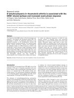
(Fig. 2). Furthermore, degranulation of mast cells in the
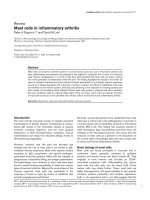
Available online />Figure 1
Mast cells within the rheumatoid synovium. Shown is fixed, paraffin-
embedded synovial tissue obtained during arthroplasty from a patient
with chronic rheumatoid arthritis. This tissue was stained with safranin-
O, which labels mast cell granule proteoglycans red, and
counterstained with hematoxylin. Note the frequent safranin-O-positive
mast cells present within the synovial sublining (several indicated with
arrows). A fold of thickened synovial lining is seen at the bottom left of
the image (outlined with a dotted line) and a blood vessel (BV) is
visible in the middle of the field, with erythrocytes staining blue.
(Section 5 µm thick; original magnification ×400.)
6
synovium is the first event observed histologically,
occurring within 1–2 hours of administration of K/BxN
serum [1]. Thus, as in antibody-mediated peritonitis,
synovial mast cells seem to act as early responders,
mobilizing the inflammatory response against a perceived
insult. In their absence, no other cell constitutively resident
within the synovium or present in the circulation seems to
have the capacity to initiate the recruitment of
inflammatory cells to the joint that characterizes arthritis in
the wild-type animal. However, details of the mechanisms
of mast cell activation as well as the relevant mast cell
effector functions in this model remain to be defined.
Mast cells and the initiation of human synovitis
The involvement of mast cells in the earliest phases of
human synovitis remains a subject for conjecture. As
described previously, mast cells can be triggered by IgG
immune complexes, complement, TLR ligands, and
microbial antigens. Each of these stimulatory pathways
may be of relevance to human arthritis. Immune complexes
are thought to cause the arthritis of serum sickness and
cryoglobulinemia but have also been documented in the
serum, synovial fluid, synovium, and cartilage of patients
with RA and are once again a field of active investigation
in the pathogenesis of RA [71–74]. Complement
activation has similarly been well documented within
rheumatoid synovium [75]. Infection with bacteria or
viruses could trigger mast cell activation by means of
TLRs and specific pathogen receptors. Even in the
absence of infection, mast cells could be stimulated via
TLRs by synovial constituents with TLR ligand activity,
including heat shock protein 60 and breakdown products
of hyaluronan, potentially amplifying any inflammatory
process within the joint [76]. Mast cell IgE receptors might
also have a role in a small subset of patients, because IgE
rheumatoid factors and IgE-containing immune complexes
have been documented in some patients with RA [77,78].
Once activated, mast cells in the synovium would be
expected to initiate inflammation through several
mechanisms; a limited number of candidate pathways are
outlined in Fig. 3. Vasoactive mediators such as histamine,
prostaglandin D
2
, and the leukotrienes increase vascular
permeability, whereas TNF, IL-1, and histamine promote
the expression of the adhesion molecules P-selectin,
E-selectin, ICAM-1, and VCAM-1 on the endothelial
surface [79,80]. Circulating leukocytes bearing appropriate
counter-receptors, such as leukocyte function-associated
antigen-1 (LFA-1) (itself of heightened affinity under the
influence of proinflammatory cytokines through ‘inside-out’
regulation), could then be recruited into the synovium
along gradients of chemotactic mast cell products such as
leukotriene B
4
, monocyte chemoattractant protein-1,
tryptases (for example mMCP6), and IL-8. Activation of
resident synovial macrophages and arriving monocytes
and neutrophils by means of interferon-γ, IL-6 and TNF
would be expected to result in further amplification of
leukocyte recruitment and an enhanced output of
proinflammatory cytokines.
Beyond the ‘jump start’: a role for mast cells in chronic
synovitis in mouse and humans?
In some murine models of bacterial and antibody-induced
disease, the physiological role of mast cells can largely be
replaced by a single administration of neutrophils or
neutrophil chemoattractants [17,31,35,38]. This observation
suggests that mast cells have no substantial continuing
role in these pathologic states. In K/BxN arthritis, and
potentially in human arthritis, is there a role for the synovial
mast cells beyond the initiation of synovitis?
An initial observation applies. In K/BxN serum transfer
arthritis, two serum injections are followed within 1–3 days
by an intense synovitis. This reaction peaks over the
course of 2 weeks but is ultimately self-limiting, resolving
within 6 weeks. Although some human joint diseases run
such a self-limited course (such as serum sickness and
postviral arthritis), many human arthritides are chronic. In
such chronic conditions, any factors inducing mast cell
activation might well be persistent. This is so in K/BxN
mice, which exhibit a progressive erosive arthritis in the
setting of persistently high levels of autoantibodies in the
serum. ‘Chronicity’ can be mimicked in wild-type mice by
means of a repeated transfer of K/BxN serum. In this
setting, synovial mast cells can undergo repetitive cycles
of activation and thus participate in ongoing disease much
more substantially than has been observed in models of
peritonitis and skin disease. Indeed, degranulating synovial
mast cells are readily observed in established K/BxN
Arthritis Research & Therapy Vol 7 No 1 Nigrovic and Lee
Figure 2
Mast cells constitute a critical pathogenic link in K/BxN serum transfer
arthritis. Compared with wild-type controls, mast-cell-deficient W/W
v
mice injected with K/BxN arthritogenic serum are resistant to the
development of arthritis. After reconstitution with cultured wild-type
mast cells, but not sham reconstitution, normal susceptibility is
restored. Error bars = SEM. (Adapted from reference [1], with
permission.)
7
arthritis [1]. Yet a functional contribution of mast cells to
continuing inflammation remains to be experimentally
determined.
In humans, given the expanded numbers of mast cells
within the joint and their enormous capacity for the
production of cytokines and chemokines, it would be
surprising indeed if they were of no consequence to the
chronic inflammatory response. The broad range of mast
cell effector functions includes the elaboration of
mediators with bioactivity directed at marrow-derived
leukocytes as well as mesenchymal tissue elements
(Fig. 3). Because the pathogenic state of inflammatory
arthritis displays prominent responses by both infiltrating
leukocytes and mesenchymal cells, in particular synovial
fibroblasts, we will examine the potential influence of mast
cells on both compartments in arthritis.
Mast cells and synovial leukocytes
The rheumatoid synovium is thick with infiltrating leukocytes.
These include T lymphocytes, B lymphocytes, macrophages,
mast cells and scattered neutrophils. Ongoing recruitment of
these cells results from the upregulation of selectins and
integrins on synovial endothelium, allowing migration up
chemotactic gradients into the joint. The composition of
inflammatory cells recruited in a continuing fashion by mast
cells, including the degree of skewing of lymphocytes toward
Th1 versus Th2 responses, might be an important
Available online />Figure 3
Candidate proinflammatory functions of mast cells in synovitis. Mast cell effector functions suggest their participation in diverse pathogenic
pathways in inflammatory arthritis, including leukocyte recruitment and activation, synovial fibroblast activation and hyperplasia, angiogenesis, and
cartilage and bone destruction. Activated mast cells elaborate mediators potently capable of enhancing vasopermeability, inducing endothelial
expression of adhesion molecules, recruiting circulating leukocytes, and activating infiltrating leukocytes as well as resident macrophages, thereby
contributing to the early phases of inflammatory arthritis. In chronic synovitis, mast cells synthesize mitogens and cytokines that activate synovial
fibroblasts, recruit macrophages, and promote the growth of new blood vessels, implicating them in synovial lining hyperplasia and pannus
formation. Further, mast cells may participate in joint destruction by the induction of matrix metalloproteinases (MMPs) from fibroblasts, by
activation of chondrocytes, and by direct and indirect promotion of osteoclast differentiation and activation. Because activated synovial fibroblasts
demonstrate enhanced stem cell factor (SCF) expression, a potentially important positive feedback loop is established in which SCF promotes
mast cell survival and proliferation, leading to the mastocytosis described in inflamed synovium. Note that the importance of these candidate
pathways in vivo remains to be established. See text for details and references. bFGF, basic fibroblast growth factor; IFN, interferon; IL, interleukin;
MCP = monocyte chemoattractant protein; M-CSF, macrophage colony-stimulating factor; MIP, macrophage inflammatory protein; PDGF, platelet-
derived growth factor; PMN, polymorphonuclear cell; RANK-L, receptor activator of NF-κB ligand; TNF, tumor necrosis factor. (Graphic design by
Steve Moskowitz.)
8
determinant of the ultimate outcome of inflammation. The
production of anti-inflammatory mediators by mast cells
remains uncharacterized [81].
Prominent within the rheumatoid synovium is a greatly
expanded population of synovial macrophages. These
cells do not proliferate locally but instead are recruited
from circulating monocytes [82]. Mast cells are potent
sources of chemokines that mediate this recruitment,
including IL-8, monocyte chemoattractant protein-1,
MIP-1α, and RANTES [3]. Mast cells might also contribute
to the activation of these macrophages through the
production of interferon-γ and IL-6. Because macrophages
are major sources of the proinflammatory cytokines TNF
and IL-1 within the joint, mast cell effects on the size and
activation state of the synovial macrophage population
might functionally modulate the course of inflammatory
arthritis.
Mast cells and the synovial mesenchyme
The synovial mesenchyme, consisting principally of
synovial fibroblasts, is prominently involved in joint
inflammation. Fibroblasts increase greatly in numbers and
assume a histological appearance suggestive of increased
synthetic activity, with expansion of the endoplasmic
reticulum and increased numbers of granules in the
cytoplasm [83]. Indeed, synovial fibroblasts make up the
shroud-like pannus characteristic of the rheumatoid joint
and are an important source of multiple mediators
implicated in arthritis. These include degradative enzymes
such as collagenase and stromelysin and proinflammatory
molecules including IL-1, IL-6, and prostaglandin E
2
(reviewed in [84]). They contribute to the differentiation
and activation of osteoclasts, the effector cell responsible
for bone erosions, through the production of macrophage
colony-stimulating factor (M-CSF) and receptor activator
of NF-κB ligand (RANKL) [85,86].
Mast cells may potently influence synovial fibroblast
biology in RA. Consistent with a proposed role in wound
healing and in multiple fibrotic disease states, mast cells
produce a range of mediators with powerful effects on
fibroblasts (Table 1) [87]. Further, synovial mast cells are
often noted in close physical proximity to synovial
fibroblasts [50]. Mast cell tryptase promotes chemotaxis
and collagen synthesis in fibroblasts, and histamine
stimulates fibroblast proliferation [88–90]. Other fibroblast
mitogens produced by mast cells include nerve growth
factor, basic fibroblast growth factor, platelet-derived
growth factor, vascular endothelial growth factor (VEGF),
and transforming growth factor-β (TGF-β) [91]. The
cytokine IL-4, produced predominantly by mast cells of a
tryptase–chymase phenotype, induces proliferation and
collagen production by fibroblasts [92], and indeed, as
noted above, MC
TC
cells tend to reside in more fibrotic
areas of the inflamed joint. Because leukotriene C
4
seems
to have antifibrotic effects, it remains possible that mast
cells can limit as well as promote fibrosis, although
scattered foci of fibrosis associated with mast cell
infiltrates in systemic mastocytosis suggest a net
profibrotic effect [91,93,94].
Mast cells may also potentiate mediator production by
synovial fibroblasts through the elaboration of cytokines
such as TNF and IL-1. IL-1 induces the elaboration of
collagenase and prostaglandin E
2
, and TNF elicits similar
responses while also inducing synovial fibroblasts to
generate IL-1 [95–97]. Indeed, the production of
collagenase and other inflammatory products of fibro-
blasts has been noted to localize to the immediate
environment of activated mast cells [98].
This communication between mast cells and synovial
fibroblasts is bidirectional. Mast cells require stimulation
by SCF for differentiation in situ as well as activation [6].
Fibroblasts in inflamed or healing tissues express higher
levels of SCF, and upregulation of SCF expression has
been noted in synovial specimens exposed to TNF
[99–101]. Indeed, such surface expression seems to be
of particular importance to mast cell development,
because Sl/Sl
d
mice unable to display surface-bound SCF
lack tissue mast cells despite an intact production of
soluble SCF [102,103]. Further, transwell experiments
demonstrate that physical contact is required for certain
stimulatory effects of fibroblasts on mast cells [104,105].
Fibroblasts might also promote the survival of mast cells
by means of SCF-independent pathways yet to be fully
defined [106].
In addition to fibroblasts, the synovial mesenchyme also
contains blood vessels. As would be expected, the
expanded cellular population in the inflamed synovium
requires an enhanced blood supply, and neoangiogenesis
has an important pathophysiological function in RA. Mast
cell mediators implicated in the promotion of angiogenesis
include heparin, vascular endothelial growth factor, TGF-β,
TNF, IL-1, and IL-18 [42,107]. Further, TNF can induce
synovial fibroblast production of another pro-angiogenic
factor, angiopoietin-1 [108]. Though the ultimate
importance of mast cells in synovial angiogenesis remains
unclear, the association of mast cells with blood vessels,
including newly developing blood vessels, makes the
promotion of angiogenesis a plausible role for mast cells
in vivo (reviewed in [109]).
Finally, some data suggest that mast cell mediators might
exert a direct effect on cartilage and bone. Thus, whereas
the coculture of chondrocytes with inactive mast cells
tends to promote the synthesis of proteoglycans, the
activation of mast cells in this context favors proteoglycan
degradation [110]. Further, the activation of chondrocytes
via IL-1, TNF, and histamine might induce the production
Arthritis Research & Therapy Vol 7 No 1 Nigrovic and Lee
9
of matrix metalloproteinases and prostaglandins [111,112].
Finally, mast cell mediators including histamine and MIP-
1α might directly promote the differentiation and activation
of osteoclasts, the final common pathway of bone
destruction in inflammatory arthritis [113–115]. Corro-
boration in vivo will be required to establish the
importance of these in vitro findings.
Conclusions
Mast cells are a normal cell population within the human
synovium, and in line with their role as sentinels they likely
have an important physiological role as an ‘early warning
system’ for infection within the vulnerable joint cavity. Data
from the K/BxN mouse model now show that mast cells
also have a critical role in the pathogenesis of inflam-
matory arthritis, in particular in arthritis induced by
autoantibody-containing immune complexes. Although a
similar mechanism remains unproven for human joint
inflammation, markers of mast cell activation are observed
in joint fluid from patients with chronic arthritis and mast
cell numbers are often greatly expanded within the
inflamed synovium. Equipped with an impressive array of
mediators, mast cells can promote synovitis by recruiting
inflammatory cells from the blood, inducing synovial
fibroblast hyperplasia and mediator production, and
fostering angiogenesis. Although much remains to be
learned about the role of the mast cell in arthritis, such a
role now seems highly likely, offering a potential new
target for therapeutic agents in the treatment of RA and
other inflammatory diseases of the joints.
Competing interests
The author(s) declare that they have no competing interests.
Acknowledgements
Supported by the Physician Scientist Development Award of the Arthri-
tis Foundation and American College of Rheumatology Research and
Education Foundation (PAN) and R01-AI059746, K08-AR02214, the
Cogan Family Foundation and the Arthritis Investigator Award of the
Arthritis Foundation, and the American College of Rheumatology
Research and Education Foundation.
References
1. Lee DM, Friend DS, Gurish MF, Benoist C, Mathis D, Brenner
MB: Mast cells: a cellular link between autoantibodies and
inflammatory arthritis. Science 2002, 297:1689-1692.
2. Corr M, Crain B: The role of Fc
γγ
R signaling in the K/B x N
serum transfer model of arthritis. J Immunol 2002, 169:6604-
6669.
3. Metcalfe DD, Baram D, Mekori YA: Mast cells. Physiol Rev 1997,
77:1033-1079.
4. Kirshenbaum AS, Kessler SW, Goff JP, Metcalfe DD: Demon-
stration of the origin of human mast cells from CD34+ bone
marrow progenitor cells. J Immunol 1991, 146:1410-1415.
5. Rodewald HR, Dessing M, Dvorak AM, Galli SJ: Identification of
a committed precursor for the mast cell lineage. Science
1996, 271:818-822.
6. Galli SJ, Tsai M, Wershil BK: The c-kit receptor, stem cell factor,
and mast cells. What each is teaching us about the others. Am
J Pathol 1993, 142:965-974.
7. Juremalm M, Olsson N, Nilsson G: Selective CCL5/RANTES-
induced mast cell migration through interactions with
chemokine receptors CCR1 and CCR4. Biochem Biophys Res
Commun 2002, 297:480-485.
8. Wang HW, Tedla N, Lloyd AR, Wakefield D, McNeil PH: Mast
cell activation and migration to lymph nodes during induction
of an immune response in mice. J Clin Invest 1998, 102:1617-
1626.
9. Friend DS, Gurish MF, Austen KF, Hunt J, Stevens RL: Senes-
cent jejunal mast cells and eosinophils in the mouse prefer-
entially translocate to the spleen and draining lymph node,
respectively, during the recovery phase of helminth infection.
J Immunol 2000, 165:344-352.
10. Gould HJ, Sutton BJ, Beavil AJ, Beavil RL, McCloskey N, Coker
HA, Fear D, Smurthwaite L: The biology of IGE and the basis of
allergic disease. Annu Rev Immunol 2003, 21:579-628.
11. Huang C, Friend DS, Qiu WT, Wong GW, Morales G, Hunt J,
Stevens RL: Induction of a selective and persistent extravasa-
tion of neutrophils into the peritoneal cavity by tryptase
mouse mast cell protease 6. J Immunol 1998, 160:1910-1919.
12. Gordon JR, Galli SJ: Mast cells as a source of both preformed
and immunologically inducible TNF-
αα
/cachectin. Nature 1990,
346:274-276.
13. Murakami M, Austen KF, Arm JP: The immediate phase of c-kit
ligand stimulation of mouse bone marrow-derived mast cells
elicits rapid leukotriene C4 generation through posttransla-
tional activation of cytosolic phospholipase A2 and 5-lipoxy-
genase. J Exp Med 1995, 182:197-206.
14. King CL, Xianli J, Malhotra I, Liu S, Mahmoud AA, Oettgen HC:
Mice with a targeted deletion of the IgE gene have increased
worm burdens and reduced granulomatous inflammation fol-
lowing primary infection with Schistosoma mansoni. J
Immunol 1997, 158:294-300.
15. Gurish MF, Bryce PJ, Tao H, Kisselgof AB, Thornton EM, Miller
HR, Friend DS, Oettgen HC: IgE enhances parasite clearance
and regulates mast cell responses in mice infected with
Trichinella spiralis. J Immunol 2004, 172:1139-1145.
16. Galli SJ, Maurer M, Lantz CS: Mast cells as sentinels of innate
immunity. Curr Opin Immunol 1999, 11:53-59.
17. Echtenacher B, Mannel DN, Hultner L: Critical protective role of
mast cells in a model of acute septic peritonitis. Nature 1996,
381:75-77.
18. Malaviya R, Ikeda T, Ross E, Abraham SN: Mast cell modulation
of neutrophil influx and bacterial clearance at sites of infec-
tion through TNF-
αα
. Nature 1996, 381:77-80.
19. Malaviya R, Ross EA, MacGregor JI, Ikeda T, Little JR, Jakschik
BA, Abraham SN: Mast cell phagocytosis of FimH-expressing
enterobacteria. J Immunol 1994, 152:1907-1914.
20. Malaviya R, Abraham SN: Role of mast cell leukotrienes in neu-
trophil recruitment and bacterial clearance in infectious peri-
tonitis. J Leukoc Biol 2000, 67:841-846.
21. McCurdy JD, Olynych TJ, Maher LH, Marshall JS: Cutting edge:
distinct Toll-like receptor 2 activators selectively induce differ-
ent classes of mediator production from human mast cells. J
Immunol 2003, 170:1625-1629.
22. Applequist SE, Wallin RP, Ljunggren HG: Variable expression of
Toll-like receptor in murine innate and adaptive immune cell
lines. Int Immunol 2002, 14:1065-1074.
23. Malaviya R, Gao Z, Thankavel K, van der Merwe PA, Abraham SN:
The mast cell tumor necrosis factor alpha response to FimH-
expressing Escherichia coli is mediated by the glycosylphos-
phatidylinositol-anchored molecule CD48. Proc Natl Acad Sci
USA 1999, 96:8110-8115.
24. Kiener HP, Baghestanian M, Dominkus M, Walchshofer S, Ghan-
nadan M, Willheim M, Sillaber C, Graninger WB, Smolen JS,
Valent P: Expression of the C5a receptor (CD88) on synovial
mast cells in patients with rheumatoid arthritis. Arthritis
Rheum 1998, 41:233-245.
25. Prodeus AP, Zhou X, Maurer M, Galli SJ, Carroll MC: Impaired
mast cell-dependent natural immunity in complement C3-
deficient mice. Nature 1997, 390:172-175.
26. Dvorak AM, McLeod RS, Onderdonk A, Monahan-Earley RA,
Cullen JB, Antonioli DA, Morgan E, Blair JE, Estrella P, Cisneros
RL, et al.: Ultrastructural evidence for piecemeal and anaphy-
lactic degranulation of human gut mucosal mast cells in vivo.
Int Arch Allergy Immunol 1992, 99:74-83.
27. Malaviya R, Abraham SN: Mast cell modulation of immune
responses to bacteria. Immunol Rev 2001, 179:16-24.
28. Goodarzi K, Goodarzi M, Tager AM, Luster AD, von Andrian UH:
Leukotriene B4 and BLT1 control cytotoxic effector T cell
recruitment to inflamed tissues. Nat Immunol 2003, 4:965-973.
Available online />10
29. Tager AM, Bromley SK, Medoff BD, Islam SA, Bercury SD,
Friedrich EB, Carafone AD, Gerszten RE, Luster AD: Leukotriene
B4 receptor BLT1 mediates early effector T cell recruitment.
Nat Immunol 2003, 4:982-990.
30. Ott VL, Cambier JC, Kappler J, Marrack P, Swanson BJ: Mast
cell-dependent migration of effector CD8
+
T cells through
production of leukotriene B4. Nat Immunol 2003, 4:974-981.
31. Malaviya R, Twesten NJ, Ross EA, Abraham SN, Pfeifer JD: Mast
cells process bacterial Ags through a phagocytic route for
class I MHC presentation to T cells. J Immunol 1996, 156:
1490-1496.
32. Bryce PJ, Miller ML, Miyajima I, Tsai M, Galli SJ, Oettgen HC:
Immune sensitization in the skin is enhanced by antigen-
independent effects of IgE. Immunity 2004, 20:1-20.
33. McLachlan JB, Hart JP, Pizzo SV, Shelburne CP, Staats HF, Gunn
MD, Abraham SN: Mast cell-derived tumor necrosis factor
induces hypertrophy of draining lymph nodes during infection.
Nat Immunol 2003, 4:1199-1205.
34. Okayama Y, Kirshenbaum AS, Metcalfe DD: Expression of a
functional high-affinity IgG receptor, Fc gamma RI, on human
mast cells: up-regulation by IFN-gamma. J Immunol 2000,
164:4332-4339.
35. Zhang Y, Ramos BF, Jakschik BA: Neutrophil recruitment by
tumor necrosis factor from mast cells in immune complex
peritonitis. Science 1992, 258:1957-1959.
36. Zhang Y, Ramos BF, Jakschik BA: Augmentation of reverse
arthus reaction by mast cells in mice. J Clin Invest 1991, 88:
841-846.
37. Ramos BF, Zhang Y, Jakschik BA: Neutrophil elicitation in the
reverse passive Arthus reaction. Complement-dependent and
-independent mast cell involvement. J Immunol 1994, 152:
1380-1384.
38. Chen R, Ning G, Zhao ML, Fleming MG, Diaz LA, Werb Z, Liu Z:
Mast cells play a key role in neutrophil recruitment in experi-
mental bullous pemphigoid. J Clin Invest 2001, 108:1151-
1158.
39. Liu Z, Giudice GJ, Swartz SJ, Fairley JA, Till GO, Troy JL, Diaz LA:
The role of complement in experimental bullous pemphigoid.
J Clin Invest 1995, 95:1539-1544.
40. Gordon JR, Galli SJ: Release of both preformed and newly syn-
thesized tumor necrosis factor
αα
(TNF-
αα
)/cachectin by mouse
mast cells stimulated via the Fc
εε
RI. A mechanism for the sus-
tained action of mast cell-derived TNF-
αα
during IgE-depen-
dent biological responses. J Exp Med 1991, 174:103-107.
41. Xiang Z, Block M, Lofman C, Nilsson G: IgE-mediated mast cell
degranulation and recovery monitored by time-lapse photog-
raphy. J Allergy Clin Immunol 2001, 108:116-121.
42. Azizkhan RG, Azizkhan JC, Zetter BR, Folkman J: Mast cell
heparin stimulates migration of capillary endothelial cells in
vitro. J Exp Med 1980, 152:931-944.
43. Nishida Y, Murase K, Isomoto H, Furusu H, Mizuta Y, Riddell RH,
Kohno S: Different distribution of mast cells and macrophages
in colonic mucosa of patients with collagenous colitis and
inflammatory bowel disease. Hepatogastroenterology 2002, 49:
678-682.
44. Boyce JA: The role of mast cells in asthma. Prostaglandins
Leukot Essent Fatty Acids 2003, 69:195-205.
45. Seibold JR, Giorno RC, Claman HN: Dermal mast cell degranu-
lation in systemic sclerosis. Arthritis Rheum 1990, 33:1702-
1709.
46. Pesci A, Bertorelli G, Gabrielli M, Olivieri D: Mast cells in fibrotic
lung disorders. Chest 1993, 103:989-996.
47. Castor W: The microscopic structure of normal human syn-
ovial tissue. Arthritis Rheum 1960, 3:140-151.
48. Crisp AJ, Chapman CM, Kirkham SE, Schiller AL, Krane SM:
Articular mastocytosis in rheumatoid arthritis. Arthritis Rheum
1984, 27:845-851.
49. Godfrey HP, Ilardi C, Engber W, Graziano FM: Quantitation of
human synovial mast cells in rheumatoid arthritis and other
rheumatic diseases. Arthritis Rheum 1984, 27:852-856.
50. Gruber B, Poznansky M, Boss E, Partin J, Gorevic P, Kaplan AP:
Characterization and functional studies of rheumatoid syn-
ovial mast cells. Activation by secretagogues, anti-IgE, and a
histamine-releasing lymphokine. Arthritis Rheum 1986, 29:
944-955.
51. Malone DG, Wilder RL, Saavedra-Delgado AM, Metcalfe DD:
Mast cell numbers in rheumatoid synovial tissues. Correla-
tions with quantitative measures of lymphocytic infiltration
and modulation by antiinflammatory therapy. Arthritis Rheum
1987, 30:130-137.
52. Gotis-Graham I, Smith MD, Parker A, McNeil HP: Synovial mast
cell responses during clinical improvement in early rheuma-
toid arthritis. Ann Rheum Dis 1998, 57:664-671.
53. Bromley M, Fisher WD, Woolley DE: Mast cells at sites of carti-
lage erosion in the rheumatoid joint. Ann Rheum Dis 1984, 43:
76-79.
54. Bromley M, Woolley DE: Histopathology of the rheumatoid
lesion. Identification of cell types at sites of cartilage erosion.
Arthritis Rheum 1984, 27:857-863.
55. Tetlow LC, Woolley DE: Distribution, activation and
tryptase/chymase phenotype of mast cells in the rheumatoid
lesion. Ann Rheum Dis 1995, 54:549-555.
56. Partsch G, Schwagerl W, Eberl R: [Histamine in rheumatic dis-
eases.] Z Rheumatol 1982, 41:19-22.
57. Frewin DB, Cleland LG, Jonsson JR, Robertson PW: Histamine
levels in human synovial fluid. J Rheumatol 1986, 13:13-14.
58. Malone DG, Irani AM, Schwartz LB, Barrett KE, Metcalfe DD:
Mast cell numbers and histamine levels in synovial fluids from
patients with diverse arthritides. Arthritis Rheum 1986, 29:956-
963.
59. Buckley MG, Walters C, Wong WM, Cawley MI, Ren S, Schwartz
LB, Walls AF: Mast cell activation in arthritis: detection of
αα
-
and
ββ
-tryptase, histamine and eosinophil cationic protein in
synovial fluid. Clin Sci (Lond) 1997, 93:363-370.
60. Lavery JP, Lisse JR: Preliminary study of the tryptase levels in
the synovial fluid of patients with inflammatory arthritis. Ann
Allergy 1994, 72:425-427.
61. Gotis-Graham I, McNeil HP: Mast cell responses in rheumatoid
synovium. Association of the MCTC subset with matrix
turnover and clinical progression. Arthritis Rheum 1997, 40:
479-489.
62. Bradding P, Okayama Y, Howarth PH, Church MK, Holgate ST:
Heterogeneity of human mast cells based on cytokine
content. J Immunol 1995, 155:297-307.
63. McNeil HP, Gotis-Graham I: Human mast cell subsets – distinct
functions in inflammation? Inflamm Res 2000, 49:3-7.
64. Gryfe A, Sanders PM, Gardner DL: The mast cell in early rat
adjuvant arthritis. Ann Rheum Dis 1971, 30:24-30.
65. MF van den Broek, WB van den Berg, LB van de Putte: The role
of mast cells in antigen induced arthritis in mice. J Rheumatol
1988, 15:544-551.
66. Kouskoff V, Korganow AS, Duchatelle V, Degott C, Benoist C,
Mathis D: Organ-specific disease provoked by systemic
autoimmunity. Cell 1996, 87:811-822.
67. Korganow AS, Ji H, Mangialaio S, Duchatelle V, Pelanda R, Martin
T, Degott C, Kikutani H, Rajewsky K, Pasquali JL, et al.: From sys-
temic T cell self-reactivity to organ-specific autoimmune
disease via immunoglobulins. Immunity 1999, 10:451-461.
68. Ji H, Ohmura K, Mahmood U, Lee DM, Hofhuis FM, Boackle SA,
Takahashi K, Holers VM, Walport M, Gerard C, et al.: Arthritis
critically dependent on innate immune system players. Immu-
nity 2002, 16:157-168.
69. Ji H, Pettit A, Ohmura K, Ortiz-Lopez A, Duchatelle V, Degott C,
Gravallese E, Mathis D, Benoist C: Critical roles for interleukin 1
and tumor necrosis factor alpha in antibody-induced arthritis.
J Exp Med 2002, 196:77-85.
70. Maccioni M, Zeder-Lutz G, Huang H, Ebel C, Gerber P, Hergueux
J, Marchal P, Duchatelle V, Degott C, van Regenmortel M, et al.:
Arthritogenic monoclonal antibodies from K/BxN mice. J Exp
Med 2002, 195:1071-1077.
71. Ruddy S, Britton MC, Schur PH, Austen KF: Complement com-
ponents in synovial fluid: activation and fixation in seroposi-
tive rheumatoid arthritis. Ann NY Acad Sci 1969, 168:
161-172.
72. Schur PH, Britton MC, Franco AE, Corson JM, Sosman JL, Ruddy
S: Rheumatoid synovitis: complement and immune com-
plexes. Rheumatology 1975, 6:34-42.
73. Firestein GS: Evolving concepts of rheumatoid arthritis. Nature
2003, 423:356-361.
74. Monach PA, Benoist C, Mathis D: The role of antibodies in
mouse models of rheumatoid arthritis, and relevance to
human disease. Adv Immunol 2004, 82:217-248.
75. Pekin TJ Jr, Zvaifler NJ: Hemolytic complement in synovial fluid.
J Clin Invest 1965, 43:1372-1382.
Arthritis Research & Therapy Vol 7 No 1 Nigrovic and Lee
11
76. Johnson GB, Brunn GJ, Platt JL: Activation of mammalian Toll-
like receptors by endogenous agonists. Crit Rev Immunol
2003, 23:15-44.
77. De Clerck LS, Struyf NJ, Bridts CH, Francx L, Van Offel JF,
Empsten FA, Westedt ML, Breedveld FC, Cats A, Stevens WJ:
Humoral immunity and composition of immune complexes in
patients with rheumatoid arthritis, with special reference to
IgE-containing immune complexes. Clin Exp Rheumatol 1989,
7:485-492.
78. Gruber B, Ballan D, Gorevic PD: IgE rheumatoid factors: quan-
tification in synovial fluid and ability to induce synovial mast
cell histamine release. Clin Exp Immunol 1988, 71:289-294.
79. Gaboury JP, Johnston B, Niu XF, Kubes P: Mechanisms underly-
ing acute mast cell-induced leukocyte rolling and adhesion in
vivo. J Immunol 1995, 154:804-813.
80. Walsh LJ, Trinchieri G, Waldorf HA, Whitaker D, Murphy GF:
Human dermal mast cells contain and release tumor necrosis
factor
αα
, which induces endothelial leukocyte adhesion mole-
cule 1. Proc Natl Acad Sci USA 1991, 88:4220-4224.
81. Lawrence T, Willoughby DA, Gilroy DW: Anti-inflammatory lipid
mediators and insights into the resolution of inflammation.
Nat Rev Immunol 2002, 2:787-795.
82. Dreher R: Origin of synovial type A cells during inflammation.
An experimental approach. Immunobiology 1982, 161:232-245.
83. Hollywell C, Morris CJ, Farr M, Walton KW: Ultrastructure of
synovial changes in rheumatoid disease and in seronegative
inflammatory arthropathies. Virchows Arch A Pathol Anat
Histopathol 1983, 400:345-355.
84. Ritchlin C: Fibroblast biology. Effector signals released by the
synovial fibroblast in arthritis. Arthritis Res 2000, 2:356-360.
85. Hamilton JA, Filonzi EL, Ianches G: Regulation of macrophage
colony-stimulating factor (M-CSF) production in cultured
human synovial fibroblasts. Growth Factors 1993, 9:157-165.
86. Gravallese EM, Manning C, Tsay A, Naito A, Pan C, Amento E,
Goldring SR: Synovial tissue in rheumatoid arthritis is a
source of osteoclast differentiation factor. Arthritis Rheum
2000, 43:250-258.
87. Noli C, Miolo A: The mast cell in wound healing. Vet Dermatol
2001, 12:303-313.
88. Abe M, Kurosawa M, Ishikawa O, Miyachi Y: Effect of mast cell-
derived mediators and mast cell-related neutral proteases on
human dermal fibroblast proliferation and type I collagen pro-
duction. J Allergy Clin Immunol 2000, 106:S78-S84.
89. Gruber BL, Kew RR, Jelaska A, Marchese MJ, Garlick J, Ren S,
Schwartz LB, Korn JH: Human mast cells activate fibroblasts:
tryptase is a fibrogenic factor stimulating collagen messenger
ribonucleic acid synthesis and fibroblast chemotaxis. J
Immunol 1997, 158:2310-2317.
90. Jordana M, Befus AD, Newhouse MT, Bienenstock J, Gauldie J:
Effect of histamine on proliferation of normal human adult
lung fibroblasts. Thorax 1988, 43:552-558.
91. Li CY, Baek JY: Mastocytosis and fibrosis: role of cytokines. Int
Arch Allergy Immunol 2002, 127:123-126.
92. Sempowski GD, Beckmann MP, Derdak S, Phipps RP: Subsets
of murine lung fibroblasts express membrane-bound and
soluble IL-4 receptors. Role of IL-4 in enhancing fibroblast
proliferation and collagen synthesis. J Immunol 1994, 152:
3606-3614.
93. Beller TC, Friend DS, Maekawa A, Lam BK, Austen KF, Kanaoka
Y: Cysteinyl leukotriene 1 receptor controls the severity of
chronic pulmonary inflammation and fibrosis. Proc Natl Acad
Sci USA 2004, 101:3047-3052.
94. Brunning RD, McKenna RW, Rosai J, Parkin JL, Risdall R: Sys-
temic mastocytosis. Extracutaneous manifestations. Am J
Surg Pathol 1983, 7:425-438.
95. Dayer JM, Beutler B, Cerami A: Cachectin/tumor necrosis
factor stimulates collagenase and prostaglandin E
2
produc-
tion by human synovial cells and dermal fibroblasts. J Exp
Med 1985, 162:2163-2168.
96. Dayer JM, de Rochemonteix B, Burrus B, Demczuk S, Dinarello
CA: Human recombinant interleukin 1 stimulates collagenase
and prostaglandin E
2
production by human synovial cells. J
Clin Invest 1986, 77:645-648.
97. Dinarello CA, Cannon JG, Wolff SM, Bernheim HA, Beutler B,
Cerami A, Figari IS, Palladino JV Jr, O’Connor MA: Tumor necro-
sis factor (cachectin) is an endogenous pyrogen and induces
production of interleukin 1. J Exp Med 1986, 163:1433-1450.
98. Tetlow LC, Woolley DE: Mast cells, cytokines, and metallopro-
teinases at the rheumatoid lesion: dual immunolocalisation
studies. Ann Rheum Dis 1995, 54:896-903.
99. Huttunen M, Naukkarinen A, Horsmanheimo M, Harvima IT: Tran-
sient production of stem cell factor in dermal cells but
increasing expression of Kit receptor in mast cells during
normal wound healing. Arch Dermatol Res 2002, 294:324-330.
100. Kiener HP, Hofbauer R, Tohidast-Akrad M, Walchshofer S,
Redlich K, Bitzan P, Kapiotis S, Steiner G, Smolen JS, Valent P:
Tumor necrosis factor alpha promotes the expression of stem
cell factor in synovial fibroblasts and their capacity to induce
mast cell chemotaxis. Arthritis Rheum 2000, 43:164-174.
101. Ceponis A, Konttinen YT, Takagi M, Xu JW, Sorsa T, Matucci-
Cerinic M, Santavirta S, Bankl HC, Valent P: Expression of stem
cell factor (SCF) and SCF receptor (c-kit) in synovial mem-
brane in arthritis: correlation with synovial mast cell hyperpla-
sia and inflammation. J Rheumatol 1998, 25:2304-2314.
102. Brannan CI, Lyman SD, Williams DE, Eisenman J, Anderson DM,
Cosman D, Bedell MA, Jenkins NA, Copeland NG: Steel-Dickie
mutation encodes a c-kit ligand lacking transmembrane and
cytoplasmic domains. Proc Natl Acad Sci USA 1991, 88:4671-
4674.
103. Kitamura Y, Go S: Decreased production of mast cells in
Sl/Sld anemic mice. Blood 1979, 53:492-497.
104. Levi-Schaffer F, Dayton ET, Austen KF, Hein A, Caulfield JP,
Gravallese PM, Liu FT, Stevens RL: Mouse bone marrow-
derived mast cells cocultured with fibroblasts. Morphology
and stimulation-induced release of histamine, leukotriene B
4
,
leukotriene C
4
, and prostaglandin D
2
. J Immunol 1987,
139:3431-3441.
105. Hogaboam C, Kunkel SL, Strieter RM, Taub DD, Lincoln P, Stan-
diford TJ, Lukacs NW: Novel role of transmembrane SCF for
mast cell activation and eotaxin production in mast cell-
fibroblast interactions. J Immunol 1998, 160:6166-6171.
106. Sellge G, Lorentz A, Gebhardt T, Levi-Schaffer F, Bektas H,
Manns MP, Schuppan D, Bischoff SC: Human intestinal fibrob-
lasts prevent apoptosis in human intestinal mast cells by a
mechanism independent of stem cell factor, IL-3, IL-4, and
nerve growth factor. J Immunol 2004, 172:260-267.
107. Koch AE: Angiogenesis as a target in rheumatoid arthritis. Ann
Rheum Dis 2003, Suppl 2:60-67.
108. Gravallese EM, Pettit AR, Lee R, Madore R, Manning C, Tsay A,
Gaspar J, Goldring MB, Goldring SR, Oettgen P: Angiopoietin-1
is expressed in the synovium of patients with rheumatoid
arthritis and is induced by tumour necrosis factor alpha. Ann
Rheum Dis 2003, 62:100-107.
109. Hiromatsu Y, Toda S: Mast cells and angiogenesis. Microsc Res
Tech 2003, 60:64-69.
110. Stevens RL, Somerville LL, Sewell D, Swafford JR, Caulfield JP,
Levi-Schaffer F, Hubbard JR, Dayton ET: Serosal mast cells
maintain their viability and promote the metabolism of carti-
lage proteoglycans when cocultured with chondrocytes.
Arthritis Rheum 1992, 35:325-335.
111. Tetlow LC, Adlam DJ, Woolley DE: Matrix metalloproteinase
and proinflammatory cytokine production by chondrocytes of
human osteoarthritic cartilage: associations with degenera-
tive changes. Arthritis Rheum 2001, 44:585-594.
112. Taylor DJ, Yoffe JR, Brown DM, Woolley DE: Histamine stimu-
lates prostaglandin E production by rheumatoid synovial cells
and human articular chondrocytes in culture. Arthritis Rheum
1986, 29:160-165.
113. Walton KJ, Duncan JM, Deschamps P, Shaughnessy SG:
Heparin acts synergistically with interleukin-11 to induce
STAT3 activation and in vitro osteoclast formation. Blood
2002, 100:2530-2536.
114. Chowdhury MH, Hamada C, Dempster DW: Effects of heparin
on osteoclast activity. J Bone Miner Res 1992, 7:771-777.
115. Scheven BA, Milne JS, Hunter I, Robins SP: Macrophage-inflam-
matory protein-1
αα
regulates preosteoclast differentiation in
vitro. Biochem Biophys Res Commun 1999, 254:773-778.
Available online />