báo cáo khoa học: "Surgical outcome of hepatocellular carcinoma patients with biliary tumor thrombi" ppt
Bạn đang xem bản rút gọn của tài liệu. Xem và tải ngay bản đầy đủ của tài liệu tại đây (266.54 KB, 5 trang )
RESEARC H Open Access
Surgical outcome of hepatocellular carcinoma
patients with biliary tumor thrombi
Wenyu Shao
1†
, Chengjun Sui
3†
, Zhenyu Liu
3
, Jiamei Yang
3
, Yanming Zhou
2,3*
Abstract
Background: To investigate the surgical outcome of hepatocellular carcinoma (HCC) patients with biliary tumor
thrombi (BTT).
Methods: Surgical outcome of 27 HCC patients with BTT (group I) were compared with randomly selected HCC
patients without BTT (group II; n = 270).
Results: One patient in group I died of hepatic failure within 30 days after resection. The 1-, 3- and 5-year
cumulative survival rates of group I were 70.3%, 25.9%, and 7.4% , respectively; these were significantly lower than
those of group II (90.6%, 54.0%, and 37.7%) (P<0.001). The rates of early recurrence (≤ 1 year) after resection were
significantly higher in group I than group II (70.3% vs. 34.8%) (P < 0.001).
Conclusion: HCC patients with BTT had a worse prognosis after resection than those without BTT. Resection
should be considered for these tumors given the lack of effective alternative therapies.
Background
Hepatocellular carcinoma (HCC), one of the most com-
mon malignancies worldwide, can frequently invade the
portal vein and cause portal vein tumor thrombus. By con-
trast, biliary tumor t hrombi (BTT) is rare, and the inci-
dence ranging from 0.53% to 12.9% in autopsy and
surgical specimens [1-4]. HCC patients with BTT have a
poor prognosis. Nonsurgical treatment modalities, such as
transcatheter arterial chemoembolization (TACE), internal
biliary stenting, radiotherapy, often has disappointing out-
comes. Surgical treatment is the only way that possibly
cures the patients. However, the role of hepatic resection
in such patients is controversial [2,5-7].
The aim of the present study was to investigate the
surgical outcome of HCC patients with BTT by compar-
ing with those without BTT.
Methods
Patients
From January 2000 to December 2006, 1246 patients
underwent hepatic resections for HCC, at the Department
of Liver Surgery, the First Affiliated Hospital of Nanjing
Medical University, and Department of Special Treatment
and Liver transplantation in Eastern Hepatobiliary Surgery
Hospital of Second Milita ry Medical University. Among
them, 27 patients were found having BTT (group I).
According to classification proposed by Esaki et al.[8],
three patients (11.1%) had microscopic BTT and 24
patients (88.9%) had macroscopic BTT. For comparison of
surgical results, 270 patients were randomly chosen from
the remaining 1,219 HCC patients without BTT and
matched 10:1 with group I by age, sex, concomitant liver
background, and resection margins (group II).
Routine imaging studies i ncluded chest radiography,
abdominal ultrasonography, and abdominal computed
tomograp hy. Endoscopic retrograde cholangiopancreato-
graphy or magnetic resonance cholangiopancreatography
were employed to evalua te the extension of a BTT. Eva-
luation of liver function included serum biochemistry,
prothrombin time. Serum hepatitis B surface antigen
(HBsAg) and hepatitis C antibody were used as the posi-
tive markers of chronic viral hepatitis infection. HCC
was diagnosed by at least two radiologic imaging show-
ing characteristic features of HCC; or one radiologic
imaging showing characteristic features of HCC asso-
ciated with alpha-fetoprotein (AFP) > 400 ng/ml; or
cytologic/histologic evidence [9]. O perative procedures
* Correspondence:
† Contributed equally
2
Department of Hepato-Biliary-Pancreato-Vascular Surgery, the First Affiliated
Hospital of Xiamen University, Xiamen, PR China
Full list of author information is available at the end of the article
Shao et al. World Journal of Surgical Oncology 2011, 9:2
/>WORLD JOURNAL OF
SURGICAL ONCOLOGY
© 2011 Shao et al; licensee BioMed Centra l Ltd. Th is is an Op en Access article distributed under th e terms of the Creati ve Commons
Attribution License (http://creativecom mons.org/licenses/by/2.0), whi ch permits unrestricted use, distribution, and reproduction in
any medium, provided the original work is properly cited.
were determined by preoperative diagnosis of location of
the primary tumors and the extension of BTT.
Tumor size was measur ed directly in the surgical spe-
cimen by pathology examination. A tumor satellite is
defined as any daughter tumor < 3 cm in size lying
with in a 3-cm zone from the dominant tumor [10]. The
histological differentiation of HCC was graded according
to the criteria of Edmondson and Steiner (G1, well dif-
ferentiated; G2, moderately differentiated; G3, poorly
differentiated; G4, undifferentiated) [11]. Macrovascular
invasion was defined as grossinvasionoftherightor
left main branches of the portal vein or the hepatic
veins [12]. Microvascular invasion indicated the pre-
sence of clusters of cancer cells floating in the vascular
space line by endothelial cells on histopathologic exami-
nation [13]. The diagnosis of liver cirrhosis was based
on the histology.
Perioperative deaths were defin ed as either within
30 days of surgery or occurring in hospital.
This retrospective study was approved by the ethics
committee of the two hospitals.
Follow-up
After discharge, patients were follow ed-up every one
month by AFP analysis and ultrasound or computed
tomography at least every three months at our outpati-
ent clinic, especially during thefirsttwoyears.Patients
who developed recurrence were treated with re-
resecti on whenever possible, or by TACE, percutaneous
ethanol injection, or radiofrequency a blation as appro-
priate. According to point of recurrenc es time from the
date of hepatectomy, recurrences were classified into
early (≤ 1 year) and late (> 1 year) recurrences [14].
Statistical analysis
Categorical and continuous variables were compared
with chi-square test and t test, respectively. Overall sur-
vival rates were estimated with the Kaplan-Meier pro-
duct-limit method and compared by log-rank test. All
statistical analyses were performed using SPSS for Win-
dows (version 11.0; SPSS Institute, Chicago, IL, USA).
P < 0.05 was considered statistically significant.
Results
Patients features
The clinical data are presented in Table 1. Compared
with groups II, gro up I patients had a higher incidence
of carbohydrate antigen19-9 (CA19-9) > 37 U/ml, higher
serum levels of total bilirubin, alanine aminotransferase
(ALT), aspartate aminotransferase (AST), g-glutamyl
transpeptidase (GGT), and alkaline phosphatase (ALP)
(P<0.001). There were no differences in age, sex, serol-
ogy for viral hepatitis, and serum albumin levels among
the two groups. Patients with serum AFP levels greater
than 400 ng/ml were found more frequently in the
group I than in the groups II, but this result did not
reach statistical significance (P = 0.058).
Pathologic features
The pathologic features of HCC patients with or with-
out BTT underwent hepatic resection are shown in
Table2.Theincidenceoftumorsize≤ 5cmtendedto
be higher in group I, but this result did not reach sta-
tistical significance (P=0.091). The incidence of
microscopic vascular invasion, tumor capsule absence,
and high Edmondson- Steiner grade in group I was
significantly higher than that in group II (P<0.05).
There were no signi ficant differences between the two
groups with respect to cirrhosis, rese ction margins,
Table 1 Clinical features between two groups
Variables Group I
(n = 27)
Group II
(n = 270)
P value
Age (years)) 47.1 ± 10.5 48.0 ±11.3 0.272
Sex (Male/Female) 24/3 232/38 0.670
HBsAg-positive 26 (96.7%) 254 (94.1%) 0.636
Anti-HCV-positive 0 2 (1.2%) 0.654
Serum AFP (> 400 ng/ml) 16 (59.3%) 109 (40.3%) 0.058
Serum CA19-9 (> 37 U/ml) 13 (48.1%) 24 (8.8%) < 0.001
Serum total bilirubin (umol/L) 116.4 ± 135.4 14.5 ± 7.8 < 0.001
Serum ALT (IU/L) 132.2 ± 107.9 59.6 ± 53.0 < 0.001
Serum AST (IU/L) 95.58 ± 51.5 60.1 ± 53.8 < 0.001
Serum albumin (g/L) 40.4 ± 4.92 41.8 ± 5.6 0.326
Serum GGT (IU/L) 583.1 ± 372.4 122.9 ± 134.1 < 0.001
Serum ALP (IU/L) 305.4 ± 148.0 132.2 ± 83.1 < 0.001
HBsAg: hepatitis B surface antigen. HCV: hepatitis C virus. AFP: alpha-
fetoprotein.
CA19-9: carbohydrate antigen19-9. BTT: biliary tumor thrombi.
ALT: alanine aminotransferase. AST: aspartate aminotransferase. GGT:
g-glutamyl transpeptidase.
ALP: alkaline phosphatase.
Table 2 Comparison of pathologic features between
group I and II
Variables Group I
(n = 27)
Group II
(n = 270)
P value
Cirrhosis 18 (66.7%) 161 (59.6%) 0.476
Tumor size ≤ 5 cm 17 (62.9%) 124 (41.4%) 0.091
Resection margins ≤ 1 cm 3 (11.1%) 32 (11.8%) 0.909
Macrovascular invasion 3 (11.1%) 26 (9.6%) 0.805
Microvascular invasion 17 (62.9%) 116 (43.0%) 0.046
Lymph node metastasis 1 (3.7%) 8 (2.9%) 0.830
Tumor satellites 5 (18.5%) 42 (15.6%) 0.688
Tumor capsule absence 21 (77.7%) 142 (52.6%) 0.012
Edmonson-Steiner grade
1-2 1 (3.7%) 56 (20.1%) 0.036
3-4 26 (96.3%) 214 (79.9%)
Shao et al. World Journal of Surgical Oncology 2011, 9:2
/>Page 2 of 5
macroscopic vascular invasion, lymph node m etastasis,
and tum or satellites.
Surgical results
The surgical procedures used in group I patients
included right anterior resection (n = 1), right posterior
resection (n = 2), right hepatectomy (n = 4), left hepa-
tectomy (n = 8), left hepatectomy w ith caudate lobect-
omy (n = 1 ), left lateral recection (n = 3), left medial
recection (n = 2), and partia l resection (n = 6). The
BTT were removed by the chole dochotomy in 18 cases,
from the cut end of the bile duct after liver resection in
five c ases, and en bloc removal with the primary tumor
in three cases. Extrahepatic bile duct resection a nd
biliaryenteric anastomosis was performed in one case.
One patient in group I died of hepatic failure within
30 days after resection. There was no perioperative mor-
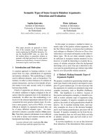
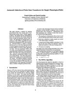
tality in group II. The 1-, 3- and 5-year cumulative sur-
vival rates of group I were 70.3%, 25.9%, and 7.4%,
respectivel y; these were significantly lower than those of
group II (90.6%, 54.0%, and 37.7%) (P<0.001) (Figure 1).
During the median follow-up period of 39 months post-
resection (range 1 to 104 months), 25 of patients (92.5%)
in group I and 218 patients (80.7%) in group II experi-
enced intrahepatic recurrence. The rates of early recur-
rence (≤1 year) after resection were significantly higher in
group I than group II (70.3% vs. 34.8%) (P<0.001).
Seven patients in group I recur renced with BTT again
and had obstructive jaundice. One patient received re-
resection, six were palliated with endoscopic stents.
Discussion
In 1949, Mallory et al. [15] described a single case of
HCC invading the gall bladder and obstructing extra-
hepatic bile d ucts. In 1975 Lin et al. [16] described
eight patients and classified icteric type hepatoma.
Since then, there are several reports concernin g HCC
with BDT have been published [ 1-7]. The outcome in
patients who received palliative treatment was poor,
withameansurvivaltimeoflessthan5months
[17-19]. Surgical treatment is the only way that possi-
bly cures the patients [19,20]. However, the resectabil-
ityrateofsuchtumorswasverylowinearlyreports,
ranged only from 2% to 13.2% [21]. In recent years,
with progress in imaging diagno sis and preoperativ e
management, the number of resectable cases were
increased remarkably [4-7,21].
HCC invades into biliary system through one of the
following three mechanisms: (1) the tumor may grow
continuously in a distal fashion, filling the entire extra-
hepatic biliary system with a solid cast of tumor; (2) a
fragment of necrotic tumor may separate from the prox-
imal intraductal growth, migrating to the distal common
bile duct and c ausing obstruction; or (3) hemorrhage
from the tumor may partially or completely fill the bili-
ary tree with blood clots [18]. Generally, the tumor
thrombus was not adherent to the bile duct wall so it
could be removed easily. Tumor thrombi rarely invade
the walls of the large bile ducts around the hepatic
hilus. Therefore, liver resection of the involved hepatic
segments with thrombectomy through a choledochot-
omy is a rational technique for curative resection [2].
The indications fo r extrahepatic bile duct resection were
macroscopic tumor invasion of the large bile ducts
around the hepatic hilus [2].
Venous inv asion is a well-established prognostic indi-
cator of HCC [22,23]. By contrast, whether BTT have a
significant impact on the prognosis of HCC remain con-
troversial. Shiomi et al. [2] reported that the 3- and
5-year survival rates were 47% and 28%, respectively, in
17 patients with BTT, s imilar to those achieved in 115
patients without BTT. Satoh et al. [5] also found that
there were no significant differences in survival between
patients with BTT and those without BTT. These data
suggest that BTT in HCC patients might have lower
aggre ssive potential and be less important as a prognos-
tic factor. Paradoxically, Yeh et al. [6] in an analysis of
17 patients who underwent resection, reported that the
overall survival was worse in patients with BTT, com-
pared with those witho ut BTT. In another recent study,
Ikenaga et al. [7] reported that the median survival time
of HCC patients with BTT after surgery was signifi-
cantly shorter than that of those without thrombi (11.4
vs. 56.1 months, P = 0.002).
Figure 1 Overall survival of patients in two groups. The overall
survival of group I was lower than group II (P < .001).
Shao et al. World Journal of Surgical Oncology 2011, 9:2
/>Page 3 of 5
Currently, there are two surgical staging systems,
which we re developed based on the analysis of patients
who received hepatic resection: one from the Liver Can-
cer Study Group of Japan (LCSGJ) and another from
the American Joint Committe e on Cancer (AJCC)/Inter-
national Union A gainst Cancer (UICC). In LCSGJ sys-
tem, presence of BTT was considered as an indicator of
“advanced stage” of HCC. A study from Japan reported
that the prognostic stratification ability of the LCSGJ
staging system is superior to that of the AJCC/UICC
staging s ystem and that the BTT is the strongest prog-
nostic factor for HCC [24]. This idea is supported by
the results in our present study showing that the resec-
tion of HCC with BTT results in a significantly worse
survival outcome compared with hepatic resection of
HCC without BTT.
In current study, the incide nce of microsc opic vascu-
lar invasion [13,23], tumor capsule absence [25,26],
high Edmondson- Steiner grade [22,27,28], and high
level of GGT [29], unfavorable clinical prognostic fac-
tors, in group I were significantly higher than the
respective factors in group II. Therefore, these factors
may account for the poor prognosis in patients with
BTT, at least in part. In addition, the incidences of
CA19-9 > 37 U/ml, elevation of total bilirubin, ALT,
AST, and ALP in group I were significantly higher
than those in group II, which may be related to
obstructive jaundice caused by BTT.
Previous studies have indicated that the HCC p atients
with BTT had smaller tumors and a higher percentage
of a tumor size ≤ 5 cm t han those without BTT [6].
Similarly, in this study, the incidence of tumor size ≤
5 cm tended to be higher in group I, despite this result
did not reach statistical significance due to the small
number of patients. Although the exact mechanism
remains unknown, some authors have suggested that a
BTTinHCCtendstogrowfasterthantheprimary
tumor itself [4].
Tumor recurrence remains the major cause of death
after resection for HCC [30,31]. Despite similar tre at-
ments, the prognosis for patients with early recurrence
was worse than that of patients with late recurrence
[14]. Qin et al. [4] reported that 14 patients with BTT
(14/28, 50.0%) were found intrah epatic HCC recurrence
with in 1 year after operation. Ikenaga et al. [7] reported
that 53% of patients suffered recurrences in the remnant
liver within 3 months after surgery. In current study,
patients wi th BTT developed early recurr ence after
resection more frequently.
We found seven patients in group I recurrenced with
BTT again. Recurrence in the liver remnant could results
from either intrahepatic metastasis from the primary
tumor or multicentric occurrence. Intrahepatic metasta-
sis is an important mechanism of early intrahepatic
recurrence after resection of HCC. Spreading via the por-
tal vein is considered the main route of intrahepatic
metastasis [14,32]. In current study, 17 patients (62.9%)
in the group I had microscopic vascular invasion. How-
ever, nine of the other ten cases without venous invasion
also developed early recurrence. Similar results had been
published previ ously by Ikenaga et al. [7], who found that
two patients with BTT without portal vein invasion suf-
fered multiple recurrences in the remnant liver. The
authors s peculate that HCC invasion biliary system may
be another route of intrahepatic metastasis.
Esaki et al. [8] reported that HCC patients with
macroscopic BTT had a bett er postoperative survival
than patients with microscopic BTT. But Ikenaga et al.
[7] found that the prognosis was similar between two
groups. In current study, statistical analysis was limited
by the too small population with microscopic BTT.
The experience wit h orthotopic liver transplantation
(OLT) in patients with BTT is limited. There were only
five cases reported in the English literatures [21,33]. Of
these patients, one died 2 0 months after OLT due to
multiple intrahepatic recurrences, one developed carci-
noma recurrence at the low er end of the common bile
duct 27 months after OLT. The other three patients
were alive without evidence of recurrence during the
follow-up period (17.6 to 28.1 months). These results
suggest that OLT may be a treatment option for HCC
with BDT in selected cases.
Conclusions
HCC patients with BTT had a worse prognosis after
resection than those without BTT. Resection should be
considered for these tumors given the lack of ef fective
alternative therapies. Further studies are needed for
understanding of molecular biology of BTT may yield
therapeutic tools that can improve the prognosis of this
subset of patients.
Consent
Written informed consent was obtained from the
patients for publication of this case series. A copy of the
written consent is available for review by th e Editor-in-
Chief of this journal.
Author details
1
Department of Liver Surgery, The First Affiliated Hospital of Nanjing Medical
University, Nanjing, PR China.
2
Department of Hepato-Biliary-Pancreato-
Vascular Surgery, the First Affiliated Hospital of Xiamen University, Xiamen,
PR China.
3
Department of Special Treatment and Liver transplantation,
Eastern Hepatobiliary Surgery Hospital, Second Military Medical University,
Shanghai, PR China.
Authors’ contributions
WS participated in the design and coordination of the study, carried out the
critical appraisal of studies and wrote the manuscript. CS, ZL, and YZ
developed the literature search, carried out the extraction of data, assisted in
Shao et al. World Journal of Surgical Oncology 2011, 9:2
/>Page 4 of 5
the critical appraisal of include d studies and assisted in writing up. WS and
CS carried out the statistical analysis of studies. JY and YZ interpreted data,
corrected and approve the manuscript. All authors read and approved the
final manuscript.
Competing interests
The authors declare that they have no competing interests.
Received: 4 September 2010 Accepted: 8 January 2011
Published: 8 January 2011
References
1. Kojiro M, Kawabata K, Kawano Y, Shirai F, Takemoto N, Nakashima T:
Hepatocellular carcinoma presenting as intrabile duct tumor growth. A
clinicopathological study of 24 cases. Cancer 1982, 49:2144-2147.
2. Shiomi M, Kamiya J, Nagino M, Uesaka K, Sano T, Hayakawa N, Kanai M,
Yamamoto H, Nimura Y: Hepatocellular carcinoma with biliary tumor
thrombi: aggressive operative approach after appropriate preoperative
management. Surgery 2001, 129:692-698.
3. Huang JF, Wang LY, Lin ZY, Chen SC, Hsieh MY, Chuang WL: Incidence and
clinical outcome of icteric type hepatocellular carcinoma. J Gastroenterol
Hepatol 2002, 17:190-195.
4. Qin LX, Ma ZC, Wu ZQ, Fan J, Zhou XD, Sun HC, Ye QH, Wang L, Tang ZY:
Diagnosis and surgical treatments of hepatocellular carcinoma with
tumor thrombosis in bile duct: experience of 34 patients. World J
Gastroenterol 2004, 10:1397-1401.
5. Satoh S, Ikai I, Honda G, Okabe H, Takeyama O, Yamamoto Y, Yamamoto N,
Iimuro Y, Shimahara Y, Yamaoka Y: Clinicopathologic evaluation of
hepatocellular carcinoma with bile duct thrombi. Surgery 2000,
128:779-783.
6. Yeh CN, Jan YY, Lee WC, Chen MF: Hepatic resection for hepatocellular
carcinoma with obstructive jaundice due to biliary tumor thrombi. World
J Surg 2004, 28:471-475.
7. Ikenaga N, Chijiiwa K, Otani K, Ohuchida J, Uchiyama S, Kondo K:
Clinicopathologic characteristics of hepatocellular carcinoma with bile
duct invasion. J Gastrointest Surg 2009, 13:492-497.
8. Esaki M, Shimada K, Sano T, Sakamoto Y, Kosuge T, Ojima H: Surgical
results for hepatocellular carcinoma with bile duct invasion: a
clinicopathologic comparison between macroscopic and microscopic
tumor thrombus. J Surg Oncol 2005, 90:226-232.
9. Bruix J, Sherman M, Llovet JM, Beaugrand M, Lencioni R, Burroughs AK,
Christensen E, Pagliaro L, Colombo M, Rodés J, EASL Panel of Experts on
HCC: Clinical management of hepatocellular carcinoma. Conclusions of
the Barcelona-2000 EASL conference. European Association for the
Study of the Liver. J Hepatol 2001, 35:421-430.
10. Liau KH, Ruo L, Shia J, Padela A, Gonen M, Jarnagin WR, Fong Y,
D’Angelica MI, Blumgart LH, DeMatteo RP: Outcome of partial
hepatectomy for large (> 10 cm) hepatocellular carcinoma. Cancer 2005,
104:1948-1955.
11. Edmondson HA, Steiner PE: Primary carcinoma of the liver. A study of
100 cases among 48,900 necropsies. Cancer 1954, 7:462-503.
12. Pawlik TM, Poon RT, Abdalla EK, Zorzi D, Ikai I, Curley SA, Nagorney DM,
Belghiti J, Ng IO, Yamaoka Y, Lauwers GY, Vauthey JN, International
Cooperative Study Group on Hepatocellular Carcinoma: Critical appraisal of
the clinical and pathologic predictors of survival after resection of large
hepatocellular carcinoma. Arch Surg 2005, 140:450-457.
13. Tsai TJ, Chau GY, Lui WY, Tsay SH, King KL, Loong CC, Hsia CY, Wu CW:
Clinical significance of microscopic tumor venous invasion in patients
with resectable hepatocellular carcinoma. Surgery 2000, 127:603-608.
14. Poon RT, Fan ST, Ng IO, Lo CM, Liu CL, Wong J:
Different risk factors and
prognosis for early and late intrahepatic recurrence after resection of
hepatocellular carcinoma. Cancer 2000, 89:500-507.
15. Mallory TB, Castleman B, Parris EE: Case records of the Massachusetts
General Hospital. N Eng Med 1947, 237:673-676.
16. Lin TY, Chen KM, Chen YR, Lin WS, Wang TH, Sung JL: Icteric type
hepatoma. Med Chi Dig 1975, 4:267-270.
17. Lai ST, Lam KT, Lee KC: Biliary tract invasion and obstruction by
hepatocellular carcinoma: report of five cases. Postgrad Med J 1992,
68:961-963.
18. Chen MF, Jan YY, Jeng LB, Hwang TL, Wang CS, Chen SC: Obstructive
jaundice secondary to ruptured hepatocellular carcinoma into the
common bile duct. Surgical experiences of 20 cases. Cancer 1994,
73:1335-1340.
19. Lau WY, Leung KL, Leung TW, Ho S, Chan M, Liew CK, Leung N, Johnson P,
Li AK: Obstructive jaundice secondary to hepatocellular carcinoma. Surg
Oncol 1995, 4:303-308.
20. Tantawi B, Cherqui D, Tran van Nhieu J, Kracht M, Fagniez PL: Surgery for
biliary obstruction by tumour thrombus in primary liver cancer. Br J Surg
1996, 83:1522-1525.
21. Peng SY, Wang JW, Liu YB, Cai XJ, Deng GL, Xu B, Li HJ: Surgical
intervention for obstructive jaundice due to biliary tumor thrombus in
hepatocellular carcinoma. World J Surg 2004, 28:43-46.
22. Haratake J, Takeda S, Kasai T, Nakano S, Tokui N: Predictable factors for
estimating prognosis of patients after resection of hepatocellular
carcinoma. Cancer 1993, 72:1178-1183.
23. Pandey D, Lee KH, Wai CT, Wagholikar G, Tan KC: Long term outcome and
prognostic factors for large hepatocellular carcinoma (10 cm or more)
after surgical resection. Ann Surg Oncol 2007, 14:2817-2823.
24. Minagawa M, Ikai I, Matsuyama Y, Yamaoka Y, Makuuchi M: Staging of
hepatocellular carcinoma: assessment of the Japanese TNM and AJCC/
UICC TNM systems in a cohort of 13,772 patients in Japan. Ann Surg
2007, 245:909-992.
25. Franco D, Capussotti L, Smadja C, Bouzari H, Meakins J, Kemeny F,
Grange D, Dellepiane M: Resection of hepatocellular carcinomas. Results
in 72 European patients with cirrhosis. Gastroenterology 1990, 98:733-738.
26. Lai EC, Ng IO, Ng MM, Lok AS, Tam PC, Fan ST, Choi TK, Wong J: Long-
term results of resection for large hepatocellular carcinoma: a
multivariate analysis of clinicopathological features. Hepatology 1990,
11:815-818.
27. Ng IO, Lai EC, Fan ST, Ng MM, So MK: Prognostic significance of
pathologic features of hepatocellular carcinoma. A multivariate analysis
of 278 patients. Cancer 1995, 76
:2443-2448.
28. Ikeda K, Saitoh S, Tsubota A, Arase Y, Chayama K, Kumada H, Watanabe G,
Tsurumaru M: Risk factors for tumor recurrence and prognosis after
curative resection of hepatocellular carcinoma. Cancer 1993, 71:19-25.
29. Zhou XD, Tang ZY, Yu YQ, Yang BH, Lu JZ, Lin ZY, Ma ZC, Zhang BH:
Characteristics and prognosis of primary liver cancer in young patients
in China. J Gastroenterol 1995, 30:632-635.
30. Cha C, Fong Y, Jarnagin WR, Blumgart LH, DeMatteo RP: Predictors and
patterns of recurrence after resection of hepatocellular carcinoma. JAm
Coll Surg 2003, 197:753-758.
31. Zhou Y, Sui C, Li B, Yin Z, Tan Y, Yang J, Liu Z: Repeat hepatectomy for
recurrent hepatocellular carcinoma: a local experience and a systematic
review. World J Surg Oncol 2010, 8:55.
32. Zhou YM, Yang JM, Li B, Yin ZF, Xu F, Wang B, Xu W, Kan T: Risk factors for
early recurrence of small hepatocellular carcinoma after curative
resection. Hepatobiliary Pancreat Dis Int 2010, 9:33-37.
33. Lee KW, Park JW, Park JB, Kim SJ, Choi SH, Heo JS, Kwon CH, Kim DJ,
Han YS, Lee SK, Joh JW: Liver transplantation for hepatocellular
carcinoma with bile duct thrombi. Transplant Proc 2006, 38:2093-2094.
doi:10.1186/1477-7819-9-2
Cite this article as: Shao et al.: Surgical outcome of hepatocellular
carcinoma patients with biliary tumor thrombi. World Journal of Surgical
Oncology 2011 9:2.
Submit your next manuscript to BioMed Central
and take full advantage of:
• Convenient online submission
• Thorough peer review
• No space constraints or color figure charges
• Immediate publication on acceptance
• Inclusion in PubMed, CAS, Scopus and Google Scholar
• Research which is freely available for redistribution
Submit your manuscript at
www.biomedcentral.com/submit
Shao et al. World Journal of Surgical Oncology 2011, 9:2
/>Page 5 of 5