báo cáo khoa học: "Clinical significance of preoperative serum interleukin-6 and C-reactive protein level in breast cancer patients" pot
Bạn đang xem bản rút gọn của tài liệu. Xem và tải ngay bản đầy đủ của tài liệu tại đây (407.72 KB, 6 trang )
RESEARCH Open Access
Clinical significance of preoperative serum
interleukin-6 and C-reactive protein level
in breast cancer patients
Praveen Ravishankaran
1*
, R Karunanithi
2
Abstract
Background: Breast cancer is a disease that continues to plague females during their entire lifetime. IL-6 and CRP
are found to be elevated in various inflammatory and malignant diseases and their levels are found to correlate
with the extent of the disease. The primary objective of this study was to determine the preoperative serum levels
of IL-6 and CRP in breast carcinoma, and to correlate them with the staging of the disease and the prognosis.
Methods: 59 female patients admitted for breast cancer were identified for the study and were subjected to
thorough evaluation. Serum levels of IL-6 were assessed via Enzyme-Linked Immuno-Sorbent Assay (ELISA), and
CRP was measured via immunoturbidimetry. Histological findings included tumour size, lymph node (LN)
metastasis, and tumour staging. Relevant investigations were made to find out the presence of distant metastasis.
Statistical analysis of the data was then processed.
Results: Increases in cancer invasion and staging are generally associated with increases in preoperative serum IL-6
levels. IL-6 and CRP levels correlated with LN metastasis (P < 0.001, P < 0.001) and TNM stage (P < 0.001, P <
0.001). Tumour invasion and the presence of distant metastasis is associated with higher IL-6 levels (P = 0.001, P =
0.009). When we established the cutoff value for IL-6 level (20.55 pg/dl) by ROC curve, we noted a significant
difference in overall survival (OS; P = 0.008). However, CRP evidenced no significance with regard to patient’sOS
levels. Serum IL-6 levels were correlated positively with CRP levels (r2 = 0.579, P < 0.01)
Conclusion: Serum levels of IL-6 correlates well with the extent of tumor invasion, LN metastasis, distant
metastasis and TNM staging thus enveloping all aspects of breast cancer.
Introduction
Breast cancer is a disease affecting millions of women as
well as men all over the w orld. The TNM system of
classification is used for staging of the disease which has
a strong infl uence on the progno sis of the patient. Wide
array of cytokines are secreted by t he breast tumours of
which IL-6 is one of them. IL-6 is a pleiotrophic cyto-
kine with a wide range functions. I L-6 binds to the IL-6
receptor, activates the Janus kinase (JAK), and subse-
quently phosphorylates the signal transducers and acti-
vators of transcription (STAT). The phosphorylated
STAT gene translocates into the nucleus and activates
the target genes like VEGF and rho which increases the
aggressiveness of the tumour. This involvement of IL-6
at a cellular level with the processes of cancer contr ol is
reflected by the results of serum studies of cancer
patients, where IL-6 may reflect prognosis and tumour
load. Elevated IL-6 levels have been associated with
advanced stage and metastasis-related morbidity [1-3]. It
has been recently repor ted that patients with metastatic
ovarian cancer and patients with metastatic renal cell
carcinoma have higher serum IL-6 levels than those
without disseminated disease [4,5]. It has also been
demonstrated that elevated IL-6 levels are associated
with a poor prognosis in tumours such as non-small-cell
lung cancer. The ontological role of IL-6 in this process
is not known [6].
C-reactive protein (CRP) is a representative marker for
inflammatory conditions, and performs a crucial anti-
infectionfunctionintheimmunesystem.Inmany
* Correspondence:
1
Department of General Surgery, Coimbatore Medical College Hospital,
Coimbatore, Tamil Nadu, India
Full list of author information is available at the end of the article
Ravishankaran and Karunanithi World Journal of Surgical Oncology 2011, 9:18
/>WORLD JOURNAL OF
SURGICAL ONCOLOGY
© 2011 Ravishankaran and Karunanithi; licensee BioMed Central Ltd. This is an Open Access article distributed under the terms of the
Creative Commons Attributio n License ( which permits unrestricted use, distribution, and
reproduction in any medium, provided the original work is properly cited.
cancers, it has been reported that chronic inflammation
is involved with malignant ch ange, and the risks of can-
cer are increased when pre-diagnostic CRP levels are
high [7]. Cancer invasion begins with inflammation
around cancer cells. Thus, it has been reported that
serum CRP levels are higher in cases of invasive cancer
than in cases of non-invasive cancer [8,9].
The principal objective of this study was to determine
the relationship between serum IL-6 and CRP levels and
staging and prognosis in breast cancer patients.
Materials and methods
Patients
Fifty nine cases of breast cancer admitted in our hospital
were selected for the study. Basic blood investigations,
chest x-ray, ECG and CT scan were done for all the
patients a nd the diagnosis was confirmed. Core n eedle
biopsy was done and the hormone recepto r status
assessed. Blood samples were drawn for IL-6 and CRP
levels on admission.
The patients were then assessed according to the
pathological TMN staging
1. Primary tu mour (T1 = ≤2cm,T2=2-5cm,T3=
>5 cm, T4 = chest wall or skin infiltration
2. Nodal staging (N1 = 1-3 nodes, N2 = 4-9 nodes, N3
= > 10 node)
3. Presence (M0) or absence (M1) of metastasis.
The patients were then subjected to surgery with or
without neo-adjuvant chemotherapy. Early invasive
breast cancer (Sta ge I, IIa and IIb) was treated wi th
mastectomy and axillary lymph node clearance followed
by adjuvant chemotherapy for all node positive breast
cancer, all cance rs that are larger than 1 cm, tumours
with high histological grade and neg ative hormone
receptor status. Advanced locoregional breast cancer
(Stage IIIa or IIIb) was t reated with modified radical
mastectomy followed by adjuva nt chemotherapy and
radiotherapy if operable and if inoperable neoadjuvant
chemotherapy was used to decrease the locoregional
cancer burden and permit subsequent surgery. For cases
with stage IV breast cancer hormone therapy was done
for hormone receptor positive tumours and chemother-
apy was given for receptor negative cancers.
Patients were asked t o come for foll ow up once every
three months for a duration of two years. Serum levels
of IL-6 and CRP were estimated every three months. All
patients provided informed consent, and the hospital
review board approved the study.
Assays for serum il-6 and crp
The blood sample for IL-6 collected using standard sam-
pling tubes were transported to the lab within 5 hrs at 20-
25°C. The samples for IL-6 were analysed using Elecsys
2010 cobas e 411 analyser by Electrochemiluminescence
immunoassay. The sandwich principle was used and the
total duration of assay took 18 minutes. The measuring
range of IL-6 is 1.5-5000 pg/ml(defined by the lower
detection limit and the maximum of the master curve).
The normal value for IL-6 in a healthy individual is
expected to be <7 pg/ml. The samples for CRP were mea-
sured immunoturbimetrically using RANDOX analyser.
Serum is used undiluted and CRP remains stable in the
serum for at least 3 days at 15 -25°C. The measuring range
of CRP is 0-220 mg/l, the normal value of CRP being <5
mg/l.
Descriptive statistical analysis
Serum levels of IL-6 and CRP were expressed as the
means ± SD. A p value of < 0.05 was considered to be
statistically significant and was calculated by one way
ANOVA. The Spearman rho correlation coefficient (r)
was employed to evaluate the correlation between the
IL-6 and CRP levels and the clinical findings. The IL-6
and CRP cut-off values for survival analysis were deter-
mined by the ROC curve. Survival durations were calcu-
lated via the Kaplan-Meier method. Statistical Pack age
for Social Sciences ( SPSS) ver. 17 software was used for
the statistical analysis.
Results
Patient characteristics
The patients were classified by their pathologic charac-
teristics, including tumor size, status of lymph node
metastasis, presence or absen ce of metastasis and TNM
staging. The patients consisted of 59 women, with a
median age of 59.11 year s (range, 36-8 5 years). In all 5
patients had stage 2A disease(8.4%), 8 patients belong to
stage 2B(13.6%) 15 patients belong to stage 3A
(25.42%),13 belongs to stage 3B(22.03%) 10 patients to
stage 3C(16.94%) and 8 to stage 4(13.56%). The other
patient characteristics are summarized in Table 1.
The relationships between IL-6, CRP levels, and clin-
ico-pathologic variables are provided by the Spearman
rho correlation coefficient (r) in Table 2.
Clinicopathological significance of IL-6 in breast cancer
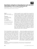
We noted that IL-6 levels were s ignificantly correlated
withthetumoursizewithhigherIL-6levelswas
detected in tumours sized ≥ 5cm(P = 0.001, r = 0.564).
Additionally, with increasing degrees of tumour inva-
sion, the median levels of IL-6 evidenced a tendency to
increase, and this difference in IL-6 levels was found to
be statistically significant (P < 0.001). In cases of LN
metastasis, we also noted a significant difference
between t he serum level of IL-6 and LN metastasis (P <
0.001, r = 0.844). The median level of IL-6 increased
proportionally with the stage of the cancer (the median
level of IL-6 in stage 2a 5.6 ± 1.5 pg/ml, stage 2b 11.7 ±
Ravishankaran and Karunanithi World Journal of Surgical Oncology 2011, 9:18
/>Page 2 of 6
4.4 pg/ml, stage 3a 16.9 ± 4.7 pg/ml, stage 3b 19.1 ± 4.8
pg/ml, stage 3c 26.3 ±7.0 pg/ml and stage 4 39.8 ±9.4
pg/ml), and thi s difference was statistically significant (P
< 0.001). Additionally, serum IL-6 levels were signifi-
cantly higher in patients with distant metastasis (39.7
±9.3 pg/ml) than i n those without dista nt metastasis
(17.3 ± 7.6 pg/ml) whose difference is also stat istically
significant(P < 0.001) (Figure 1).
(A) IL-6 levels according to tumor depth. (B) IL-6
levels according to LN metastasis. (C) IL-6 levels
acco rding to the metastasis. (D) IL-6 levels according to
TNM staging.
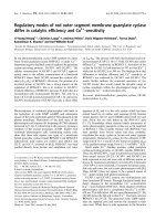
The patients were divided into two groups on the basis
of an IL-6 cutoff value of 20.3 pg/ml by the ROC curve
with a sensitivity of 88.6% and a specificity of 54.1%.
We noted significant differences in the Overall Survi-
val between the two groups (82.7% versus 97.2%; P =
0.008) See Figur e 2 for O verall survival curve according
to IL-6 (Interleukin-6) levels.
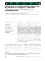
Clinicopathological significance of CRP
We noted that CRP levels did not diff er significantly
with tumour size (r = 0.374, P =0.304).Howeverwe
noted significant differences in serum CRP levels
between patients with lymph node metastasis and those
without lymph node metastasis (r = 0.690, P =0.000).
The median levels of CRP increased with increasing
stage, and we also noted significant differences between
Table 1 Patient characteristics
No. of patients %
Total number of patients 59
Age
Median(Range) 59.11(36-85)
Depth of tumor invasion
pT1 7 11.9
pT2 15 25.4
pT3 14 23.7
pT4 23 38.9
LN metastasis
N0 5 8.6
N1 17 28.8
N2 21 35.6
N3 16 27.1
Distant metastasis
Metastasis(-) 51 86.4
Metastasis(+) 8 13.6
Table 2 Correlation between the IL-6, CRP and
clinicopathological parameters
IL-6 CRP
Median ±
SD
rPMedian ±
SD
RP
Total pg/ml (mg/dl)
Tumor depth
pT1 8.1 ± 5.7 0.564 0.001 8.4 ± 3.1 0.374 0.304
pT2 17.8 ± 8.6 10.5 ± 2.7
pT3 19.2 ± 10.0 9.0 ± 2.4
pT4 26.2 ± 11.2 16.0 ± 3.3
LN meta
N1 11.6 ± 4.8 0.844 0.000 9.6 ± 4.1 0.690 0.000
N2 20.7 ± 6.9 15.7 ± 9.0
N3 32.1 ± 10.7 29.4 ± 15.6
Distant meta
Metastasis
(-)
17.3 ±7.6 0.773 0.009 13.9 ± 8.5 0.175 0.061
Metastasis
(+)
39.7 ± 9.3 37.4 ± 16.0
TNM stage
2A 5.6 ± 1.5 0.702 0.000 10.1 ± 3.9 0.463 0.000
2B 11.7 ± 4.4 9.2 ± 4.6
3A 16.9 ± 4.7 13.8 ± 7.2
3B 19.1 ± 4.8 12.8 ± 9.2
3C 26.3 ± 7.0 21.5 ± 9.9
4 39.8 ± 9.4 37.5 ± 16.0
Figure 1 IL-6 and the characteristics of breast tumour. (A) IL-6
levels according to tumor depth. (B) IL-6 levels according to LN
metastasis. (C) IL-6 levels according to the metastasis. (D) IL-6 levels
according to TNM staging.
Ravishankaran and Karunanithi World Journal of Surgical Oncology 2011, 9:18
/>Page 3 of 6
the CRP level and cancer stage (the median level of CRP
in stage 2a 10.1 ± 3.9 mg/dl, stage 2b 9.2 ± 4. 6 mg/dl,
stage 3a 13.8 ± 7.2 mg/dl, stage 3b 12.8 ± 9.2 mg/dl,
stage3c21.5±9.9mg/dlandstage437.5±16.0;P <
0.001). The CRP levels did not differ significantly in
patients with metastasis (37.4 ± 16.0 mg/dl) as com-
pared to those without metastasis (13.9 ± 8.5 mg/dl, P =
0.061). Figure 3 shows (A) CRP levels according to
tumor depth. (B) CRP levels according to LN metastasis.
(C) CRP levels ac cording to distant metastasis. ( D) CRP
levels according to TNM staging.
15.5 mg/dl was taken as the cutoff value of CRP by
ROC curve, after which the patients were divided into
two groups. The sen sitivity and specificity of 15.5 mg/dl
as the cutoff value were 62.1% and 75.3% on OS. We
noted no significant difference in the OS values (84.4%
vs 92.3%, P = 0.197) among the groups.
Association between IL-6 and CRP
Serum IL-6 levels also correlated positively with that of
CRP (r
2
= 0.579, p < 0.01) thus proving a positive asso-
ciation between the two variable (Figure 4).
Discussion
It has been long established that the pathologic variables
of tumour size, lymph node status, and histologic
tumour grade are significant prognostic indicators in
breast carcinoma [10-13]. More recently , biomarkers of
prognosis have been identified [14-16] and a radiological
predictor of survival has been discovered [17], but the
value of t umour size, lymph node status, and tumour
grade as powerful predictors of survival remains [18].
In this study, the serum levels of both IL-6 and CRP
evidenced statistically significant differences related to
the stage of LN metastasis. The serum levels of IL-6 evi-
denced statistically significant differences with relation
to changes in tumor size. As the stage of the disease
Figure 2 Overall survival curve according to IL-6 (Interleukin-6)
levels.
Figure 3 CRP and the characteristics of breast tumour. (A) CRP
levels according to tumor depth. (B) CRP levels according to LN
metastasis. (C) CRP levels according to distant metastasis. (D) CRP
levels according to TNM staging.
Figure 4 Correlation between IL-6 and CRP in breast cancer.
Ravishankaran and Karunanithi World Journal of Surgical Oncology 2011, 9:18
/>Page 4 of 6
increased, serum IL-6 and CRP levels increased propor-
tionately. Additionally, the median levels of IL-6 were
significa ntly higher in the patients with distant metasta-
sis than in those without distant metastasi s, but in CRP,
this was not proven. We also noted a significant associa-
tion between the levels of IL-6 and CRP (p < 0.01).
Thus the levels of IL-6 correlates with all the aspects
of breast cancer like tumour size lymph node involve-
ment, distant metastasis and the final T NM staging of
the disease. The overall survival of the patient also
seems to be affected in patients with el evated levels of
IL-6. The levels of CRP correlated only with lymph
node metastasis and not with tumour size and distant
metastasis. CRP also does not correlate with the overall
survival of the patient.
It has been proved that TNM staging correlates with
the prognosis of patients with breast cancer. As IL-6 has
a direct correlation with the stage of the disease it may
indirectly correlate with the prognosis of the patient
unlike that of CRP.
Interleukin-6 (IL-6) is a multi-poietic cytokine that
induces the growth and differentiation of immune cells,
the production and expression of other cytokines, and
acute-phase protein synthesis. IL-6 also exerts several
effects on cancer cells[19,20]. In the development and
progression of cancer, angiogenesis is a crucial and
essential proces s. IL-6 is associated with angiogenesis by
virtue of its ability to induce the mRNA of vascular
endothelial growth factor (VEGF), which is typically a
direct angiogen [19]. Additionally, IL-6 activates the Rho
protein, which is associated with cell-cell adhesion and
invasion in malignancy [21]. Toge ther these factors
increase t he aggressiveness of the tumour. It has been
indicated in this study that IL-6 level increases as the
aggressive behaviour of the tumour increases (IL-6 levels
increase as the stage of the cancer increases).
CRP is generated by the liver and other organs in
response to the release of IL-6 by monocytes and other
immune cells. Thus, when IL-6 levels increased, CRP
levels also increased. This has been proven by the posi-
tive association between IL-6 and CRP in this study.
Conclusion
Thus the levels of IL-6 has a positive correlation with
TNM staging system of breast cancer thus indirectly
correlating with the prognosis of the patient. CRP esti-
mation does not seem to be very useful in e valuating
the patient with breast cancer, though its level correlates
with that of IL-6.
Limitations of the study
1) A larger sample size needs to be evaluated to reach a
definite conclusion.
2) A longer follow up of t he patient is also essential
for completeness and is currently underway.
Acknowledgements
We wish to acknowledge the help rendered by Dr.Ravindra Bhat and Dr.
Rajasabapathy of Ganga hospital, Coimbatore in helping us bring out this
paper.
Conflict of interest
The authors declare that they have no competing interests.
Author details
1
Department of General Surgery, Coimbatore Medical College Hospital,
Coimbatore, Tamil Nadu, India.
2
Department of orthopaedics and spine
surgery, Ganga Hospital, Coimbatore, Tamil Nadu, India.
Authors’ contributions
PR conceived the study, collected the data and drafted the manuscript. KR
participated in the design of the study and performed the statistical analysis.
Both the authors read and approved the final manuscript.
Received: 20 November 2010 Accepted: 6 February 2011
Published: 6 February 2011
References
1. Adler HL, McCurdy MA, Kattan MW, Timme TL, Scardino PT, Thompson TC:
Elevated levels of circulating interleukin-6 and transforming growth
factor-beta1 in patients with metastatic prostatic carcinoma. J Urol 1999,
161:182-187.
2. Nakashima J, Tachibana M, Horiguchi Y, Oya M, Ohigashi T, Asakura H,
Murai M: Serum interleukin 6 as a prognostic factor in patients with
prostate cancer. Clin Cancer Res 2000, 6:2702-2706.
3. Wise GJ, Marella VK, Talluri G, Shirazian D: Cytokine variations in patients
with hormone treated prostate cancer. J Urol 2000, 164:722-725.
4. Blay JY, Negrier S, Combaret V, Attali S, Goillot E, Merrouche Y, Mercatello A,
Ravault A, Tourani JM, Moskovtchenko JF: Serum level of interleukin 6 as a
prognosis factor in metastatic renal cell carcinoma. Cancer Res 1992,
52:3317-3322.
5. Scambia G, Testa U, Benedetti Panici P, Foti E, Martucci R, Gadducci A,
Perillo A, Facchini V, Peschle C, Mancuso S: Prognostic significance of
interleukin 6 serum levels in patients with ovarian cancer. Br J Cancer
1995, 71:354-356.
6. De Vita F, Orditura M, Auriemma A, Infusino S, Roscigno A, Catalano G:
Serum levels of interleukin 6 as a prognostic factor in advanced non-
small cell lung cancer. Oncol Rep 1998, 5:649-652.
7. Erlinger TP, Platz EA, Rifai N, Helzlsouer KJ: C-reactive protein and the risk
of incident colorectal cancer. JAMA 2004, 291:585-590.
8. Nozoe T, Mori E, Takahashi I, Ezaki T: Preoperative elevation of serum C-
reactive protein as an independent prognostic indicator of colorectal
carcinoma. Surg Today 2008, 38:597-602.
9. Polterauer S, Grimm C, Tempfer C, Sliutz G, Speiser P, Reinthaller A,
Hefler LA: C-reactive protein is a prognostic parameter in patients with
cervical cancer. Gynecol Oncol 2007, 107:114-117.
10. Haybittle JL, Blamey RW, Elston CW, Johnson J, Doyle PJ, Campbell FC,
Nicholson RI, Griffiths K: A prognostic index in primary breast cancer. Br J
Cancer 1982, 45:361-366.
11. Duffy SW, Taba’r L, Fagerberg G, Gad A, South MC, Day NE: Breast
screening, prognostic factors and survival–results from the Swedish two
county study. Br J Cancer 1991, 64:1133-1138.
12. Bloom HJG, Richardson WW: Histological grading and prognosis in breast
cancer: a study of 1409 cases of which 539 have been followed up for
15 years. Br J Cancer 1957, 11:359-377.
13. Todd JH, Dowle C, Williams MR, Elston CW, Ellis IO, Hinton CP, Blamey RW,
Haybittle JL: Confirmation of a prognostic index in primary breast cancer.
Br J Cancer 1987, 56:489-492.
14. Lonn U, Lonn S, Nilsson B, Stenkvist B:
Breast cancer: prognostic
significance
of c-erb-B2 and int-2 amplification compared with DNA
ploidy, S-phase fraction, and conventional clinicopathological features.
Breast Cancer Res Treat 1994, 29:237-245.
Ravishankaran and Karunanithi World Journal of Surgical Oncology 2011, 9:18
/>Page 5 of 6
15. Hensel M, Schneeweiss A, Sinn HP, Egerer G, Solomayer E, Haas R, Bastert G,
Ho AD: P53 is the strongest predictor of survival in high-risk primary
breast cancer patients undergoing high-dose chemotherapy with
autologous blood stem cell support. Int J Cancer 2002, 100:290-296.
16. Malmstrom P, Bendahl PO, Boiesen N, Brünner N, Idvall I, Fernö M: S-phase
fraction and urokinase plasminogen activator are better markers for
distant recurrences than Nottingham Prognostic Index and histological
grade in a prospective study of premenopausal lymph node-negative
breast cancer. J Clin Oncol 2000, 19:2010-2019.
17. Taba’r L, Chen HH, Duffy SW, Yen MF, Chiang CF, Dean PB, Smith RA: A
novel method for prediction of long-term outcome of women with T1a,
T1b, and 10-14 mm invasive breast cancers: a prospective study. Lancet
2000, 355:429-433.
18. Taba’r L, Duffy SW, Vitak B, Chen HH, Prevost TC: The natural history of
breast carcinoma: what have we learned from screening? Cancer 1999,
86:449-462.
19. Cohen T, Nahari D, Cerem LW, Neufeld G, Levi BZ: Interleukin 6 induces
the expression of vascular endothelial growth factor. J Biol Chem 1996,
271:736-741.
20. Thong-Ngam D, Tangkijvanich P, Lerknimitr R, Mahachai V,
Theamboonlers A, Poovorawan Y: Diagnostic role of serum interleukin-18
in gastric cancer patients. World J Gastroenterol 2006, 12:4473-4477.
21. Lin MT, Lin BR, Chang CC, Chu CY, Su HJ, Chen ST, Jeng YM, Kuo ML: IL-6
induces AGS gastric cancer cell invasion via activation of the c-Src/
RhoA/ROCK signaling pathway. Int J Cancer 2007, 120:2600-2608.
doi:10.1186/1477-7819-9-18
Cite this article as: Ravishankaran and Karunanithi: Clinical significance of
preoperative serum interleukin-6 and C-reactive protein level in breast
cancer patients. World Journal of Surgical Oncology 2011 9:18.
Submit your next manuscript to BioMed Central
and take full advantage of:
• Convenient online submission
• Thorough peer review
• No space constraints or color figure charges
• Immediate publication on acceptance
• Inclusion in PubMed, CAS, Scopus and Google Scholar
• Research which is freely available for redistribution
Submit your manuscript at
www.biomedcentral.com/submit
Ravishankaran and Karunanithi World Journal of Surgical Oncology 2011, 9:18
/>Page 6 of 6